

NCBI Bookshelf. A service of the National Library of Medicine, National Institutes of Health.
Varki A, Cummings RD, Esko JD, et al., editors. Essentials of Glycobiology [Internet]. 3rd edition. Cold Spring Harbor (NY): Cold Spring Harbor Laboratory Press; 2015-2017. doi: 10.1101/glycobiology.3e.055


Essentials of Glycobiology [Internet]. 3rd edition.
Show details
The use of chemical tools to inhibit glycosylation provides a powerful approach for studying glycan functions and serves as a starting point for drug discovery. This chapter discusses various types of inhibitors, including natural products, substrate-based tight-binding inhibitors, glycoside primers, inhibitors found through screening chemical libraries, and examples of rationally designed inhibitors based on three-dimensional structures of enzymes.
ADVANTAGES OF INHIBITORS
Chapters 44, 45, and 49 describe various natural and induced mutants with defects in glycosylation. These mutants have helped to define genes that encode various transferases and glycosidases, and in some cases alternate biosynthetic pathways have been uncovered. Mutants also provide insights into the function of glycosylation in cells and tissues and models for human inborn errors in metabolism and disease. However, one limitation of studying mutants is that the analyses are usually restricted to the cell or organism from which the mutant strain was isolated. Additionally, many mutations are lethal in animals, which makes the study of the gene in adult animals more difficult.
Inhibitors of glycosyltransferases and glycosidases provide another approach for studying glycosylation in cells, tissues, and whole organisms that avoids some of the problems associated with studying mutants. Many of these compounds are small molecules that are taken up readily by a variety of cell types and some can be absorbed through the gut, providing an opportunity for designing drugs to treat human diseases and disorders correlated with altered glycosylation (Chapter 57). Because the field is quite large, only a selection of inhibitors that act on specific enzymes or metabolic pathways and that illustrate certain basic concepts are discussed here (Table 55.1). Agents that block protein/carbohydrate interactions are surveyed in Chapter 29, and the aminoglycoside antibiotics are discussed briefly in Chapter 57.
TABLE 55.1.
Classes of inhibitors
METABOLIC INHIBITORS
A number of inhibitors have been described that block glycosylation by interfering with the metabolism of common precursors or intracellular transport activities. Some of these compounds act indirectly by impeding the transit of proteins between the endoplasmic reticulum (ER), Golgi, and trans-Golgi network. For example, the fungal metabolite brefeldin A causes retrograde transport of Golgi components located proximal to the trans-Golgi network back to the ER. Thus, treating cells with brefeldin A separates enzymes located in the trans-Golgi network from those found in the ER and Golgi, and uncouples the assembly of the core structures of some glycans from later reactions, such as sialylation or sulfation. The drug can be used to examine if two pathways reside in the same compartment or share the same enzymes. Because the localization and array of the enzymes vary considerably in different cell types, extrapolating the effects of brefeldin A from one system to another is often difficult.
Some inhibitors act at key steps in intermediary metabolism in which precursors involved in glycosylation are formed. For example, a glutamine analog, 6-diazo-5-oxo-L-norleucine (DON), blocks glutamine: fructose-6-phosphate amidotransferase, the enzyme of the hexosamine biosynthetic pathway that forms glucosamine from fructose and glutamine (Chapter 5). Depressing glucosamine production in this way has a pleiotropic effect on glycan assembly because all of the major families contain N-acetylglucosamine or N-acetylgalactosamine. DON also affects other glutamine using enzymes and, therefore, care should be taken to limit nonspecific side effects. Chlorate is another type of general inhibitor that blocks sulfation. The chlorate anion (ClO4−) is an analog of sulfate (SO42−) and it forms an abortive complex with the sulfurylase involved in the formation of phosphoadenosine-5′-phosphosulfate (PAPS), the active sulfate donor for all known sulfation reactions. Thus, treating cells with chlorate (usually 10–30 mm) inhibits sulfation by >90%, but the effect is not specific for any particular class of glycan or sulfation reaction (e.g., tyrosine sulfation is also affected).
A number of sugar analogs have been made with the hope that they might show selective inhibition of glycosylation. 2-Deoxyglucose and fluorinated analogs of sugars (3-deoxy-3-fluoroglucosamine, 4-deoxy-4-fluoroglucosamine, 6-deoxy-6-fluoro-N-acetylglucosamine, 2-deoxy-2-fluoroglucose, 2-deoxy-2-fluoromannose, 2-deoxy-2-fluorofucose, and 3-fluorosialic acid) inhibit glycoprotein biosynthesis, but the mechanism underlying the inhibitory effect is unclear in many cases. Early studies of 2-deoxyglucose showed that the analog was converted to UDP-2-deoxyglucose as well as to GDP-2-deoxyglucose and dolichol-P-2-deoxyglucose. Inhibition of glycoprotein formation apparently occurs as a result of accumulation of various dolichol oligosaccharides containing 2-deoxyglucose, which cannot be elongated or transferred to glycoproteins normally. Similarly, 4-deoxy-N-acetylglucosamine is converted to UDP-4-deoxy-N-acetylglucosamine, resulting in inhibition of heparan sulfate formation without incorporation of the analog. 4-Deoxy-xylose also inhibits glycosaminoglycan assembly presumably by competing with naturally occurring xylosylated substrates. Care must be taken in interpreting the results of experiments using these compounds because they may have pleiotropic effects on glycan assembly caused by overlap of nucleotide precursors.
TUNICAMYCIN: INHIBITION OF DOLICHOL-PP-GlcNAc ASSEMBLY
A number of natural products have been found to alter glycosylation. Tunicamycin belongs to a class of nucleoside antibiotics composed of uridine, an 11-carbon disaccharide called aminodeoxydialdose (tunicamine), and a fatty acid of variable length (13 to 17 carbons), branching, and unsaturation (Figure 55.1). Tunicamycin was first identified in Streptomyces lysosuperificus, and related compounds were found later in other microorganisms. It derives its name from its antiviral activity, which occurs by inhibiting viral coat (or “tunica”) formation.
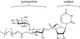
FIGURE 55.1.
Structure of tunicamycin, which consists of uridine conjugated to the disaccharide, tunicamine.
Tunicamycin inhibits N-glycosylation in eukaryotes by blocking the transfer of N-acetylglucosamine-1-phosphate (GlcNAc-1-P) from UDP-GlcNAc to dolichol-P (catalyzed by GlcNAc phosphotransferase; GPT), thereby decreasing the formation of dolichol-PP-GlcNAc (Chapter 9). Other GlcNAc transferase reactions are not inhibited (e.g., GlcNAcTI–V), but the transfer of GlcNAc-1-P to undecaprenyl-P and the formation of undecaprenyl-PP-MurNAc pentapeptide (which is involved in bacterial peptidoglycan biosynthesis) are sensitive to tunicamycin (Chapter 21). Tunicamycin acts as a tight-binding competitive inhibitor, presumably because it resembles the donor nucleotide sugar. The Ki value for tunicamycin is ∼5 × 10⁻⁸ m, whereas the Km value for UDP-GlcNAc is ∼3 × 10⁻⁶ m. The actual amount of tunicamycin needed to inhibit glycosylation varies in different cells (0.1–10 µg/mL), possibly because of variable uptake and culture conditions or differences in the level of expression of the phosphotransferase. Given the key role of N-glycosylation in protein folding and quality control in the ER (Chapter 39), it is not suprising that tunicamycin is cytotoxic to cells, and that resistant mutants overproduce GPT. Similarly, transfection of cells with the cloned GPT confers resistance, suggesting that the variable dose of inhibitor required in different cells may reflect variation in enzyme levels.
Tunicamycin has been used extensively for studying the role of N-glycans in glycoprotein maturation, secretion, and function, since its first discovery in 1973. The drug induces apoptosis preferentially in cancer cells, presumably because of alterations in glycosylation of various cell-surface receptors and signaling molecules and by inducing ER stress (Chapter 39). Thus, inhibition of N-glycan formation could be useful for treating cancer patients. Other potential applications include substrate reduction therapy for treatment of lysosomal storage disorders (Chapter 44), congenital disorders of glycosylation (Chapter 45), or naturally occurring mutations that create N-glycosylation sites in cell-surface receptors (gain-of-glycosylation mutants; Chapter 45).
Amphomycin, a lipopeptide, inhibits dolichol-P-mannose synthesis by apparently forming complexes with the carrier lipid dolichol-P. Other lipophilic compounds that bind lipid intermediates in bacterial cell wall synthesis also have also been studied (Chapter 21).
PLANT ALKALOIDS: NATURAL INHIBITORS OF GLYCOSIDASES
Plant alkaloids block N-linked glycosylation by inhibiting the processing glycosidases (α-glucosidases and α-mannosidases) involved in trimming nascent chains (Table 55.2). Unlike tunicamycin, which blocks glycosylation of glycoproteins entirely, the alkaloids inhibit the trimming reactions that occur after the Glc3Man9GlcNAc2 oligosaccharide is attached to a glycoprotein (Chapter 9), resulting in the appearance of glycoproteins on the cell surface lacking the characteristic termini found on mature N-glycans (Chapter 14). α-Glucosidase inhibitors involved in the initial processing of N-glycans and in quality control of protein folding (Chapter 39) include castanospermine (from the seed of the Australian chestnut tree, Castanosperum australe), which inhibits α-glucosidases I and II, australine (also from C. australe), which preferentially inhibits α-glucosidase I, and deoxynojirimycin (from Streptomyces species), which preferentially inhibits α-glucosidase II (Table 55.2). Castanospermine and australine cause accumulation of fully glucosylated chains, whereas deoxynojirimycin results in chains containing one to two glucose residues. Unexpectedly, treating cells with these inhibitors revealed that some trimming of the mannose residues could occur independently of removal of the glucose residues (Chapter 9).
Table 55.2.
Examples of alkaloids that inhibit glycosidases involved in N-linked glycan biosynthesis
Swainsonine was first discovered in plants from the western United States (Astragalus species, also known as locoweed) and Australia (Swainsona canescens), and was later found in the fungus Rhizoctonia leguminocola that infects red clover. Consumption of these plants by animals causes a severe abnormality called locoism and accumulation of glycoproteins in the lymph nodes. Swainsonine inhibits α-mannosidase II, causing the accumulation of paucimannose oligosaccharides (Man4GlcNAc2 and Man5GlcNAc2) and hybrid-type chains at the expense of complex oligosaccharides. In addition, swainsonine also inhibits the lysosomal α-mannosidase. Mannostatin A works in a similar way, but differs significantly in structure from swainsonine (Table 55.2). Other mannosidase inhibitors include deoxymannojirimycin and kifunensin, which selectively inhibit α-mannosidase I. These agents cause the accumulation of Man7–9GlcNAc2 oligosaccharides on glycoproteins.
All of the above listed inhibitors have in common polyhydroxylated ring systems that mimic the orientation of hydroxyl groups in the natural substrates, but a strict correlation between stereochemistry and enzyme target (α-glucosidase vs. α-mannosidase) does not exist. The compounds contain nitrogen, usually in place of the ring oxygen. One idea is that the nitrogen in the protonated state may mimic the positive charge on the ring oxygen that arises from delocalization of charge from the tentative carbocation at C-1 generated during the hydrolysis reaction. Crystal structures for the α-mannosidase are available with bound inhibitors.
Alkylated and acylated analogs of the alkaloids have interesting and useful properties. N-Butylation of deoxynojirimycin actually converts the glucosidase inhibitor into an inhibitor of glycolipid biosynthesis. Alkylation of the amino group or acylation of the hydroxyl groups can improve the potency of the compound, presumably by facilitating uptake across the plasma and Golgi membranes. Some of these compounds have shown positive effects for treating diabetes, lysosomal storage diseases, cancer, and HIV infection, but also induces male sterility (see Chapter 57).
INHIBITION OF O-GalNAc INITIATION OF MUCIN-TYPE GLYCANS
Few inhibitors are available that block O-linked glycans compared with N-linked glycan biosynthesis. Mucin-type O-linked glycan biosynthesis is initiated by polypeptidyl N-acetylgalactosaminyltransferases (ppGalNAcTs), a large family of enzymes that use UDP-GalNAc as a common donor and various glycoprotein acceptors (Chapter 10). Screening a synthetic library of uridine analogs against members of the ppGalNAcT family yielded two compounds that disrupt O-GalNAc addition (Figure 55.2). These compounds have Ki values of ∼8 µm with respect to UDP-GalNAc. Like tunicamycin, these inhibitors suppress glycosylation without selectivity for different glycoprotein targets. These first-generation enzyme inhibitors work on O-linked glycans and raise the possibility that inhibitors of specific ppGalNAcT isoforms as well as other types of O-linked glycans, such as O-xylose (Chapter 17), O-glucose (Chapter 18), and O-GlcNAc (Chapter 19) may be developed eventually.
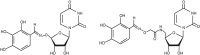
FIGURE 55.2.
Broad-spectrum inhibitors of the ppGalNAcTs identified from screening a uridine-based library.
INHIBITION OF O-GlcNAc MODIFICATION
The importance of O-GlcNAc addition to many cytoplasmic and nuclear proteins (Chapter 19) has stimulated great interest in developing agents to inhibit its addition by O-GlcNAc transferase (OGT) or its removal by O-GlcNAc-specific β-hexosaminidase (O-GlcNAcase). Alloxan and streptozotocin affect O-GlcNAc addition, but these compounds lack specificity. The first potentially useful OGT inhibitors were obtained by screening chemical libraries for compounds that displaced a fluorescent derivative of the donor sugar, UDP-GlcNAc. The active compounds do not block other N-acetylglucosamine addition reactions—for example, one involved in formation of the polysaccharide backbone of bacterial peptidoglycan (Chapter 21). In addition, 5-S-GlcNAc is another inhibitor of OGT (Figure 55.3). Its peracetylated form (Ac-5SGlcNAc), as well as a similar chemical inhibitor, peracetyl 4-thio-GlcNAc (Ac-4SGlcNAc), crosses cell membranes and undergoes deacetylation by nonspecific esterases to generate active inhibitors.
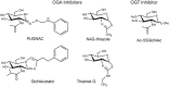
FIGURE 55.3.
Inhibitors of O-GlcNAc-specific β-hexosaminidase (OGA) and O-GlcNAc transferase (OGT).
Several O-GlcNAcase inhibitors are based on N-acetylglucosamine. The first compound in this class, PUGNAc (O-[2-acetamido-2-deoxy-D-glucopyranosylidene]amino-N-phenylcarbamate; Figure 55.3) inhibits O-GlcNAcase at nanomolar concentrations, but also inhibits lysosomal β-hexosaminidases (HexA and HexB; Chapter 44). Thiamet-G and the related N-acetylglucosamine-thiazoline (NAG-thiazoline) are more specific and inhibit at lower concentrations than PUGNAc. A rationally designed glucoimidazole, GlcNAcstatin, inhibits O-GlcNAcase with a Ki of 4.6 pm and shows 105-fold selectivity over HexA and HexB. These compounds inhibit the enzyme in cells and tissues, providing new tools to study the function of O-GlcNAc, and are potential candidates for drug therapy.
SUBSTRATE ANALOGS: DIRECTED SYNTHESIS OF INHIBITORS
A number of inhibitors of specific transferases have been developed based on the concept that substrate analogs might act as tight-binding inhibitors. The general strategy is to modify the hydroxyl group that acts as the nucleophile during formation of the glycosidic bond or groups in its immediate vicinity (Table 55.3). Many designer compounds lack inhibitory activity, because modification of the targeted hydroxyl group prevents binding of the analog to the enzyme by interfering with hydrogen bonding networks that position the substrate. In other cases, the analogs show Ki values in the approximate range of the Km values for the unmodified substrate. As one might expect, the analogs usually act competitively with respect to the unmodified substrate, but in a few cases the inhibition pattern is more complex, suggesting possible binding outside the active site.
TABLE 55.3.
Synthetic substrate-based inhibitors of glycosyltransferases
Nucleotide sugar analogs provide opportunities for blocking classes of enzymes that use a common donor (e.g., all fucosyltransferases use GDP-fucose). A large number of nucleotide sugar derivatives have been made (e.g., N- and O-substituted analogs of UDP-GalNAc) and several inhibit the enzymes in vitro, but have proven less useful in living cells because of poor uptake. “Bisubstrate” analogs consist of the nucleoside sugar donor or PAPS covalently linked to the acceptor substrate by way of a neutral bridging group. This arrangement may generate inhibitors whose binding characteristics reflect the product of the affinity constants for donor and acceptor (approximated by the product of the individual Km values). Bisubstrates that have been made have Ki values in the range of the Km values for the nucleotide donors, suggesting that the correct geometry for the bridging group may have not been attained or that the analog binds in ways that differ from the natural substrates.
The search for active compounds often benefits from serendipidy and the synthesis of di-, tri-, and tetrasaccharides with the desired modifications is rather labor intensive. Nevertheless, the approach has yielded insights into the binding and reactivity of the enzymes, and substrate analogs with selectivity for particular enzymes have been developed in this way. Because many of the transferases have now been purified and cloned, we can look forward to more detailed kinetic and crystallographic studies, which will provide clues for deriving mechanism-based inhibitors in the future (Chapter 6).
GLYCOSIDE PRIMERS: MIMICKING WHAT ALREADY WORKS
The utility of any glycosyltransferase inhibitor ultimately depends on its ability to cross the plasma membrane and enter the Golgi where the glycosyltransferases reside. Unfortunately, many of the compounds described above lack activity in live cells, presumably because their polarity and charge prevents their uptake. More than 40 years ago, Okayama and colleagues found that D-xylose in β-linkage to a hydrophobic aglycone (the noncarbohydrate portion of a glycoside; e.g., p-nitrophenol) was taken up rather efficiently and inhibited the assembly of glycosaminoglycans on proteoglycans. Xylosides mimic the natural substrate, xylosylated serine residues in proteoglycan core proteins, and thus act as a substrate. “Priming” of chains occurs on the added xyloside, which diverts the assembly process from the endogenous core proteins and causes inhibition of proteoglycan formation. In general, cells incubated with xylosides secrete large amounts of individual glycosaminoglycan chains and accumulate proteoglycans containing truncated chains. The enormous success of β-D-xylosides in altering proteoglycan biosynthesis suggested that other glycosides might act as “primers” as well (Table 55.4). Subsequent studies have shown that β-N-acetylgalactosaminides prime oligosaccharides found on mucins and inhibit O-glycosylation of glycoproteins. Other active glycosides include β-glucosides, β-galactosides, β-N-acetylglucosaminides, and even disaccharides and trisaccharides. These latter compounds require conjugation to appropriate aglycones and acetylation to neutralize the polar hydroxyl groups on the sugars. Cells contain several carboxyesterases that remove the acetyl groups and render the compounds available to the transferases in the Golgi.
TABLE 55.4.
Examples of glycoside primers
Priming by glycosides occurs in a concentration-dependent manner, but the efficiency varies widely among different compounds and cell types. These variations may relate to the relative abundance of endogenous substrates, enzyme concentration and composition, the solubility of different glycosides, their susceptibility to hydrolysis, their uptake across the plasma membrane and into the Golgi, and their relative affinity for the glycosyltransferases. The type of chain made on a given primer also depends on concentration and aglycone structure, which may reflect selective partitioning of primers into different intracellular compartments or into different branches of biosynthetic pathways. Like priming, inhibition of glycoprotein, glycolipid, or proteoglycan formation occurs in a dose-dependent fashion, but the blockade is rarely complete, probably because of the inability of glycosides to mimic the entire endogenous substrates.
Primers represent starting points for making analogs with tight-binding inhibitor properties described above. Many of the compounds described in Table 55.3 could be converted to permeable acylated glycosides and tested in live cells for inhibitory activity. Active compounds could potentially become carbohydrate-based drugs to treat glycosylation-dependent diseases. Oligosaccharide priming may have beneficial effects as well. Xylosides, for example, can be absorbed through the gut, and when consumed at sufficient concentration, they show antithrombotic activity. Many glycosides occur naturally, because various organisms (especially plants) produce hydrophobic compounds as part of chemical defense and conjugate them to sugars to render them soluble. Thus, the human diet may contain various types of glycosides with interesting (and unknown) biological activities.
Care must be taken in interpreting the results of experiments using glycoside primers. For example, β-D-xylosides also prime glycans related in structure to glycosphingolipids and HNK-1. In some cases priming per se is not the mechanism responsible for inhibition of glycosylation, but rather inhibition occurs because of competitive binding of the primer to a target enzyme. Finally, primers could deplete cells of nucleotide sugars and have multiple effects on glycosylation. For example, 4-methyl-umbelliferone is often used to block hyaluronan biosynthesis. The precise mechanism of action is unknown, but is thought to involve depletion of UDP-GlcA because of glucuronidation of the compound. Reduction of UDP-GlcA in turn could affect formation of sulfated glycosaminoglycans and other glucuronic acid–containing glycans and alter the pools of other nucleotide sugars, such as UDP-Xyl.
INHIBITORS OF GLYCOLIPIDS AND GPI ANCHORS
Reagents that can alter the assembly of glycolipids in cells have been described. Xylosides have a mild effect on glycolipid formation, possibly because of the similarity between xylose and glucose and the assembly of a GM3-like compound (Neu5Acα2-3Galβ1-4Xylβ-O-R) on the primer. Because cells will take up intermediates in glycolipid biosynthesis, they behave like synthetic glycoside primers. For example, glucosylceramide will give rise to complex glycolipids when fed to cells. On the basis of this observation, an analog containing a reactive exocyclic epoxide group was prepared. The compound inhibits glycolipid formation (IC50 ∼ 8 µm), presumably by reaction of the epoxide with a nucleophile in the active site of lactosylceramide synthase. D-threo-1-phenyl-2-decanoylamino-3-morpholino-1-propanol (D-PDMP), an analog of glucosylceramide, was synthesized originally to inhibit glucosylceramide synthesis in patients with Gaucher disease. However, this compound directly inhibits the activity of purified lactosylceramide synthase.
As mentioned above, the α-glucosidase inhibitor, N-butyldeoxynojirimycin, has been used to inhibit glycosphingolipid formation because it blocks glucosylceramide synthesis. This compound is now in clinical use for treating type I Gaucher's disease, a lysosomal storage disorder in which glucocerebrosidase is missing (Chapter 44). Its beneficial activity occurs through “substrate deprivation” by blocking synthesis of glycosphingolipids, thereby “depriving” the lysosome of substrate. Sphingolipid analogs (Figure 55.4) are more potent inhibitors. Lengthening the hydrocarbon chain from 10 to 16 carbons further enhances efficacy. These compounds can almost completely deplete glycolipids in cells, and require only micromolar concentrations for activity.
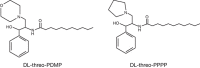
FIGURE 55.4.
Inhibitors of glycosphingolipid formation. These analogs are potent inhibitors of the glucosyltransferase that initiates glycosphingolipid formation.
Inhibitors of GPI anchor formation have also been described. Mannose analogs (2-deoxy-2-fluoro-D-glucose and 2-deoxy-D-glucose) inhibit the formation of dolichol-P-mannose in vivo and thus inhibit GPI biosynthesis. However, they lack specificity because these agents also can affect other pathways dependent on dolichol-P-mannose. Mannosamine inhibits GPI anchor formation both in Trypanosoma brucei and in mammalian cells by the formation of ManNH2-Man-GlcN-PI. Apparently, mannosamine in its activated form (GDP-ManNH2) is used as a substrate in the second mannosyltransferase reaction, but the ManNH2-Man-GlcN-PI intermediate will not act as a substrate for the next α2-mannosyl-transferase (Chapter 12). GlcNR-phosphatidylinositols with different substituents (R) act as substrate analogs and some act as suicide inhibitors in vitro. Another class of inhibitors is based on fatty acid analogs that only trypanosomes incorporate into GPI anchors. Trypanosomes, unlike their mammalian hosts, incorporate myristic acid into GPI anchors by exchanging myristic acid for other fatty acids in the phosphatidylinositol moiety. By making a series of analogs, an inhibitor was found that is highly toxic to trypanosomes in culture and nontoxic to mammalian cells (10-[propoxy] decanoic acid). Such reagents are drug candidates for treating trypanosomiasis, which is endemic in sub-Saharan regions of Africa.
RATIONAL DESIGN USING CRYSTAL STRUCTURES
Neuraminidase (Sialidase) Inhibitors
Studies of influenza neuraminidase exemplify the power of rationally designed drugs. The crystal structure for influenza neuraminidase was obtained in 1983, and since then many other enzymes have been characterized from other sources. Even before the crystal structure had been obtained, a neuraminidase inhibitor was deduced by assuming that the hydrolysis reaction probably involved a transition state with a carbocation intermediate at C-2. This would result in C-2 and C-3 adopting a trigonal planar (sp2) configuration, and therefore compounds that mimicked this geometry were hoped to have inhibitory activity. Indeed, Neu5Ac-2-ene (DANA; Figure 55.5) has a micromolar Ki value. Interestingly, this compound works on most sialidases, but not on the trypanosome trans-sialidase and only weakly on bacterial sialidases.
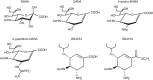
FIGURE 55.5.
Structure of influenza neuraminidase inhibitors. Chemical structure of Neu5Ac, NANA; 2-deoxy-2,3-dehydro-N-acetyl neuraminic acid, DANA; 4-amino-DANA; 4-guanidino-DANA (Relenza, zanamivir); (3R, 4R, 5S)-4-acetamido-5-amino-3-(1-ethylpropoxyl)-1-cyclohexane-1-carboxylic (more...)
Visual inspection of influenza neuraminidase with the inhibitor bound showed that two glutamate residues lined a pocket near carbon 4 of the sialic acid analog (Figure 55.6). The pocket was fairly open, suggesting that a bulkier substituent at this position might be tolerated, at least sterically. A substrate analog was produced containing a positively charged guanidinium group instead of the hydroxyl at carbon 4 (4-guanidino-DANA; Figure 55.5), creating an analog with remarkable influenza neuraminidase inhibitor potency (Ki value of 10−11 m). The higher affinity was presumably due to an additional salt bridge formed between the charged guanidinium group and the carboxylates lining the pocket. The analog is nearly a million times less potent on human sialidases, leading to its use as the anti-influenza drug Relenza (Chapter 57). It does not work on bacterial sialidases, however, because the equivalent pocket is filled with an arginine group.
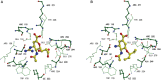
FIGURE 55.6.
Crystallographic structures of influenza virus neuraminidase (N9 subtype) with two different rationally designed inhibitors bound in the active site. The inhibitors are shown as colored ball and stick models, where yellow is carbon, blue is nitrogen, (more...)
Subsequent studies substituted a cyclohexene to mimic the planar ring of the proposed intermediate in hydrolysis. Good inhibitors were found and an orally active analog is widely used, the oral anti-influenza drug Tamiflu (Figure 55.5; Chapter 57). As the crystal structures for other sialidases are solved, the design of species-specific analogs may be possible. This rational approach to inhibitor design holds great potential, not only for neuraminidase inhibitors, but also for the design of compounds that might block the activity of various glycosyltransferases.
Sulfotransferase Inhibitors
A large family of Golgi sulfotransferases installs sulfate esters on a variety of glycans using PAPS as the active sulfate donor. High-resolution crystal structures are now available for both glycan-modifying sulfotransferases as well as soluble drug detoxifying sulfotransferases. These enzymes have in common a conserved fold that binds PAPS (see Chapter 6). A series of small aromatic compounds (such as 2,6-dichloro-4-nitrophenol and pentachlorophenol) and several disaccharide analogs of GlcNAc-6-sulfotransferase substrates have inhibitory activity. Screening targeted libraries of purine derivatives yielded compounds with high selectivity towards individual sulfotransferases, suggesting that subtle differences in the PAPS-binding sites can be exploited.
The ability to screen large libraries against sulfotransferases has been facilitated by the development of high-throughput assays. Most low-throughput assays measure the transfer of [35S] from [35S]PAPS or measure the radiolabel transfer to a carbohydrate substrate bearing a hydrophobic tail that can easily be isolated using a reverse-phase cartridge. Some sulfotransferases will catalyze the reverse reaction in the presence of high concentrations of a sulfated donor. For example, β-arylsulfotransferase IV (β-AST-IV) will catalyze the reverse transfer of the sulfuryl group from p-nitrophenol sulfate to PAP, generating PAPS and p-nitrophenolate ion. When coupled to another sulfotransferase of interest, β-AST-IV regenerates PAPS and stoichiometric amounts of the ion, which can be monitored by UV absorbance. The enzyme also will transfer sulfate to water. A library of 35,000 compounds with purine and pyrimidine scaffolds was screened using β-AST-IV and a fluorescence assay that measured desulfation of 4-methylumbelliferone sulfate. Multiple hits were obtained with moderate inhibition, and subsequent structure elaboration of the library resulted in the generation of a very tight binding small molecule inhibitor with a Km value five orders of magnitude lower than the natural substrate. In theory, this approach can be exploited for other enzymes for which high-throughput assays can be developed.
ACKNOWLEDGMENTS
The authors appreciate helpful comments and suggestions from Jarrod W. Barnes and Marilda Lisboa.
FURTHER READING
- Chapman E, Best MD, Hanson SR, Wong CH. 2004. Sulfotransferases: Structure, mechanism, biological activity, inhibition, and synthetic utility. Angew Chem Int Ed Engl 43: 3526–3548. [PubMed: 15293241]
- Dorfmueller HC, Borodkin VS, Schimpl M, Shepherd SM, Shpiro NA, van Aalten DM. 2006. GlcNAcstatin: A picomolar, selective O-GlcNAcase inhibitor that modulates intracellular O-glcNAcylation levels. J Am Chem Soc 128: 16484–16485. [PMC free article: PMC7116141] [PubMed: 17177381]
- Brown JR, Crawford BE, Esko JD. 2007. Glycan antagonists and inhibitors: A fount for drug discovery. Crit Rev Biochem Mol Biol 42: 481–515. [PubMed: 18066955]
- Chaudhary PM, Tupe SG, Deshpande MV. 2013. Chitin synthase inhibitors as antifungal agents. Mini Rev Med Chem 13: 222–236. [PubMed: 22512590]
- Tu Z, Lin YN, Lin CH. 2013. Development of fucosyltransferase and fucosidase inhibitors. Chem Soc Rev 42: 4459–4475. [PubMed: 23588106]
- Galley NF, O'Reilly AM, Roper DI. 2014. Prospects for novel inhibitors of peptidoglycan transglycosylases. Bioorg Chem 55: 16–26. [PMC free article: PMC4126109] [PubMed: 24924926]
- Gouin SG. 2014. Multivalent inhibitors for carbohydrate-processing enzymes: Beyond the “lock-and-key” concept. Chemistry 20: 11616–11628. [PubMed: 25081380]
- Kallemeijn WW, Witte MD, Wennekes T, Aerts JM. 2014. Mechanism-based inhibitors of glycosidases: Design and applications. Adv Carbohydr Chem Biochem 71: 297–338. [PubMed: 25480507]
- Kim EJ, Bond MR, Love DC, Hanover JA. 2014. Chemical tools to explore nutrient-driven O-GlcNAc cycling. Crit Rev Biochem Mol Biol 49: 327–342. [PMC free article: PMC6396312] [PubMed: 25039763]
- Shayman JA, Larsen SD. 2014. The development and use of small molecule inhibitors of glycosphingolipid metabolism for lysosomal storage diseases. J Lipid Res 55: 1215–1225. [PMC free article: PMC4076080] [PubMed: 24534703]
- ADVANTAGES OF INHIBITORS
- METABOLIC INHIBITORS
- TUNICAMYCIN: INHIBITION OF DOLICHOL-PP-GlcNAc ASSEMBLY
- PLANT ALKALOIDS: NATURAL INHIBITORS OF GLYCOSIDASES
- INHIBITION OF O-GalNAc INITIATION OF MUCIN-TYPE GLYCANS
- INHIBITION OF O-GlcNAc MODIFICATION
- SUBSTRATE ANALOGS: DIRECTED SYNTHESIS OF INHIBITORS
- GLYCOSIDE PRIMERS: MIMICKING WHAT ALREADY WORKS
- INHIBITORS OF GLYCOLIPIDS AND GPI ANCHORS
- RATIONAL DESIGN USING CRYSTAL STRUCTURES
- ACKNOWLEDGMENTS
- FURTHER READING
- Review Chemical Tools for Inhibiting Glycosylation.[Essentials of Glycobiology. 2009]Review Chemical Tools for Inhibiting Glycosylation.Esko JD, Bertozzi CR. Essentials of Glycobiology. 2009
- Review Chemical Tools for Inhibiting Glycosylation.[Essentials of Glycobiology. 2022]Review Chemical Tools for Inhibiting Glycosylation.Vocadlo DJ, Lowary TL, Bertozzi CR, Schnaar RL, Esko JD. Essentials of Glycobiology. 2022
- An inhibitor of the human UDP-GlcNAc 4-epimerase identified from a uridine-based library: a strategy to inhibit O-linked glycosylation.[Chem Biol. 2002]An inhibitor of the human UDP-GlcNAc 4-epimerase identified from a uridine-based library: a strategy to inhibit O-linked glycosylation.Winans KA, Bertozzi CR. Chem Biol. 2002 Jan; 9(1):113-29.
- Discovery of novel inhibitors of a disintegrin and metalloprotease 17 (ADAM17) using glycosylated and non-glycosylated substrates.[J Biol Chem. 2012]Discovery of novel inhibitors of a disintegrin and metalloprotease 17 (ADAM17) using glycosylated and non-glycosylated substrates.Minond D, Cudic M, Bionda N, Giulianotti M, Maida L, Houghten RA, Fields GB. J Biol Chem. 2012 Oct 19; 287(43):36473-87. Epub 2012 Aug 27.
- Review Anti-cancer glycosidase inhibitors from natural products: a computational and molecular modelling perspective.[Anticancer Agents Med Chem. 2015]Review Anti-cancer glycosidase inhibitors from natural products: a computational and molecular modelling perspective.Singh A, Mhlongo N, Soliman ME. Anticancer Agents Med Chem. 2015; 15(8):933-46.
- Chemical Tools for Inhibiting Glycosylation - Essentials of GlycobiologyChemical Tools for Inhibiting Glycosylation - Essentials of Glycobiology
Your browsing activity is empty.
Activity recording is turned off.
See more...