Summary
Clinical characteristics.
Branchiooculofacial syndrome (BOFS) is characterized by branchial (cervical or infra- or supra-auricular) skin defects that range from barely perceptible thin skin or hair patch to erythematous "hemangiomatous" lesions to large weeping erosions; ocular anomalies that can include microphthalmia, anophthalmia, coloboma, cataract, and nasolacrimal duct stenosis/atresia; and facial anomalies that can include dolichocephaly, hypertelorism or telecanthus, broad nasal tip, upslanted palpebral fissures, cleft lip or prominent philtral pillars that give the appearance of a repaired cleft lip (formerly called "pseudocleft lip") with or without cleft palate, upper lip pits, and lower facial weakness (asymmetric crying face or partial weakness of cranial nerve VII). Malformed and prominent pinnae and hearing loss from inner ear and/or petrous bone anomalies are common. Intellect is usually normal.
Diagnosis/testing.
The diagnosis of BOFS is established in a proband with characteristic clinical findings and a heterozygous pathogenic variant in TFAP2A identified by molecular genetic testing.
Management.
Treatment of manifestations: In general, children with BOFS should be managed by a multispecialty team including craniofacial specialists, plastic surgeons, otolaryngologists, and speech-language therapists. Small, linear, or superficial branchial skin defects may heal spontaneously; however, some require surgical intervention. Treatment of ophthalmic manifestations is per pediatric ophthalmologist. Nasolacrimal duct stenosis or atresia often requires surgery. Anophthalmia or severe microphthalmia may require a conformer (a structure, usually plastic, inserted into the eye socket to encourage its growth). It is recommended that cleft lip be repaired by an experienced pediatric plastic surgeon. Nasal tip abnormalities, lesser forms of cleft lip ("pseudocleft"), and malformed pinnae may need surgical correction. Standard treatments for hearing loss, renal malformations, dental manifestations, and congenital heart defects. Treatment of sensory, psychologic, and developmental challenges with supportive therapies.
Surveillance: Ophthalmology examination and vision assessment as recommended by ophthalmologist; audiology evaluation as recommended by otolaryngologist and/or audiologist; at each visit assess for a recurrent urinary tract infection suggestive of vesicoureteral reflux, assess teeth for size, number, carries, and malocclusion, and assess for new cysts; developmental and behavioral assessment annually or as needed; monitor for signs of low self-esteem and other psychological issues at each visit in older children as they enter adolescence.
Genetic counseling.
BOFS is inherited in an autosomal dominant manner. De novo pathogenic variants are observed in 50%-60% of affected individuals. Each child of an individual with BOFS has a 50% chance of inheriting the pathogenic variant. Once the TFAP2A pathogenic variant has been identified in an affected family member, prenatal and preimplantation genetic testing are possible.
Diagnosis
There are no formal diagnostic guidelines for branchiooculofacial syndrome (BOFS) developed by consensus panels, algorithms using a hierarchy of clinical findings, or evidence-based test standards. Diagnostic criteria have been proposed (see Table I in Milunsky et al [2011]).
Suggestive Findings
BOFS should be suspected in probands with findings in two or three of the following categories.
Branchial (cutaneous) defects. Cervical or infra- or supra-auricular skin defects:
- Vary from barely perceptible thin skin or hair patch to erythematous "hemangiomatous" lesions to large weeping erosions;
- Are most distinctive when they are bilateral and anterior cervical in location, and may be described as "cutis aplasia" [Wurfbain et al 2023];
- Differ from the punctuate sinus tracts of the branchiootorenal (BOR) syndrome;
- If very mild, may be unrecognized and heal spontaneously, but tend to "weep."
Ocular anomalies
- Microphthalmia, anophthalmia
- Coloboma
- Cataract
- Ptosis
- Nasolacrimal duct stenosis/atresia
- Strabismus
Facial anomalies
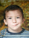
- Characteristic appearance with dolichocephaly, hypertelorism or telecanthus, broad nasal tip, and upslanted palpebral fissures (See Figure 1.)
- Cleft lip or prominent philtral pillars (technically known as a lesser-form cleft lip [formerly "pseudocleft lip"]), with or without cleft palate (Isolated cleft palate has not been reported.)
- Upper lip pits
- Lower facial nerve and/or muscle hypoplasia (asymmetric crying face, partial weakness of cranial nerve VII)
- Inner ear and petrous bone anomalies such as cochlear dysplasia, Mondini dysplasia, and enlarged vestibular aqueduct
- Malformed and prominent pinnae
- Hearing loss (conductive, sensorineural, mixed)
Establishing the Diagnosis
The clinical diagnosis of BOFS can be established in a proband based on proposed clinical diagnostic criteria [Milunsky et al 2011], or the molecular diagnosis can be established in a proband with suggestive findings and a heterozygous pathogenic (or likely pathogenic) variant in TFAP2A identified by molecular genetic testing (see Table 1).
Note: (1) Per ACMG/AMP variant interpretation guidelines, the terms "pathogenic variant" and "likely pathogenic variant" are synonymous in a clinical setting, meaning that both are considered diagnostic and can be used for clinical decision making [Richards et al 2015]. Reference to "pathogenic variants" in this section is understood to include likely pathogenic variants. (2) Identification of a heterozygous TFAP2A variant of uncertain significance does not establish or exclude the diagnosis.
Clinical Diagnosis
All three of the main features are present:
- Branchial (cutaneous) skin defect
- Ocular anomaly
- Facial anomalies (characteristic facial appearance)
OR
Two of the three main features plus one of the following are present:
- Affected first-degree relative, independently diagnosed
- Ectopic thymus (dermal)
Molecular Diagnosis
Molecular genetic testing approaches can include a combination of gene-targeted testing (single-gene testing, multigene panel) and comprehensive genomic testing (exome sequencing, genome sequencing). Gene-targeted testing requires that the clinician determine which gene(s) are likely involved (see Option 1), whereas comprehensive genomic testing does not (see Option 2).
Option 1
- Single-gene testing. Sequence analysis of TFAP2A is performed first to detect missense, nonsense, and splice site variants and small intragenic deletions/insertions. Note: Depending on the sequencing method used, single-exon, multiexon, or whole-gene deletions/duplications may not be detected. If no variant is detected by the sequencing method used, the next step is to perform gene-targeted deletion/duplication analysis to detect exon and whole-gene deletions or duplications.
- A multigene panel that includes TFAP2A and other genes of interest (see Differential Diagnosis) may also be considered to identify the genetic cause of the condition while limiting identification of variants of uncertain significance and pathogenic variants in genes that do not explain the underlying phenotype. Note: (1) The genes included in the panel and the diagnostic sensitivity of the testing used for each gene vary by laboratory and are likely to change over time. (2) Some multigene panels may include genes not associated with the condition discussed in this GeneReview. (3) In some laboratories, panel options may include a custom laboratory-designed panel and/or custom phenotype-focused exome analysis that includes genes specified by the clinician. (4) Methods used in a panel may include sequence analysis, deletion/duplication analysis, and/or other non-sequencing-based tests.
Option 2
Comprehensive genomic testing does not require the clinician to determine which gene is likely involved. Exome sequencing is most commonly used; genome sequencing is also possible.
For an introduction to comprehensive genomic testing click here. More detailed information for clinicians ordering genomic testing can be found here.
Table 1.
Molecular Genetic Testing Used in Branchiooculofacial Syndrome
Clinical Characteristics
Clinical Description
Most individuals with branchiooculofacial syndrome (BOFS) can be diagnosed in infancy on the basis of their clinical features. Females and males are affected equally. Although the facial features are generally recognizable, some individuals may have subtle differences [Authors, personal observation].
Classic BOFS Findings
Branchial (cutaneous) defects occur in a cervical (90%) or infra- or supra-auricular (60%) location.
- Defects vary from barely perceptible thin skin or hair patch to erythematous "hemangiomatous" lesions to large weeping erosions.
- The mildest defects may be unrecognized and in rare cases heal completely spontaneously. There may be a small residual sinus or tract that may appear to "weep," revealing the patency.
Ocular anomalies include the following:
- Structural eye malformations can include microphthalmia, anophthalmia, coloboma, or cataract.
- Periorbital abnormalities include nasolacrimal duct stenosis/atresia leading to weeping eyes and ptosis.
- Visual concerns include strabismus and significant visual impairment.
- Lam et al [2023] reviewed the ocular findings in 172 previously reported individuals and one additional individual with BOFS; the most common findings included nasolacrimal duct stenosis (57%), coloboma (46%), anophthalmia/microphthalmia (37%), cataract (16%), strabismus (14%), and myopia (12%).
Facial anomalies. Characteristic facial appearance includes dolichocephaly, hypertelorism or telecanthus, broad nasal tip, and upslanted palpebral fissures (see Figure 1). Other findings may include:
- Cleft lip or prominent philtral pillars (technically known as a lesser-form cleft lip [formerly "pseudocleft lip"])
- Occurring with or without cleft palate (99%)
- No instances of isolated cleft palate reported
- Upper lip pits
- Lower facial nerve and/or muscle hypoplasia (asymmetric crying face, partial weakness of cranial nerve VII)
- Ear anomalies
- Malformed and prominent pinnae
- Inner ear and petrous bone anomalies such as cochlear dysplasia, Mondini dysplasia, and enlarged vestibular aqueduct
- Hearing loss (70%) (conductive, sensorineural, mixed)
- Broad nose with full nasal tip, which is distinct from the appearance of the nose in other individuals with cleft lip
Additional Findings Observed in BOFS
Immune system. Thymic anomalies (ectopic, dermal) (~35%), typically bilateral with normal thymic function
Renal system
- Structural anomalies (35%) (e.g., dysplastic, absent, multicystic)
- Vesicoureteral reflux
Ectodermal (hair, teeth, nails)
- Premature hair graying, poliosis (forelock or patchy) (35%)
- Hypoplastic teeth
- Dysplastic nails
- Cysts, subcutaneous (dermoid-like, often on the scalp; less commonly other areas of the head and neck)
Psychomotor development (typically normal)
- Visual and hearing handicaps (frequent)
- Autism spectrum disorder, intellectual disability (rare)
Growth restriction. Uncommon
Miscellaneous and rare (<5 individuals each)
- Heterochromia irides
- Congenital heart defect (atrial septal defect, tetralogy of Fallot)
- Polydactyly (bilateral, usually postaxial)
- Medulloblastoma (1 individual) [Milunsky et al 2008]
- Trigonocephaly (1 individual) [Wurfbain et al 2023]
Genotype-Phenotype Correlations
No clear genotype-phenotype correlation exists.
Significant inter- and intrafamilial variability have been observed with the same pathogenic variants [Milunsky et al 2011]. Missense, frameshift, and splicing variants along with more complex rearrangements [Tekin et al 2009, Milunsky et al 2011] throughout the gene result in similar phenotypes.
The majority of individuals with a deletion involving TFAP2A appear to have an abnormally prominent philtrum that may be on the spectrum of microform cleft lip [Lin et al 2009]. LeBlanc et al [2013] described an infant and mother with a 593-kb deletion including TFAP2A and five additional genes. Neither is reported to have any type of cleft or abnormal philtrum. Otherwise, the marked inter- and intrafamilial variability appear similar to that observed with intragenic pathogenic variants.
Penetrance
BOFS has shown almost complete penetrance. Careful examination of individuals identified in a family with BOFS with a TFAP2A pathogenic variant is necessary to reveal subtle findings including premature graying (individuals may have dyed their hair), faint hair on the neck, or heterochromia of the irides.
Prevalence
The prevalence of BOFS is not known. It is a rare condition, with fewer than 150 individuals having a well-described clinical and/or molecular diagnosis. An informal survey of clinical geneticists who attended a 2017 dysmorphology conference identified an additional 27 unpublished individuals (18 with a clinical diagnosis and nine with a molecular diagnosis). While these numbers are insufficient to calculate a population-based prevalence, they support the impression that BOFS remains a rare disorder.
Genetically Related (Allelic) Disorders
No phenotypes other than those discussed in this GeneReview are known to be associated with germline pathogenic variants in TFAP2A.
Differential Diagnosis
The branchiooculofacial syndrome phenotype is distinctive and can typically be differentiated on a clinical basis from disorders with overlapping features (see Table 2).
Table 2.
Disorders of Interest in the Differential Diagnosis of Branchiooculofacial Syndrome
Management
Evaluations Following Initial Diagnosis
To establish the extent of disease and needs of an individual diagnosed with branchiooculofacial syndrome (BOFS), the evaluations summarized in Table 3 (if not performed as part of the evaluation that led to the diagnosis) are recommended.
Table 3.
Branchiooculofacial Syndrome: Recommended Evaluations Following Initial Diagnosis
Note: (1) Motor delays are not part of BOFS; thus, physical and occupational therapy is not anticipated. (2) The role of cancer surveillance is not established.
Treatment of Manifestations
Milunsky et al [2011] provided management guidelines that remain clinically useful and have not been updated. In general, children with BOFS and multiple anomalies should be followed in a setting in which multispecialty care can be provided by a team including, for example, craniofacial specialists, plastic surgeons, otolaryngologists, and speech-language therapists (see Table 4). Ideally, multispecialty evaluations and surgery should be performed within a craniofacial clinic.
Table 4.
Branchiooculofacial Syndrome: Treatment of Manifestations
Surveillance
To monitor existing manifestations and the individual's response to supportive care, the evaluations summarized in Table 5 are recommended.
Table 5.
Branchiooculofacial Syndrome: Recommended Surveillance
Evaluation of Relatives at Risk
It is appropriate to evaluate apparently asymptomatic older and younger at-risk relatives of an affected individual in order to identify as early as possible those who would benefit from surveillance and treatment of hearing, vision, renal, and other manifestations. Evaluations can include:
- Molecular genetic testing if the pathogenic variant in the family is known;
- A careful physical examination to look for subtle physical findings of BOFS if the pathogenic variant in the family is not known.
See Genetic Counseling for issues related to testing of at-risk relatives for genetic counseling purposes.
Therapies Under Investigation
Search ClinicalTrials.gov in the US and EU Clinical Trials Register in Europe for information on clinical studies for a wide range of diseases and conditions.
Genetic Counseling
Genetic counseling is the process of providing individuals and families with information on the nature, mode(s) of inheritance, and implications of genetic disorders to help them make informed medical and personal decisions. The following section deals with genetic risk assessment and the use of family history and genetic testing to clarify genetic status for family members; it is not meant to address all personal, cultural, or ethical issues that may arise or to substitute for consultation with a genetics professional. —ED.
Mode of Inheritance
Branchiooculofacial syndrome (BOFS) is inherited in an autosomal dominant manner.
Risk to Family Members
Parents of a proband
- Approximately 40%-50% of individuals diagnosed with BOFS have an affected parent [Milunsky et al 2011].
- Approximately 50%-60% of individuals diagnosed with BOFS have the disorder as the result of a de novo TFAP2A pathogenic variant [Milunsky et al 2011].
- If a molecular diagnosis has been established in the proband and the proband appears to be the only affected family member (i.e., a simplex case), molecular genetic testing is recommended for the parents of the proband to confirm their genetic status and to allow reliable recurrence risk counseling.
- If the pathogenic variant identified in the proband is not identified in either parent and parental identity testing has confirmed biological maternity and paternity, the following possibilities should be considered:
- The proband has a de novo pathogenic variant.
- The proband inherited a pathogenic variant from a parent with germline (or somatic and germline) mosaicism.* Note: Testing of parental leukocyte DNA may not detect all instances of somatic mosaicism and will not detect a pathogenic variant that is present in the germ cells only.* A parent with somatic and germline mosaicism for a TFAP2A pathogenic variant may be mildly/minimally affected [Milunsky et al 2011].
- The family history of some individuals diagnosed with BOFS may appear to be negative because of failure to recognize the disorder in family members or a milder phenotypic presentation. Therefore, an apparently negative family history cannot be confirmed without molecular genetic testing to establish that neither parent is heterozygous for the pathogenic variant identified in the proband.
Sibs of a proband. The risk to the sibs of the proband depends on the genetic status of the proband's parents:
- If a parent of the proband is affected and/or is known to have the pathogenic variant identified in the proband, the risk to the sibs is 50%. BOFS is associated with almost complete penetrance; however, significant intrafamilial variability has been observed [Milunsky et al 2011].
- If the proband has a known TFAP2A pathogenic variant that cannot be detected in the leukocyte DNA of either parent, the recurrence risk to sibs is slightly greater than that of the general population because of the possibility of parental mosaicism [Milunsky et al 2011].
- If the parents appear to be clinically unaffected but their genetic status is unknown, the risk to the sibs of a proband appears to be low but increased over that of the general population because of the possibility of a milder phenotypic presentation in a heterozygous parent and the possibility of parental mosaicism.
Offspring of a proband. Each child of an individual with BOFS has a 50% chance of inheriting the TFAP2A pathogenic variant.
Other family members. The risk to other family members depends on the status of the proband's parents: if a parent has the TFAP2A pathogenic variant, the parent's family members may be at risk.
Related Genetic Counseling Issues
See Management, Evaluation of Relatives at Risk for information on evaluating at-risk relatives for the purpose of early diagnosis and treatment.
Family planning
- The optimal time for determination of genetic risk and discussion of the availability of prenatal/preimplantation genetic testing is before pregnancy.
- It is appropriate to offer genetic counseling (including discussion of potential risks to offspring and reproductive options) to young adults who are affected.
DNA banking. Because it is likely that testing methodology and our understanding of genes, pathogenic mechanisms, and diseases will improve in the future, consideration should be given to banking DNA from probands in whom a molecular diagnosis has not been confirmed (i.e., the causative pathogenic mechanism is unknown). For more information, see Huang et al [2022].
Prenatal Testing and Preimplantation Genetic Testing
Once the TFAP2A pathogenic variant has been identified in an affected family member, prenatal and preimplantation genetic testing are possible.
Differences in perspective may exist among medical professionals and within families regarding the use of prenatal testing. While most centers would consider use of prenatal testing to be a personal decision, discussion of these issues may be helpful.
Resources
GeneReviews staff has selected the following disease-specific and/or umbrella support organizations and/or registries for the benefit of individuals with this disorder and their families. GeneReviews is not responsible for the information provided by other organizations. For information on selection criteria, click here.
- American Cleft Palate-Craniofacial AssociationPhone: 919-933-9044
- Face Equality InternationalUnited Kingdom
Molecular Genetics
Information in the Molecular Genetics and OMIM tables may differ from that elsewhere in the GeneReview: tables may contain more recent information. —ED.
Table A.
Branchiooculofacial Syndrome: Genes and Databases
Table B.
OMIM Entries for Branchiooculofacial Syndrome (View All in OMIM)
Molecular Pathogenesis
TFAP2A is a retinoic acid-responsive member of the AP-2 family of transcription factors that regulate gene expression during embryogenesis of the eye, ear, face, body wall, limbs, and neural tube [Schorle et al 1996, Zhang et al 1996, Ahituv et al 2004, Nelson & Williams 2004]. TFAP2A is also involved in tumorigenesis, with protein expression levels affecting cell transformation, tumor growth, metastasis, and survival [Jean et al 1998, Heimberger et al 2005, Orso et al 2007]. Numerous gene interactions likely underlie the variability in phenotype resulting from molecular defects involving TFAP2A. TFAP2A is known to be expressed in premigratory and migratory neural crest cells [Hilger-Eversheim et al 2000, Li & Cornell 2007] and is required for early morphogenesis of the lens [Gestri et al 2009].
Although the pathogenic variants occur throughout the gene, a hot spot region in exons 6 and 7 that contains missense variants in about 90% of probands/families with BOFS has been identified [Milunsky et al 2011].
Li et al [2013] demonstrated that several pathogenic variants in the DNA-binding domain can have dominant-negative activity on wild type AP-2α protein. Hence, differences in activity due to null, hypomorphic, or antimorphic alleles may lead to the phenotypic variability characteristic of BOFS.
Mechanism of disease causation. Loss of function
Chapter Notes
Author Notes
As of January 2018, there is no disease advocacy organization ("support group") for BOFS. Through Dr Lin, several parents of children with BOFS have reached out to the families of newly diagnosed individuals.
Acknowledgments
We thank the many families and international colleagues who have supported our research.
Revision History
- 28 September 2023 (sw) Comprehensive update posted live
- 29 March 2018 (ha) Comprehensive update posted live
- 31 May 2011 (me) Review posted live
- 11 January 2011 (al) Original submission
References
Published Guidelines / Consensus Statements
- Milunsky JM, Maher TM, Zhao G, Wang Z, Mulliken JB, Chitayat D, Clemens M, Stalker HJ, Bauer M, Burch M, Chénier S, Cunningham ML, Drack AV, Janssens S, Karlea A, Klatt R, Kini U, Klein O, Lachmeijer AM, Megarbane A, Mendelsohn NJ, Meschino WS, Mortier GR, Parkash S, Ray CR, Roberts A, Roberts A, Reardon W, Schnur RE, Smith R, Splitt M, Tezcan K, Whiteford ML, Wong DA, Zori R, Lin AE. Genotype-phenotype analysis of the branchio-oculo-facial syndrome. Am J Med Genet A. 2011;155A:22-32. [PubMed]
Literature Cited
- Ahituv N, Erven A, Fuchs H, Guy K, Ashery-Padan R, Williams T, de Angelis MH, Avraham KB, Steel KP. An ENU-induced mutation in AP-2alpha leads to middle ear and ocular defects in Doarad mice. Mamm Genome. 2004;15:424-32. [PubMed: 15181535]
- Gestri G, Osborne RJ, Wyatt AW, Gerrelli D, Gribble S, Stewart H, Fryer A, Bunyan DJ, Prescott K, Collin JR, Fitzgerald T, Robinson D, Carter NP, Wilson SW, Ragge NK. Reduced TFAP2A function causes variable optic fissure closure and retinal defects and sensitizes eye development to mutations in other morphogenetic regulators. Hum Genet. 2009;126:791-803. [PMC free article: PMC3083835] [PubMed: 19685247]
- Heimberger AB, McGary EC, Suki D, Ruiz M, Wang H, Fuller GN, Bar-Eli M. Loss of the AP-2alpha transcription factor is associated with the grade of human gliomas. Clin Cancer Res. 2005;11:267-72. [PubMed: 15671555]
- Hilger-Eversheim K, Moser M, Schorle H, Buettner R. Regulatory roles of AP-2 transcription factors in vertebrate development, apoptosis and cell-cycle control. Gene. 2000;260:1-12. [PubMed: 11137286]
- Huang SJ, Amendola LM, Sternen DL. Variation among DNA banking consent forms: points for clinicians to bank on. J Community Genet. 2022;13:389-97. [PMC free article: PMC9314484] [PubMed: 35834113]
- Jean D, Gershenwald JE, Huang S, Luca M, Hudson MJ, Tainsky MA, Bar-Eli M. Loss of AP-2 results in up-regulation of MCAM/MUC18 and an increase in tumor growth and metastasis of human melanoma cells. J Biol Chem. 1998;273:16501-8. [PubMed: 9632718]
- Lam K, Cassidy B, Arreola R, Al Saif H, King K, Couser NL. A new case and comprehensive review of the ophthalmic manifestations of 172 individuals with branchio-oculo-facial syndrome. J Pediatr Ophthalmol Strabismus. 2023;60:295-301. [PubMed: 36263936]
- LeBlanc SK, Yu S, Barnett CP. 6p.24 microdeletion involving TFAP2A without classic features of branchio-oculo-facial syndrome. Am J Med Genet. 2013;161A:901-4. [PubMed: 23495225]
- Li H, Sheridan R, Williams T. Analysis of TFAP2A mutations in branchio-oculo-facial syndrome indicated functional complexity within the AP-2α DNA-binding domain. Hum Mol Genet. 2013;22:3195-206. [PMC free article: PMC3723307] [PubMed: 23578821]
- Li W, Cornell RA. Redundant activities of Tfap2a and Tfap2c are required for neural crest induction and development of other non-neural ectoderm derivatives in zebrafish embryos. Dev Biol. 2007;304:338-54. [PMC free article: PMC1904501] [PubMed: 17258188]
- Lin AE, Yuzuriha S, McLean S, Mulliken JB. Lesser forms of cleft lip associated with the branchio-oculo-facial syndrome. J Craniofac Surg. 2009;20:608-11. [PubMed: 19795528]
- Milunsky JM, Maher TA, Zhao G, Roberts AE, Stalker HJ, Zori RT, Burch MN, Clemens M, Mulliken JB, Smith R, Lin AE. TFAP2A mutations result in branchio-oculo-facial syndrome. Am J Hum Genet. 2008;82:1171-7. [PMC free article: PMC2427243] [PubMed: 18423521]
- Milunsky JM, Maher TM, Zhao G, Wang Z, Mulliken JB, Chitayat D, Clemens M, Stalker HJ, Bauer M, Burch M, Chénier S, Cunningham ML, Drack AV, Janssens S, Karlea A, Klatt R, Kini U, Klein O, Lachmeijer AM, Megarbane A, Mendelsohn NJ, Meschino WS, Mortier GR, Parkash S, Ray CR, Roberts A, Roberts A, Reardon W, Schnur RE, Smith R, Splitt M, Tezcan K, Whiteford ML, Wong DA, Zori R, Lin AE. Genotype-phenotype analysis of the branchio-oculo-facial syndrome. Am J Med Genet A. 2011;155A:22-32. [PubMed: 21204207]
- Nelson DK, Williams T. Frontonasal process-specific disruption of AP-2alpha results in postnatal midfacial hypoplasia, vascular anomalies, and nasal cavity defects. Dev Biol. 2004;267:72-92. [PubMed: 14975718]
- Orso F, Fassetta M, Penna E, Solero A, De Filippo K, Sismondi P, De Bortoli M, Taverna D. The AP-2alpha transcription factor regulates tumor cell migration and apoptosis. Adv Exp Med Biol. 2007;604:87-95. [PubMed: 17695722]
- Raveh E, Papsin BC, Forte V. Branchio-oculo-facial syndrome. Int J Pediatr Otorhinolaryngol. 2000;53:149-56. [PubMed: 10906521]
- Richards S, Aziz N, Bale S, Bick D, Das S, Gastier-Foster J, Grody WW, Hegde M, Lyon E, Spector E, Voelkerding K, Rehm HL, et al. Standards and guidelines for the interpretation of sequence variants: a joint consensus recommendation of the American College of Medical Genetics and Genomics and the Association for Molecular Pathology. Genet Med. 2015;17:405-24. [PMC free article: PMC4544753] [PubMed: 25741868]
- Schorle H, Meier P, Buchert M, Jaenisch R, Mitchell PJ. Transcription factor AP-2 essential for cranial closure and craniofacial development. Nature. 1996;381:235-8. [PubMed: 8622765]
- Stoetzel C, Riehm S, Bennouna Greene V, Pelletier V, Vigneron J, Leheup B, Marion V, Hellé S, Danse JM, Thibault C, Moulinier L, Veillon F, Dollfus H. Confirmation of TFAP2A gene involvement in branchio-oculo-facial syndrome (BOFS) and report of temporal bone anomalies. Am J Med Genet A. 2009;149A:2141-6. [PubMed: 19764023]
- Tekin M, Sirmaci A, Yüksel-Konuk B, Fitoz S, Sennaroğlu L. A complex TFAP2A allele is associated with branchio-oculo-facial syndrome and inner ear malformation in a deaf child. Am J Med Genet A. 2009;149A:427-30. [PubMed: 19206157]
- Wurfbain LF, Cox IL, van Dooren MF, Lachmeijer AMA, Verhoeven VJM, van Hagen JM, Heijligers M, Klein Wassink-Ruiter JS, Koene S, Maas SM, Veenstra-Knol HE, Ploos van Amstel JK, Massink MPG, Mink van der Molen AB, van den Boogaard MH. Diagnostic gene panel testing in (non)-syndromic patients with cleft lip, alveolus and/or palate in the Netherlands. Mol Syndromol. 2023;14:270-82. [PMC free article: PMC10425715] [PubMed: 37589029]
- Zhang J, Hagopian-Donaldson S, Serbedzija G, Elsemore J, Plehn-Dujowich D, McMahon AP, Flavell RA, Williams T. Neural tube, skeletal and body wall defects in mice lacking transcription factor AP-2. Nature. 1996;381:238-41. [PubMed: 8622766]
Publication Details
Author Information and Affiliations
Massachusetts General Hospital for Children
Boston, Massachusetts
Asheville, North Carolina
Senior Director, Molecular Genetics
Co-Director, Center for Human Genetics, Inc
Cambridge, Massachusetts
Publication History
Initial Posting: May 31, 2011; Last Update: September 28, 2023.
Copyright
GeneReviews® chapters are owned by the University of Washington. Permission is hereby granted to reproduce, distribute, and translate copies of content materials for noncommercial research purposes only, provided that (i) credit for source (http://www.genereviews.org/) and copyright (© 1993-2024 University of Washington) are included with each copy; (ii) a link to the original material is provided whenever the material is published elsewhere on the Web; and (iii) reproducers, distributors, and/or translators comply with the GeneReviews® Copyright Notice and Usage Disclaimer. No further modifications are allowed. For clarity, excerpts of GeneReviews chapters for use in lab reports and clinic notes are a permitted use.
For more information, see the GeneReviews® Copyright Notice and Usage Disclaimer.
For questions regarding permissions or whether a specified use is allowed, contact: ude.wu@tssamda.
Publisher
University of Washington, Seattle, Seattle (WA)
NLM Citation
Lin AE, Haldeman-Englert CR, Milunsky JM. Branchiooculofacial Syndrome. 2011 May 31 [Updated 2023 Sep 28]. In: Adam MP, Feldman J, Mirzaa GM, et al., editors. GeneReviews® [Internet]. Seattle (WA): University of Washington, Seattle; 1993-2024.
