NCBI Bookshelf. A service of the National Library of Medicine, National Institutes of Health.

Adam MP, Feldman J, Mirzaa GM, et al., editors. GeneReviews® [Internet]. Seattle (WA): University of Washington, Seattle; 1993-2024.

GRIN2B-Related Neurodevelopmental Disorder

Synonym: GRIN2B Encephalopathy
, MD
Institute of Human Genetics
University of Leipzig Hospitals and Clinics
Leipzig, Germany
, MD
Institute of Human Genetics
University of Leipzig Hospitals and Clinics
Leipzig, Germany

Initial Posting: ; Last Revision: March 25, 2021.

Estimated reading time: 20 minutes

Summary

Clinical characteristics.

GRIN2B-related neurodevelopmental disorder is characterized by mild to profound developmental delay / intellectual disability (DD/ID) in all affected individuals. Muscle tone abnormalities (spasticity and/or hypotonia, occasionally associated with feeding difficulties), as well as epilepsy and autism spectrum disorder (ASD) / behavioral issues, are common. Other infantile- or childhood-onset findings include microcephaly; dystonic, dyskinetic, or choreiform movement disorder; and/or cortical visual impairment. Brain MRI reveals a malformation of cortical development in a minority of affected individuals. To date, fewer than 100 individuals with GRIN2B-related neurodevelopmental disorder have been reported.

Diagnosis/testing.

The diagnosis of a GRIN2B-related neurodevelopmental disorder is established in a proband by identification of either a heterozygous pathogenic variant or exon or whole-gene deletion of GRIN2B on molecular genetic testing.

Management.

Treatment of manifestations: DD/ID, muscle tone abnormalities (spasticity, hypotonia, and feeding difficulties), epilepsy, ASD/behavioral issues, movement disorders, and/or cortical visual impairment are treated as per standard practice.

Surveillance: Of clinical manifestations as clinically indicated.

Genetic counseling.

GRIN2B-related neurodevelopmental disorder is inherited in an autosomal dominant manner. All probands reported to date with a GRIN2B-related neurodevelopmental disorder whose parents have undergone molecular genetic testing have the disorder as a result of a de novo GRIN2B pathogenic variant or deletion. If the proband represents a simplex case (i.e., the only affected family member) and the GRIN2B pathogenic variant found in the proband cannot be detected in the leukocyte DNA of either parent, the recurrence risk to sibs is estimated to be 1% because of the theoretic possibility of parental mosaicism. Given this risk, prenatal testing and preimplantation genetic testing may be considered.

Diagnosis

Formal diagnostic criteria for GRIN2B-related neurodevelopmental disorder have not been established.

Suggestive Findings

GRIN2B-related neurodevelopmental disorder should be considered in individuals with the following clinical and/or brain MRI findings.

Clinical findings

  • Mild-to-profound developmental delay (DD) or intellectual disability (ID); AND
  • Any of the following features presenting in infancy or childhood:
    • Epilepsy
    • Autism spectrum disorder / behavioral issues
    • Microcephaly
    • Muscle tone abnormalities such as hypotonia (occasionally associated with feeding difficulties) and spasticity
    • Dystonic, dyskinetic, or choreiform movement disorder
    • Cortical visual impairment

Brain MRI findings. MRI reveals a malformation of cortical development (MCD) consisting of diffuse cortical dysplasia including polymicrogyria (see Polymicrogyria Overview), hypoplastic corpus callosum, enlarged/dysplastic basal ganglia, and hippocampal dysplasia. The MCD can also resemble the tubulinopathies spectrum (see Tubulinopathies Overview).

Establishing the Diagnosis

The diagnosis of a GRIN2B-related neurodevelopmental disorder is established in a proband by identification of either a heterozygous pathogenic (or likely pathogenic) variant or exon or whole-gene deletion of GRIN2B on molecular genetic testing (see Table 1).

Note: (1) Larger contiguous-gene deletions including but not limited to GRIN2B are not discussed in this GeneReview. (2) Per ACMG variant interpretation guidelines, the terms "pathogenic variants" and "likely pathogenic variants" are synonymous in a clinical setting, meaning that both are considered diagnostic and both can be used for clinical decision making. Reference to "pathogenic variants" in this section is understood to include any likely pathogenic variants.

Molecular genetic testing approaches can include use of a multigene panel, chromosomal microarray analysis, and/or more comprehensive genomic testing.

Gene-targeted testing requires that the clinician determine which gene(s) are likely involved, whereas genomic testing does not. Because the phenotypes of many disorders with intellectual disability overlap, most children with GRIN2B-related neurodevelopmental disorder are diagnosed by genomic testing. Note: Single-gene testing (sequence analysis of GRIN2B, followed by gene-targeted deletion/duplication analysis) is rarely useful and typically NOT recommended.

  • An intellectual disability multigene panel that includes GRIN2B and other genes of interest (see Differential Diagnosis) typically provides the best opportunity to identify the genetic cause of the condition while limiting identification of pathogenic variants in genes that do not explain the underlying phenotype. Note: (1) The genes included in the panel and the diagnostic sensitivity of the testing used for each gene vary by laboratory and are likely to change over time. (2) Some multigene panels may include genes not associated with the condition discussed in this GeneReview. (3) Methods used in a panel may include sequence analysis, deletion/duplication analysis, and/or other non-sequencing-based tests. For GRIN2B-related disorder a multigene panel that also includes deletion/duplication analysis is recommended (see Table 1).
    For an introduction to multigene panels click here. More detailed information for clinicians ordering genetic tests can be found here.
  • Chromosomal microarray analysis (CMA) uses oligonucleotide or SNP arrays to detect genome-wide large deletions/duplications (including GRIN2B) that cannot be detected by sequence analysis.
  • Comprehensive genomic testing does not require the clinician to determine which gene(s) are likely involved. Exome sequencing is most commonly used; genome sequencing is also possible.
    For an introduction to comprehensive genomic testing click here. More detailed information for clinicians ordering genomic testing can be found here.

Table 1.

Molecular Genetic Testing Used in GRIN2B-Related Neurodevelopmental Disorder

Gene 1MethodProportion of Probands with a Pathogenic Variant 2 Detectable by Method
GRIN2B Sequence analysis 382/86 4
Gene-targeted deletion/duplication analysis 5 or chromosomal microarray (CMA) 64/86 4
1.

See Table A. Genes and Databases for chromosome locus and protein.

2.

See Molecular Genetics for information on allelic variants detected in this gene.

3.

Sequence analysis detects variants that are benign, likely benign, of uncertain significance, likely pathogenic, or pathogenic. Variants may include small intragenic deletions/insertions and missense, nonsense, and splice site variants; typically, exon or whole-gene deletions/duplications are not detected. For issues to consider in interpretation of sequence analysis results, click here.

4.

For references, see Molecular Pathogenesis, Pathogenic variants. Note: Three additional individuals with contiguous gene deletions (not included in these calculations) have been reported: two with chromosome translocations and one with a chromosome inversion disrupting GRIN2B [Endele et al 2010, Talkowski et al 2012].

5.

Gene-targeted deletion/duplication analysis detects intragenic deletions or duplications. Methods used may include a range of techniques such as quantitative PCR, long-range PCR, multiplex ligation-dependent probe amplification (MLPA), and a gene-targeted microarray designed to detect single-exon deletions or duplications.

6.

Chromosomal microarray analysis (CMA) uses oligonucleotide or SNP arrays to detect genome-wide large deletions/duplications (including GRIN2B) that cannot be detected by sequence analysis. The ability to determine the size of the deletion/duplication depends on the type of microarray used and the density of probes in the 12p13.1 region (which includes GRIN2B). CMA designs in current clinical use target the 12p13.1 region.

Clinical Characteristics

Clinical Description

GRIN2B-related neurodevelopmental disorder is characterized in all affected individuals by mild to profound developmental delay / intellectual disability (DD/ID). Epilepsy (seen in 51%) and autism spectrum disorder (ASD) and autistic-like behaviors (26%) are common. Other infantile- or childhood-onset findings include microcephaly; muscle tone abnormalities (hypotonia, spasticity); dystonic, dyskinetic, or choreiform movement disorder; and/or cortical visual impairment. To date, fewer than 100 individuals with GRIN2B-related neurodevelopmental disorder have been reported in cohorts of individuals with DD/ID/ASD, early-onset epilepsy, and malformations of cortical development (MCD).

Unless otherwise noted, the information in this section is based on extended data of Platzer et al [2017]. Detailed clinical assessment was available for 61 patients, with specification of ID in 54. Brain MRI was performed in 47 patients.

DD/ID

The degree of DD/ID can be severe or profound (61%, 33/54), moderate (24%, 13/54), or mild (15%, 8/54) using standard assessments of psychomotor development or IQ testing.

Signs of developmental regression have been noted in four children (7%, 4/61), one of whom had transient regression of language skills at age six years with improvement beginning at age eight years and another who had recurrent periods of global regression starting at age three years. No detailed information is available for the other two children.

Muscle Tone Abnormalities

Hypotonia has been reported in more than half the patients (56%, 34/61). Five (15% of those with muscular hypotonia) required tube feeding. All five of these individuals had severe ID.

Spasticity was seen in 14 (23%) of 61 patients, all with severe ID.

Epilepsy

Epilepsy is present in 31 (51%) of 61 of individuals and characterized by the following.

Features

  • Onset is from birth to age nine years.
  • Seizure frequency ranges from multiple episodes per day to a few seizures per year.
  • Seizures are refractory to anti-seizure medication in approximately half of individuals treated.

Seizure types

  • Seizures may be generalized (58%, 18/31) and/or focal (48%, 15/31) and/or epileptic spasms (35%, 11/31) with some patients displaying multiple seizure types over time.
  • EEG patterns comprise generalized, focal, and multifocal epileptiform activity and/or hypsarrhythmia.

Syndromes. Most children with epileptic spasms also show hypsarrhythmia or hypsarrhythmia-like EEG patterns and fulfill diagnostic criteria for West syndrome.

ASD

Autistic features were seen in 16 (26%) of 61 individuals. In addition, in one study of the behavioral phenotype of five individuals with GRIN2B-related neurodevelopmental disorder without ASD, the authors observed hyperactivity, impulsivity, distractibility, stereotypies, short attention span, sleeping problems, and social behavior that is friendly but lacking boundaries [Freunscht et al 2013].

Other

Microcephaly occurred in 11 (18%) of 61 individuals; all 11 had severe ID. Three of these also had an MCD.

Movement disorders (10%, 6/61) included involuntary dystonic, dyskinetic, and/or choreiform movements.

Cortical visual impairment (CVI) (8%, 5/61) has been reported in four patients: three also had an MCD, and the fourth, who had a normal brain MRI, was identified in a cohort of individuals with ID and CVI [Bosch et al 2016].

Note: A report of an individual with approximately 50% mosaicism for a GRIN2B pathogenic missense variant in blood (no other tissues were tested) did not provide sufficient clinical information to allow comparison of the phenotype with individuals with a heterozygous germline pathogenic variant [Stosser et al 2018].

Brain Imaging

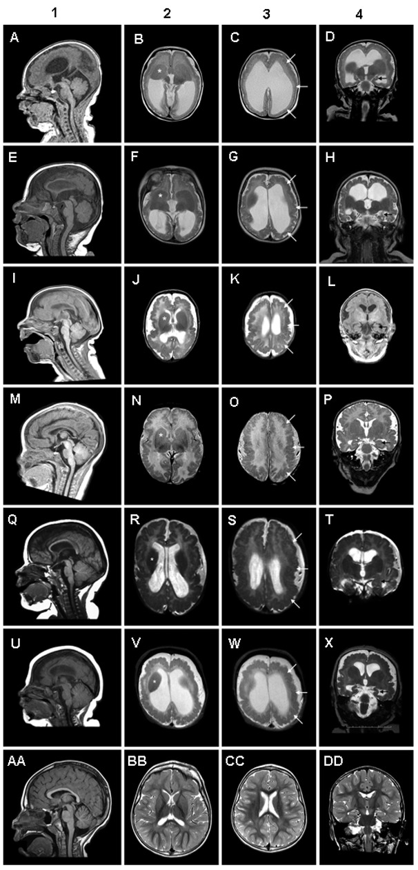
A malformation of cortical development (MCD) has been seen in six (13%) of 47 individuals; the diffuse cortical dysplasia was consistent with that of polymicrogyria (see Polymicrogyria Overview). Cortical findings included a mixture of large and small gyri separated by shallow sulci (Figure 1). The gray-white border appeared smooth.

Other findings included the following:

  • Hypoplastic corpus callosum of varying degrees
  • Enlarged and mildly dysplastic basal ganglia
  • Hippocampal dysplasia with thick leaves and open hilus
  • Enlarged tecta
  • Absent septum pellucidum

The malformation of cortical development is also consistent with that of tubulinopathies (see Tubulinopathies Overview). The identified individuals with MCD display a very similar degree of severity, and there are no reports of affected individuals with less pronounced malformations of cortical development.

Generalized cerebral volume loss indicating cerebral atrophy was seen in four other individuals (9%, 4/47).

Genotype-Phenotype Correlations

Variant class and intellectual outcome show a significant correlation: heterozygotes for a GRIN2B pathogenic variant resulting in a null allele (e.g., nonsense or frameshift variants, deletion involving whole exons or the entire gene, translocation and inversion disrupting GRIN2B) tended to display mild or moderate ID, while heterozygotes for pathogenic missense variants displayed severe ID (Fisher's exact test, p=0.0079) [Platzer et al 2017].

Missense variants in GRIN2B that cause a malformation of cortical development are located in transmembrane domain M3, in the ligand-binding domain S2, and in the linker between S2 and the transmembrane domain M4, a finding consistent with GRIN1 variants causing an MCD [Fry et al 2018] (see GRIN1-Related Neurodevelopmental Disorder).

Penetrance

Penetrance of GRIN2B-related neurodevelopmental disorder is thought to be 100%.

Prevalence

The prevalence of GRIN2B-related neurodevelopmental disorder in the general population is unknown. To date, fewer than 100 individuals have been reported.

The prevalence of GRIN2B-related neurodevelopmental disorder among individuals with neurodevelopmental disorders and/or childhood-onset epilepsy is around 0.2% [Platzer et al 2017].

Differential Diagnosis

Phenotypic features associated with heterozygous GRIN2B pathogenic variants are not sufficient to diagnose GRIN2B-related neurodevelopmental disorder.

All genes known to be associated with ID, early-onset epileptic encephalopathy, and malformations of cortical development (especially diffuse polymicrogyria and tubulinopathies) should be included in the differential diagnosis of GRIN2B-related neurodevelopmental disorder (see Table 2) as individuals with GRIN2B-related neurodevelopment disorder can present with a combination of clinically unspecific phenotypes such as DD/ID/ASD and/or epilepsy. The underlying genetic causes of these phenotypes comprise a very heterogeneous group of disorders, as is the case with tubulinopathies, polymicrogyria, and their differential diagnoses.

Table 2.

Genes to Consider in the Differential Diagnosis of GRIN2B-Related Neurodevelopmental Disorder

PhenotypeGenes 1GeneReview/OMIM
Intellectual disability>180Autosomal dominant: OMIM PS156200
Autosomal recessive: OMIM PS249500
Nonsyndromic, X-linked: OMIM PS309530
Syndromic, X-linked: OMIM PS309510
Early-onset epileptic encephalopathy>50OMIM PS308350
Polymicrogyria~50 Polymicrogyria Overview
Tubulinopathies TUBA1A
TUBA8
TUBB
TUBG1
TUBB2A
TUBB2B
TUBB3
Tubulinopathies Overview
1.

See linked GeneReview or OMIM phenotypic series entry for further information.

Management

Evaluations Following Initial Diagnosis

To establish the extent of disease and needs in an individual diagnosed with GRIN2B-related neurodevelopmental disorder, the evaluations summarized in Table 3 (if not performed as part of the evaluation that led to diagnosis) are recommended.

Table 3.

Recommended Evaluations Following Initial Diagnosis in Individuals with GRIN2B-Related Neurodevelopmental Disorder

System/ConcernEvaluationComment
Ocular OphthalmologicAssess for cortical visual impairment.
Gastrointestinal/
Feeding
Feeding, nutrition status, weight gainDetermine if tube feeding is required.
Musculoskeletal Clinical eval for tone abnormalitiesAssess for muscular hypotonia &/or spasticity.
Neurologic NeurologicIncl clinical eval for movement disorders, EEG, brain MRI
Psychiatric/
Behavioral
NeuropsychiatricFor persons age >12 mos: screen for behavior concerns incl sleep disturbances, ADHD, anxiety, &/or traits suggestive of ASD.
Miscellaneous/
Other
Developmental assessmentIncl motor, speech/language, general cognitive, vocational skills
Consultation w/clinical geneticist &/or genetic counselor

ADHD = attention-deficit/hyperactivity disorder; ASD = autism spectrum disorder

Treatment of Manifestations

Table 4.

Treatment of Manifestations in Individuals with GRIN2B-Related Neurodevelopmental Disorder

Manifestation/ConcernTreatmentConsiderations/Other
Abnormal vision &/or strabismus Standard treatment(s) as recommended by experienced ophthalmologist
Seizures Standard treatment w/ASM by experienced neurologist 1Many ASMs may be effective; none has been demonstrated effective specifically for this disorder.
Hypotonia, spasticity, & movement disorder Standard treatment(s) as recommended by experienced neurologist

ASM = anti-seizure medication

1.

Education of parents regarding common seizure presentations is appropriate. For information on non-medical interventions and coping strategies for parents or caregivers of children diagnosed with epilepsy, see Epilepsy Foundation Toolbox.

Developmental Delay / Intellectual Disability Management Issues

The following information represents typical management recommendations for individuals with developmental delay / intellectual disability in the United States; standard recommendations may vary from country to country.

Ages 0-3 years. Referral to an early intervention program is recommended for access to occupational, physical, speech, and feeding therapy. In the United States, early intervention is a federally funded program available in all states.

Ages 3-5 years. In the US, developmental preschool through the local public school district is recommended. Before placement, an evaluation is made to determine needed services and therapies and an individualized education plan (IEP) is developed.

Ages 5-21 years

  • In the US, an IEP based on the individual's level of function should be developed by the local public school district. Affected children are permitted to remain in the public school district until age 21.
  • Discussion about transition plans including financial, vocation/employment, and medical arrangements should begin at age 12 years. Developmental pediatricians can provide assistance with transition to adulthood.

All ages. Consultation with a developmental pediatrician is recommended to ensure the involvement of appropriate community, state, and educational agencies and to support parents in maximizing quality of life.

Consideration of private supportive therapies based on the affected individual's needs is recommended. Specific recommendations regarding type of therapy can be made by a developmental pediatrician.

In the US:

  • Developmental Disabilities Administration (DDA) enrollment is recommended. DDA is a public agency that provides services and support to qualified individuals. Eligibility differs by state but is typically determined by diagnosis and/or associated cognitive/adaptive disabilities.
  • Families with limited income and resources may also qualify for supplemental security income (SSI) for their child with a disability.

Motor Dysfunction

Gross motor dysfunction

  • Physical therapy is recommended to maximize mobility.
  • Consider use of durable medical equipment as needed (e.g., wheelchairs, walkers, bath chairs, orthotics, adaptive strollers).

Fine motor dysfunction. Occupational therapy is recommended for difficulty with fine motor skills that affect adaptive function such as feeding, grooming, dressing, and writing.

Oral motor dysfunction. Assuming that the individual is safe to eat by mouth, feeding therapy, typically from an occupational or speech therapist is recommended for affected individuals who have difficulty feeding as a result of poor oral motor control.

Communication issues. Consider evaluation for alternative means of communication (e.g., augmentative and alternative communication [AAC]) for individuals who have expressive language difficulties.

Social/Behavioral Concerns

Children may qualify for and benefit from interventions used in treatment of autism spectrum disorder, including applied behavior analysis (ABA). ABA therapy is targeted to the individual child's behavioral, social, and adaptive strengths and weaknesses and is typically performed one on one with a board-certified behavior analyst.

Consultation with a developmental pediatrician may be helpful in guiding parents through appropriate behavior management strategies or providing prescription medications (e.g., medication used to treat attention-deficit/hyperactivity disorder) when necessary.

Concerns about serious aggressive or destructive behavior can be addressed by a pediatric psychiatrist.

Surveillance

Table 5.

Recommended Surveillance for Individuals with GRIN2B-Related Neurodevelopmental Disorder

System/ConcernEvaluationFrequency
Ocular OphthalmologicAs clinically indicated
Gastrointestinal Feeding, nutrition status, weight gain
Musculoskeletal Monitor gross & fine motor development in those w/tone abnormalities.
Neurologic Monitor treatment effectiveness in those w/seizures, movement disorders, &/or spasticity.
Psychiatric Behavioral assessment for anxiety, attention, & aggressive or self-injurious behavior
Miscellaneous/
Other
Monitor developmental progress & educational needs.

Evaluation of Relatives at Risk

See Genetic Counseling for issues related to testing of at-risk relatives for genetic counseling purposes.

Therapies Under Investigation

In vitro studies on oocytes of Xenopus laevis suggest a beneficial treatment response of pathogenic missense GRIN2B gain-of-function variants to blockers of the N-methyl D-aspartate receptor (e.g., memantine, radiprodil) [Lemke et al 2014, Mullier et al 2017, Platzer et al 2017]. However, a significant clinical benefit from treatment with such compounds has not yet been demonstrated [Platzer et al 2017].

Search ClinicalTrials.gov in the US and EU Clinical Trials Register in Europe for access to information on clinical studies for a wide range of diseases and conditions. Note: There may not be clinical trials for this disorder.

Genetic Counseling

Genetic counseling is the process of providing individuals and families with information on the nature, mode(s) of inheritance, and implications of genetic disorders to help them make informed medical and personal decisions. The following section deals with genetic risk assessment and the use of family history and genetic testing to clarify genetic status for family members; it is not meant to address all personal, cultural, or ethical issues that may arise or to substitute for consultation with a genetics professional. —ED.

Mode of Inheritance

GRIN2B-related neurodevelopmental disorders are inherited in an autosomal dominant manner.

Risk to Family Members

Parents of a proband

  • To date all probands with a GRIN2B-related neurodevelopmental disorder whose parents have undergone molecular genetic testing have the disorder as a result of a de novo GRIN2B pathogenic variant or GRIN2B exon or whole-gene deletion.
  • Molecular genetic testing is recommended for the parents of a proband with an apparent de novo genetic alteration.
  • If the pathogenic variant found in the proband cannot be detected in the leukocyte DNA of either parent, the pathogenic variant most likely occurred de novo in the proband. Another possible explanation is that the proband inherited a genetic alteration from a parent with germline mosaicism. Although parental germline mosaicism has not been reported to date, molecular genetic tests sensitive enough to detect low-level germline mosaicism (e.g., allele-specific PCR, next-generation sequencing methods) may be considered.
  • Theoretically, if the parent is the individual in whom the GRIN2B genetic alteration first occurred, the parent may have somatic mosaicism for the variant and may be mildly/minimally affected [Stosser et al 2018].

Sibs of a proband

  • The risk to the sibs of the proband depends on the genetic status of the proband's parents: if the proband represents a simplex case (i.e., the only affected family member) and the GRIN2B pathogenic variant found in the proband cannot be detected in the leukocyte DNA of either parent, the recurrence risk to sibs is estimated to be 1% because of the theoretic possibility of parental mosaicism [Rahbari et al 2016].
  • In a study assessing mosaicism in the apparently asymptomatic parents of children with developmental and epileptic encephalopathy, the frequency of parental somatic and (inferred) germline mosaicism was found to be 10% [Myers et al 2018].

Offspring of a proband. Individuals with a GRIN2B-related neurodevelopmental disorder are not known to reproduce.

Other family members. Given that all probands with a GRIN2B-related neurodevelopmental disorder reported to date have the disorder as a result of a de novo genetic alteration, the risk to other family members is presumed to be low.

Related Genetic Counseling Issues

Family planning

  • The optimal time for determination of genetic risk and discussion of the availability of prenatal/preimplantation genetic testing is before pregnancy.
  • It is appropriate to offer genetic counseling (including discussion of potential risks to offspring and reproductive options) to parents of affected individuals.

Prenatal Testing and Preimplantation Genetic Testing

Risk to future pregnancies is presumed to be low, as the proband most likely has a de novo GRIN2B pathogenic variant or deletion of GRIN2B. However, based on the theoretic possibility of parental mosaicism (reported to be 10% in one study on apparently asymptomatic parents of children with developmental and epileptic encephalopathy [Myers et al 2018]), the recurrence risk to sibs is estimated to be 1% [Rahbari et al 2016]. Given this risk, prenatal testing and preimplantation genetic testing may be considered.

Differences in perspective may exist among medical professionals and within families regarding the use of prenatal testing, particularly if the testing is being considered for the purpose of pregnancy termination rather than early diagnosis. While most centers would consider use of prenatal testing to be a personal decision, discussion of these issues may be helpful.

Resources

GeneReviews staff has selected the following disease-specific and/or umbrella support organizations and/or registries for the benefit of individuals with this disorder and their families. GeneReviews is not responsible for the information provided by other organizations. For information on selection criteria, click here.

Molecular Genetics

Information in the Molecular Genetics and OMIM tables may differ from that elsewhere in the GeneReview: tables may contain more recent information. —ED.

Table A.

GRIN2B-Related Neurodevelopmental Disorder: Genes and Databases

Data are compiled from the following standard references: gene from HGNC; chromosome locus from OMIM; protein from UniProt. For a description of databases (Locus Specific, HGMD, ClinVar) to which links are provided, click here.

Table B.

OMIM Entries for GRIN2B-Related Neurodevelopmental Disorder (View All in OMIM)

138252GLUTAMATE RECEPTOR, IONOTROPIC, N-METHYL-D-ASPARTATE, SUBUNIT 2B; GRIN2B
613970INTELLECTUAL DEVELOPMENTAL DISORDER, AUTOSOMAL DOMINANT 6, WITH OR WITHOUT SEIZURES; MRD6
616139DEVELOPMENTAL AND EPILEPTIC ENCEPHALOPATHY 27; DEE27

Molecular Pathogenesis

N-methyl-D-aspartate receptors (NMDARs) are ligand-gated ion channels expressed throughout the brain mediating excitatory neurotransmission. Signaling via NMDAR plays an important role in brain development, learning, memory, and other higher cognitive functions. NMDAR are diheterotetramers or triheterotetramers composed of two glycine-binding GluN1subunits (encoded by GRIN1) and two glutamate-binding GluN2 subunits (GRIN2A through GRIN2D) [Traynelis et al 2010]. Simultaneous binding of both agonists activates the NMDAR, which opens a cation-selective pore leading to an influx of Ca2+ and depolarization.

Compared with the ubiquitously expressed GluN1 subunits, the GluN2 subunits show specific spatiotemporal expression profiles throughout the central nervous system [Paoletti et al 2013]. The GluN2B and GluN2D subunits are expressed prenatally, whereas expression of the GluN2A and GluN2C subunits significantly increases shortly after birth. Over time, postnatal expression of GluN2B is progressively restricted to the forebrain in rat and mouse models.

Gene structure. GRIN2B spans about 400 kb of genomic DNA and comprises 13 exons (transcript NM_000834.4). For a detailed summary of gene and protein information, see Table A, Gene.

Pathogenic variants. Missense, nonsense, frameshift, and splicing pathogenic variants have been reported (see Table A, Locus-Specific Databases) [Endele et al 2010, Tarabeux et al 2011, de Ligt et al 2012, O'Roak et al 2012, Allen et al 2013, Dimassi et al 2013, Freunscht et al 2013, Adams et al 2014, Hamdan et al 2014, Kenny et al 2014, Lemke et al 2014, O'Roak et al 2014, Deciphering Developmental Disorders Study Group 2015, Grozeva et al 2015, Yavarna et al 2015, Zhang et al 2015, Zhu et al 2015, Bosch et al 2016, Retterer et al 2016, Smigiel et al 2016, Platzer et al 2017].

Of note, discussion of the phenotypes associated with large rearrangements at 12p13.1 that involve GRIN2B – and often contiguous genes – have not been included in this GeneReview because the observed clinical findings cannot be attributed with assurance solely to GRIN2B. Nonetheless, these deletions [Dimassi et al 2013], translocations, apparently balanced chromosome rearrangements, and inversions [Endele et al 2010, Talkowski et al 2012] are mentioned here for completeness. Three interstitial deletions in 12p13.1 involving parts of GRIN2B are non-recurrent and range in size from 0.58 Mb to 4.1 Mb [Dimassi et al 2013]. All deletions involve neighboring genes: the shortest one resulted in a deletion of the first exon of GRIN2B and the neighboring gene ATF7IP, which corresponds to the minimal region of overlap; the other two deletions comprise 29 and 21 neighboring genes.

Normal gene product. The protein consists of 1,484 amino acids and contains an amino-terminal domain, two ligand-binding domains (S1 and S2), four transmembrane domains (M1-M4), and a C-terminal domain.

Abnormal gene product. Pathogenic missense variants cluster within or in very close proximity to the ligand-binding domains S1 and S2, as well as the transmembrane domains M1-M4 [Platzer et al 2017].

Chapter Notes

Author Notes

Konrad Platzer, MD
Institute of Human Genetics
University of Leipzig Hospitals and Clinics
Philipp-Rosenthal-Str. 55
04103 Leipzig, Germany

Johannes R Lemke, MD
Institute of Human Genetics
University of Leipzig Hospitals and Clinics
Philipp-Rosenthal-Str. 55
04103 Leipzig, Germany

Institute of Human Genetics

GRIN Database

Revision History

  • 25 March 2021 (aa) Revision: incorporated parental mosaicism data from Myers et al [2018]
  • 31 May 2018 (bp) Review posted live
  • 30 October 2017 (jrl) Original submission

References

Literature Cited

  • Adams DR, Yuan H, Holyoak T, Arajs KH, Hakimi P, Markello TC, Wolfe LA, Vilboux T, Burton BK, Fajardo KF, Grahame G, Holloman C, Sincan M, Smith AC, Wells GA, Huang Y, Vega H, Snyder JP, Golas GA, Tifft CJ, Boerkoel CF, Hanson RW, Traynelis SF, Kerr DS, Gahl WA. Three rare diseases in one sib pair: RAI1, PCK1, GRIN2B mutations associated with Smith-Magenis syndrome, cytosolic PEPCK deficiency and NMDA receptor glutamate insensitivity. Mol Genet Metab. 2014;113:161–70. [PMC free article: PMC4219933] [PubMed: 24863970]
  • Allen AS, Berkovic SF, Cossette P, Delanty N, Dlugos D, Eichler EE, Epstein MP, Glauser T, Goldstein DB, Han Y, Heinzen EL, Hitomi Y, Howell KB, Johnson MR, Kuzniecky R, Lowenstein DH, Lu YF, Madou MR, Marson AG, Mefford HC, Esmaeeli Nieh S, O'Brien TJ, Ottman R, Petrovski S, Poduri A, Ruzzo EK, Scheffer IE, Sherr EH, Yuskaitis CJ, Abou-Khalil B, Alldredge BK, Bautista JF, Berkovic SF, Boro A, Cascino GD, Consalvo D, Crumrine P, Devinsky O, Dlugos D, Epstein MP, Fiol M, Fountain NB, French J, Friedman D, Geller EB, Glauser T, Glynn S, Haut SR, Hayward J, Helmers SL, Joshi S, Kanner A, Kirsch HE, Knowlton RC, Kossoff EH, Kuperman R, Kuzniecky R, Lowenstein DH, McGuire SM, Motika PV, Novotny EJ, Ottman R, Paolicchi JM, Parent JM, Park K, Poduri A, Scheffer IE, Shellhaas RA, Sherr EH, Shih JJ, Singh R, Sirven J, Smith MC, Sullivan J, Lin Thio L, Venkat A, Vining EP, Von Allmen GK, Weisenberg JL, Widdess-Walsh P, Winawer MR, et al. De novo mutations in epileptic encephalopathies. Nature. 2013;501:217–21. [PMC free article: PMC3773011] [PubMed: 23934111]
  • Bosch DG, Boonstra FN, de Leeuw N, Pfundt R, Nillesen WM, de Ligt J, Gilissen C, Jhangiani S, Lupski JR, Cremers FP, de Vries BB. Novel genetic causes for cerebral visual impairment. Eur J Hum Genet. 2016;24:660–5. [PMC free article: PMC4930090] [PubMed: 26350515]
  • Deciphering Developmental Disorders Study Group. Large-scale discovery of novel genetic causes of developmental disorders. Nature. 2015;519:223–8. [PMC free article: PMC5955210] [PubMed: 25533962]
  • de Ligt J, Willemsen MH, van Bon BW, Kleefstra T, Yntema HG, Kroes T, Vulto-van Silfhout AT, Koolen DA, de Vries P, Gilissen C, et al. Diagnostic exome sequencing in persons with severe intellectual disability. N Engl J Med. 2012;367:1921–9. [PubMed: 23033978]
  • Dimassi S, Andrieux J, Labalme A, Lesca G, Cordier MP, Boute O, Neut D, Edery P, Sanlaville D, Schluth-Bolard C. Interstitial 12p13.1 deletion involving GRIN2B in three patients with intellectual disability. Am J Med Genet A. 2013;161A:2564–9. [PubMed: 23918416]
  • Endele S, Rosenberger G, Geider K, Popp B, Tamer C, Stefanova I, Milh M, Kortüm F, Fritsch A, Pientka FK, Hellenbroich Y, Kalscheuer VM, Kohlhase J, Moog U, Rappold G, Rauch A, Ropers HH, von Spiczak S, Tönnies H, Villeneuve N, Villard L, Zabel B, Zenker M, Laube B, Reis A, Wieczorek D, Van Maldergem L, Kutsche K. Mutations in GRIN2A and GRIN2B encoding regulatory subunits of NMDA receptors cause variable neurodevelopmental phenotypes. Nat Genet. 2010;42:1021–6. [PubMed: 20890276]
  • Freunscht I, Popp B, Blank R, Endele S, Moog U, Petri H, Prott EC, Reis A, Rübo J, Zabel B, Zenker M, Hebebrand J, Wieczorek D. Behavioral phenotype in five individuals with de novo mutations within the GRIN2B gene. Behav Brain Funct. 2013;9:20. [PMC free article: PMC3685602] [PubMed: 23718928]
  • Fry AE, Fawcett KA, Zelnik N, Yuan H, Thompson BAN, Shemer-Meiri L, Cushion TD, Mugalaasi H, Sims D, Stoodley N, Chung SK, Rees MI, Patel CV, Brueton LA, Layet V, Giuliano F, Kerr MP, Banne E, Meiner V, Lerman-Sagie T, Helbig KL, Kofman LH, Knight KM, Chen W, Kannan V, Hu C, Kusumoto H, Zhang J, Swanger SA, Shaulsky GH, Mirzaa GM, Muir AM, Mefford HC, Dobyns WB, Mackenzie AB, Mullins JGL, Lemke JR, Bahi-Buisson N, Traynelis SF, Iago HF, Pilz DT. De novo mutations in GRIN1 cause extensive bilateral polymicrogyria. Brain. 2018;141:698–712. [PMC free article: PMC5837214] [PubMed: 29365063]
  • Grozeva D, Carss K, Spasic-Boskovic O, Tejada MI, Gecz J, Shaw M, Corbett M, Haan E, Thompson E, Friend K, Hussain Z, Hackett A, Field M, Renieri A, Stevenson R, Schwartz C, Floyd JA, Bentham J, Cosgrove C, Keavney B, Bhattacharya S, et al. Targeted next-generation sequencing analysis of 1,000 individuals with intellectual disability. Hum Mutat. 2015;36:1197–204. [PMC free article: PMC4833192] [PubMed: 26350204]
  • Hamdan FF, Srour M, Capo-Chichi JM, Daoud H, Nassif C, Patry L, Massicotte C, Ambalavanan A, Spiegelman D, Diallo O, Henrion E, Dionne-Laporte A, Fougerat A, Pshezhetsky AV, Venkateswaran S, Rouleau GA, Michaud JL. De novo mutations in moderate or severe intellectual disability. PLoS Genet. 2014;10:e1004772. [PMC free article: PMC4214635] [PubMed: 25356899]
  • Kenny EM, Cormican P, Furlong S, Heron E, Kenny G, Fahey C, Kelleher E, Ennis S, Tropea D, Anney R, Corvin AP, Donohoe G, Gallagher L, Gill M, Morris DW. Excess of rare novel loss-of-function variants in synaptic genes in schizophrenia and autism spectrum disorders. Mol Psychiatry. 2014;19:872–9. [PubMed: 24126926]
  • Lemke JR, Hendrickx R, Geider K, Laube B, Schwake M, Harvey RJ, James VM, Pepler A, Steiner I, Hörtnagel K, Neidhardt J, Ruf S, Wolff M, Bartholdi D, Caraballo R, Platzer K, Suls A, De Jonghe P, Biskup S, Weckhuysen S. GRIN2B mutations in West syndrome and intellectual disability with focal epilepsy. Ann Neurol. 2014;75:147–54. [PMC free article: PMC4223934] [PubMed: 24272827]
  • Mullier B, Wolff C, Sands ZA, Ghisdal P, Muglia P, Kaminski RM, André VM. GRIN2B gain of function mutations are sensitive to radiprodil, a negative allosteric modulator of GluN2B-containing NMDA receptors. Neuropharmacology. 2017;123:322–31. [PubMed: 28533163]
  • Myers CT, Hollingsworth G, Muir AM, Schneider AL, Thuesmunn Z, Knupp A, King C, Lacroix A, Mehaffey MG, Berkovic SF, Carvill GL, Sadleir LG, Scheffer IE, Mefford HC. Parental mosaicism in "de novo" epileptic encephalopathies. N Engl J Med. 2018;378:1646–8. [PMC free article: PMC5966016] [PubMed: 29694806]
  • O'Roak BJ, Stessman HA, Boyle EA, Witherspoon KT, Martin B, Lee C, Vives L, Baker C, Hiatt JB, Nickerson DA, Bernier R, Shendure J, Eichler EE. Recurrent de novo mutations implicate novel genes underlying simplex autism risk. Nat Commun. 2014;5:5595. [PMC free article: PMC4249945] [PubMed: 25418537]
  • O'Roak BJ, Vives L, Fu W, Egertson JD, Stanaway IB, Phelps IG, Carvill G, Kumar A, Lee C, Ankenman K, Munson J, Hiatt JB, Turner EH, Levy R, O'Day DR, Krumm N, Coe BP, Martin BK, Borenstein E, Nickerson DA, Mefford HC, Doherty D, Akey JM, Bernier R, Eichler EE, Shendure J. Multiplex targeted sequencing identifies recurrently mutated genes in autism spectrum disorders. Science. 2012;338:1619–22. [PMC free article: PMC3528801] [PubMed: 23160955]
  • Paoletti P, Bellone C, Zhou Q. NMDA receptor subunit diversity: impact on receptor properties, synaptic plasticity and disease. Nat Rev Neurosci. 2013;14:383–400. [PubMed: 23686171]
  • Platzer K, Yuan H, Schütz H, Winschel A, Chen W, Hu C, Kusumoto H, Heyne HO, Helbig KL, Tang S, Willing MC, Tinkle BT, Adams DJ, Depienne C, Keren B, Mignot C, Frengen E, Strømme P, Biskup S, Döcker D, Strom TM, Mefford HC, Myers CT, Muir AM, LaCroix A, Sadleir L, Scheffer IE, Brilstra E, van Haelst MM, van der Smagt JJ, Bok LA, Møller RS, Jensen UB, Millichap JJ, Berg AT, Goldberg EM, De Bie I, Fox S, Major P, Jones JR, Zackai EH, Abou Jamra R, Rolfs A, Leventer RJ, Lawson JA, Roscioli T, Jansen FE, Ranza E, Korff CM, Lehesjoki AE, Courage C, Linnankivi T, Smith DR, Stanley C, Mintz M, McKnight D, Decker A, Tan WH, Tarnopolsky MA, Brady LI, Wolff M, Dondit L, Pedro HF, Parisotto SE, Jones KL, Patel AD, Franz DN, Vanzo R, Marco E, Ranells JD, Di Donato N, Dobyns WB, Laube B, Traynelis SF, Lemke JR. GRIN2B encephalopathy: novel findings on phenotype, variant clustering, functional consequences and treatment aspects. J Med Genet. 2017;54:460–70. [PMC free article: PMC5656050] [PubMed: 28377535]
  • Rahbari R, Wuster A, Lindsay SJ, Hardwick RJ, Alexandrov LB, Turki SA, Dominiczak A, Morris A, Porteous D, Smith B, Stratton MR, Hurles ME, et al. Timing, rates and spectra of human germline mutation. Nat Genet. 2016;48:126–33. [PMC free article: PMC4731925] [PubMed: 26656846]
  • Retterer K, Juusola J, Cho MT, Vitazka P, Millan F, Gibellini F, Vertino-Bell A, Smaoui N, Neidich J, Monaghan KG. Clinical application of whole-exome sequencing across clinical indications. Genet Med. 2016;18:696–704. [PubMed: 26633542]
  • Smigiel R, Kostrzewa G, Kosinska J, Pollak A, Stawinski P, Szmida E, Bloch M, Szymanska K, Karpinski P, Sasiadek MM, Ploski R. Further evidence for GRIN2B mutation as the cause of severe epileptic encephalopathy. Am J Med Genet A. 2016;170:3265–70. [PubMed: 27605359]
  • Stosser MB, Lindy AS, Butler E, Retterer K, Piccirillo-Stosser CM, Richard G, McKnight DA. High frequency of mosaic pathogenic variants in genes causing epilepsy-related neurodevelopmental disorders. Genet Med. 2018;20:403–10. [PMC free article: PMC5895461] [PubMed: 28837158]
  • Talkowski ME, Rosenfeld JA, Blumenthal I, Pillalamarri V, Chiang C, Heilbut A, Ernst C, Hanscom C, Rossin E, Lindgren AM, Pereira S, Ruderfer D, Kirby A, Ripke S, Harris DJ, Lee JH, Ha K, Kim HG, Solomon BD, Gropman AL, Lucente D, Sims K, Ohsumi TK, Borowsky ML, Loranger S, Quade B, Lage K, Miles J, Wu BL, Shen Y, Neale B, Shaffer LG, Daly MJ, Morton CC, Gusella JF. Sequencing chromosomal abnormalities reveals neurodevelopmental loci that confer risk across diagnostic boundaries. Cell. 2012;149:525–37. [PMC free article: PMC3340505] [PubMed: 22521361]
  • Tarabeux J, Kebir O, Gauthier J, Hamdan FF, Xiong L, Piton A, Spiegelman D, Henrion É, Millet B, Fathalli F, Joober R, Rapoport JL, DeLisi LE, Fombonne É, Mottron L, Forget-Dubois N, Boivin M, Michaud JL, Drapeau P, Lafrenière RG, Rouleau GA, Krebs MO, et al. Rare mutations in N-methyl-D-aspartate glutamate receptors in autism spectrum disorders and schizophrenia. Transl Psychiatry. 2011;1:e55. [PMC free article: PMC3309470] [PubMed: 22833210]
  • Traynelis SF, Wollmuth LP, McBain CJ, Menniti FS, Vance KM, Ogden KK, Hansen KB, Yuan H, Myers SJ, Dingledine R. Glutamate receptor ion channels: structure, regulation, and function. Pharmacol Rev. 2010;62:405–96. [PMC free article: PMC2964903] [PubMed: 20716669]
  • Yavarna T, Al-Dewik N, Al-Mureikhi M, Ali R, Al-Mesaifri F, Mahmoud L, Shahbeck N, Lakhani S, AlMulla M, Nawaz Z, Vitazka P, Alkuraya FS, Ben-Omran T. High diagnostic yield of clinical exome sequencing in Middle Eastern patients with Mendelian disorders. Hum Genet. 2015;134:967–80. [PubMed: 26077850]
  • Zhang Y, Kong W, Gao Y, Liu X, Gao K, Xie H, Wu Y, Zhang Y, Wang J, Gao F, Wu X, Jiang Y. Gene mutation analysis in 253 Chinese children with unexplained epilepsy and intellectual/developmental disabilities. PLoS ONE. 2015;10:e0141782. [PMC free article: PMC4636363] [PubMed: 26544041]
  • Zhu X, Petrovski S, Xie P, Ruzzo EK, Lu YF, McSweeney KM, Ben-Zeev B, Nissenkorn A, Anikster Y, Oz-Levi D, Dhindsa RS, Hitomi Y, Schoch K, Spillmann RC, Heimer G, Marek-Yagel D, Tzadok M, Han Y, Worley G, Goldstein J, Jiang YH, Lancet D, Pras E, Shashi V, McHale D, Need AC, Goldstein DB. Whole-exome sequencing in undiagnosed genetic diseases: interpreting 119 trios. Genet Med. 2015;17:774–81. [PMC free article: PMC4791490] [PubMed: 25590979]

Figures

Figure 1.

Figure 1.

MRI of patients with malformation of cortical development

A-D: patient 1; E-H: patient 2; I-L: patient 3; M-P: patient 4; Q-T: patient 5; U-X: patient 6; AA-DD: normal control

Column 1. T1-weighted midsagittal images. All are normal except for mildly low forehead in individuals A, I, U; however, several are slightly off the midline.

Column 2. T2-weighted axial images through the basal ganglia showing relatively large and mildly dysplastic basal ganglia (asterisks in B, F, J, N, R, and V) compared with normal.

Column 3. T2-weighted axial images through the higher lateral ventricles showing a diffuse irregular gyral pattern. White arrows indicate small gyri and limited intracortical microgyri in the left hemisphere, an appearance intermediate between typical polymicrogyria and the cortical appearance of tubulinopathies.

Column 4. T2-weighted coronal images through the hippocampus. Black arrows indicate hippocampal dysplasia with thick leaves and open hilus, ranging from severe (D, H, X) to moderate (L, P).

Reproduced with permission from Platzer et al [2017]

Copyright © 1993-2024, University of Washington, Seattle. GeneReviews is a registered trademark of the University of Washington, Seattle. All rights reserved.

GeneReviews® chapters are owned by the University of Washington. Permission is hereby granted to reproduce, distribute, and translate copies of content materials for noncommercial research purposes only, provided that (i) credit for source (http://www.genereviews.org/) and copyright (© 1993-2024 University of Washington) are included with each copy; (ii) a link to the original material is provided whenever the material is published elsewhere on the Web; and (iii) reproducers, distributors, and/or translators comply with the GeneReviews® Copyright Notice and Usage Disclaimer. No further modifications are allowed. For clarity, excerpts of GeneReviews chapters for use in lab reports and clinic notes are a permitted use.

For more information, see the GeneReviews® Copyright Notice and Usage Disclaimer.

For questions regarding permissions or whether a specified use is allowed, contact: ude.wu@tssamda.
Bookshelf ID: NBK501979PMID: 29851452