Tubulinopathies Overview
Nadia Bahi-Buisson, MD, PhD and Camille Maillard, PhD.
Author Information and AffiliationsInitial Posting: March 24, 2016; Last Update: September 16, 2021.
Estimated reading time: 17 minutes
Summary
The purpose of this overview is to increase the clinician's awareness of the clinical phenotypes, genetic causes, and management of tubulinopathies, a wide, overlapping range of brain malformations caused by pathogenic variants of genes encoding different isotypes of tubulin.
The following are the goals of this overview.
Goal 2.
Review the genetic causes of tubulinopathies.
Goal 5.
Review general medical management of tubulinopathies.
1. Clinical Characteristics of Tubulinopathies
Tubulinopathies (or tubulin-related cortical dysgenesis) comprise a wide and overlapping range of brain malformations as well as other clinical features caused by pathogenic variants in genes encoding different isotypes of tubulin [Romero et al 2018].
Brain Malformations
Lissencephaly ranges from a thickened cortex and complete absence of sulci (agyria) to a thickened cortex and a few shallow sulci (pachygyria) [Di Donato et al 2017].
Classic lissencephaly is characterized by marked thickening of the cortex with a posterior-to-anterior gradient of severity (i.e., more severe involvement posteriorly [parietal and occipital lobes] than anteriorly [orbitofrontal and anterior temporal regions]). Most often, cerebellar structure is normal and the basal ganglia appear normal except that the anterior limb of the internal capsule is usually not visible.
Lissencephaly with cerebellar hypoplasia. Some rare forms of lissencephaly are associated with a disproportionately small cerebellum.
Lissencephaly with agenesis of the corpus callosum. The corpus callosum in living individuals is commonly
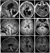
dysmorphic (missing rostrum plus flat genu and anterior body), hypoplastic, or partially absent. In contrast, all prenatally diagnosed cases show complete agenesis ( and ).
Centrally predominant pachygyria is characterized by pachygyria involving the insula and the frontal, temporal, and parietal opercula [
Bahi-Buisson et al 2008]. All have abnormalities of the basal ganglia that appear as large round structures in which the caudate, putamen, and globus pallidus are indistinguishable. Hypoplasia of the anterior limbs of the internal capsule is a major feature. Common findings are vermian hypoplasia and brain stem hypoplasia. The corpus callosum is commonly
dysmorphic or hypoplastic; less frequently, it is partially or completely absent ().
Representative images of TUBA1A-related tubulinopathies A-C. Infant age four months with classic lissencephaly. Other features include dysmorphic corpus callosum, dysmorphic/dysplastic internal capsules, and hypoplasia of the cerebellar vermis.
Microlissencephaly refers to the most severe of the cortical dysplasias that combines extreme microcephaly and lissencephaly with hemispheres lacking primary fissures and olfactory sulci. Mainly diagnosed by prenatal MRI, findings at a median age of 25 weeks' gestation include microcephaly, absent-to-poorly operculized cortex, virtually no visible gyration, severe cerebellar hypoplasia, absent or severely hypoplastic basal ganglia, and usually complete agenesis of the corpus callosum. Associated malformations include pontocerebellar hypoplasia [Fallet-Bianco et al 2008, Lecourtois et al 2010, Fallet-Bianco et al 2014].
Dysgyria describes a cortex of normal thickness but with an abnormal gyral pattern characterized by abnormalities of sulcal depth or orientation: obliquely oriented sulci directed radially toward the center of the cerebrum and narrow gyri separated by abnormally deep or shallow sulci without imaging evidence of lissencephaly, pachygyria, cobblestone cortex, polymicrogyria, or other cortical abnormalities [Mutch et al 2016, Blumkin et al 2020]. This pattern tends to predominate in the perisylvian areas.
MRI reveals a "coarse" appearance with a thick cortex and irregular surfaces on both the pial and grey-white junction sides (, , ). Dysgyria can range in severity and distribution.
Representative images of TUBB2B-related tubulinopathies A-C. Child age three years with cortical dysgyria resembling polymicrogyria. The cortex has a coarse appearance with excessively folded gyri (arrowheads in B). Cortical dysgyria resembling polymicrogyria (more...)
Representatives images of TUBB3-related tubulinopathies A-C. Child age three years with diffuse cortical dysgyria. The cortex shows a coarse appearance, with excessively folded gyri (arrowhead in B). Associated malformations include complete agenesis (more...)
Note: Dysgyria was previously referred to as "simplified gyral pattern" or "polymicrogyria-like cortical dysplasia"; these terms are potentially confusing.
Basal ganglia, thalami, and corpus callosum. One of the key feature of tubulinopathies is dysmorphic basal ganglia. This pattern is usually asymmetric with bulbous appearance of the caudate and the thalami, with diffuse, branched or absent anterior limb of the internal capsule. The lateral ventricles have an irregular contour and abnormal rounding of the frontal horns likely related to the basal ganglia dysplasia.
The corpus callosum is variably affected, ranging from almost complete agenesis to normal.
Recognizable cerebellar and brain stem abnormalities. The most characteristic tubulinopathy-related cerebellar malformation is dysplasia of the superior cerebellum, especially the vermis (with "diagonal" folia – i.e., folia crossing the midline at an oblique angle). Less commonly, the vermis is hypoplastic with the anterior vermis more severely affected. The cerebellar hemispheres are either normal in size or mildly hypoplastic with mild asymmetry.
A large majority of affected individuals show brain stem hypoplasia that is usually asymmetric with a midline ventral indentation and asymmetric inferior and middle cerebellar peduncles [Oegema et al 2015].
Clinical Features of the Tubulinopathies
The clinical features of the tubulinopathies include motor and intellectual disabilities and epilepsy.
Motor and cognitive and impairments, present in almost all individuals with a tubulinopathy, correlate with the severity of brain malformations.
Lissencephaly, microlissencephaly, and generalized severe dysgyria: spastic tetraplegia and virtually no voluntary motor control and absent eye contact
Mild-to-moderate dysgyria: mild motor disability and intellectual disabilities
While most affected individuals have severe-to-profound intellectual disability, a minority have less extensive cortical malformations that result in only moderate intellectual disability, and a few have limited malformations that allow near-normal cognitive abilities. In the latter instance, the cortical malformation is typically less severe and less extensive on MRI.
Epilepsy varies significantly among affected individuals and is not necessarily determined by the severity of the cortical malformation, the gene involved, or the causative pathogenic variant [Romaniello et al 2019]. However:
Additional findings. Facial diplegia and strabismus suggestive of pseudobulbar palsy are often observed in central pachygyria and various forms of dysgyria [Bahi-Buisson et al 2008].
Prognosis. Individuals with the milder forms of tubulinopathies survive into adulthood, while those with the most severe forms may die at a young age as a result of complications such as seizures or pneumonia.
2. Genetic Causes of Tubulinopathies
The genetic causes of tubulinopathies and their associated complex cortical malformations are summarized in Table 1.
Table 1.
Tubulinopathies: Molecular Genetics and Complex Cortical Malformations
View in own window
- 1.
Genes are in alphabetic order.
- 2.
- 3.
At the extreme severe end of the spectrum, only one fetus was reported with microlissencephaly and corpus callosum agenesis, severe brain stem and cerebellar hypoplasia, and dysmorphic basal ganglia [Poirier et al 2010] ().
3. Differential Diagnosis of Tubulinopathies
Tubulinopathies need to be distinguished clinically from other brain malformations that may resemble them (Table 2).
Table 2.
Differential Diagnosis of Tubulinopathies
View in own window
- 1.
- 2.
As currently defined, Miller-Dieker syndrome is associated with deletions that include both PAFAH1B1 (LIS1) and YWHAE (a region of ~1.3 Mb harboring many genes) in 17p13.3 [Pilz et al 1998, Cardoso et al 2003].
- 3.
4. Evaluation Strategies to Identify the Genetic Cause of Tubulinopathy in a Proband
Establishing a specific genetic cause of a tubulinopathy:
Can aid in discussions of prognosis (which are beyond the scope of this
GeneReview) and
genetic counseling;
Family history. A three-generation family history should be taken, with attention to relatives with manifestations of a tubulinopathy and documentation of relevant findings through direct examination or review of medical records, including results of molecular genetic testing.
Molecular genetic testing approaches can include a combination of gene-targeted testing (multigene panel) and comprehensive genomic testing (exome sequencing, genome sequencing). Gene-targeted testing requires the clinician to hypothesize which gene(s) are likely involved, whereas genomic testing does not.
A multigene panel that includes some or all of the genes listed in
Table 1 is most likely to identify the genetic cause of the condition while limiting identification of variants of
uncertain significance and pathogenic variants in genes that do not explain the underlying
phenotype. Note: (1) The genes included in the panel and the diagnostic
sensitivity of the testing used for each
gene vary by laboratory and are likely to change over time. (2) Some multigene panels may include genes not associated with the condition discussed in this
GeneReview. (3) In some laboratories, panel options may include a custom laboratory-designed panel and/or custom phenotype-focused
exome analysis that includes genes specified by the clinician. (4) Methods used in a panel may include
sequence analysis,
deletion/duplication analysis, and/or other non-sequencing-based tests.
For an introduction to multigene panels click
here. More detailed information for clinicians ordering genetic tests can be found
here.
Comprehensive
genomic testing (which does not require the clinician to determine which
gene[s] are likely involved) may be considered.
Exome sequencing is most commonly used;
genome sequencing is also possible.
For an introduction to comprehensive
genomic testing click
here. More detailed information for clinicians ordering genomic testing can be found
here.
5. General Medical Management of Tubulinopathies
A pediatric neurologist with expertise in the management of children with multiple disabilities and medically refractory epilepsy is recommended for long-term management.
Supportive management, including an individualized therapy plan that includes physical therapy to manage the complications of spasticity, occupational therapy, speech therapy, and vision therapy for oculomotor deficits and/or strabismus should begin at the time of diagnosis to ensure the best possible functionality and developmental outcome. Of note, it is appropriate to institute measures early on to manage potential complications of spasticity (e.g., joint contractures or reduced range of motion), which can increase the risk for decubitus ulcers as well as affect mobility and hygiene.
Those with congenital fibrosis of the extraocular muscles may require nonsurgical and/or surgical treatment.
Nutritional needs in infants with the more severe brain malformations (e.g., lissencephaly, generalized polymicrogyria) are usually managed by nasogastric tube feedings, followed by gastrostomy tube placement as needed.
Seizures are treated with anti-seizure medications based on the specific seizure type. In general, seizures should be treated promptly by specialists, as poor seizure control frequently worsens feeding and increases both the likelihood that a gastrostomy tube will be needed and the risk for aspiration.
Education of parents regarding common seizure presentations is appropriate. For information on non-medical interventions and coping strategies for parents or caregivers of children diagnosed with epilepsy, see Epilepsy Foundation Toolbox.
For individuals with severe cortical malformations (lissencephalies, polymicrogyria-like cortical dysplasia, microlissencephaly), it is usually appropriate to discuss the level of care to be provided in the event of a severe intercurrent illness.
6. Genetic Counseling of Family Members of an Individual with a Tubulinopathy
Genetic counseling is the process of providing individuals and families with
information on the nature, mode(s) of inheritance, and implications of genetic disorders to help them
make informed medical and personal decisions. The following section deals with genetic
risk assessment and the use of family history and genetic testing to clarify genetic
status for family members; it is not meant to address all personal, cultural, or
ethical issues that may arise or to substitute for consultation with a genetics
professional. —ED.
Mode of Inheritance
Tubulinopathies caused by pathogenic variants in TUBA1A, TUBB2A, TUBB2B, TUBB3, TUBB (TUBB5), or TUBG1 are inherited in an autosomal dominant manner.
Risk to Family Members
Parents of a proband
More than 95% of individuals diagnosed with a tubulinopathy have a
de novo pathogenic variant in
TUBA1A,
TUBB2A,
TUBB2B,
TUBB3,
TUBB (
TUBB5), or
TUBG1.
Rarely, an individual diagnosed with a tubulinopathy has an affected parent. These individuals generally have either a
TUBB3 or (less frequently)
TUBB2B pathogenic variant.If the
pathogenic variant identified in the
proband is not identified in either parent, the following possibilities should be considered:
The
proband has a
de novo pathogenic variant. Note: A pathogenic variant is reported as "
de novo" if: (1) the pathogenic variant found in the proband is not detected in parental DNA; and (2) parental identity testing has confirmed biological maternity and paternity. If parental identity testing is not performed, the variant is reported as "assumed
de novo" [
Richards et al 2015].
The family history of some individuals diagnosed with a tubulinopathy may appear to be negative because of failure to recognize the disorder in family members or reduced
penetrance. Therefore, an apparently negative family history cannot be confirmed unless
molecular genetic testing has demonstrated that neither parent is
heterozygous for the
pathogenic variant identified in the
proband.
Sibs of a proband. The risk to the sibs of the proband depends on the genetic status of the proband's parents:
If a parent of the
proband is affected and/or is known to have the
pathogenic variant identified in the proband, the risk to the sibs of inheriting the pathogenic variant is 50%.
If the parents have not been tested for the
pathogenic variant identified in the
proband but are clinically unaffected, the risk to the sibs of the proband appears to be low. However, sibs of a proband with clinically unaffected parents are still presumed to be at increased risk for a tubulinopathy because of the possibility of the possibility of parental
germline mosaicism.
Offspring of a proband. Each child of an individual with a tubulinopathy has a 50% chance of inheriting the pathogenic variant.
Other family members
The risk to other family members depends on the status of the
proband's parents: if a parent is affected, the parent's family members may be at risk.
Prenatal Testing and Preimplantation Genetic Testing
Once the tubulinopathy-related pathogenic variant has been identified in an affected family member, prenatal testing for a pregnancy at increased risk and preimplantation genetic testing are possible.
Differences in perspective may exist among medical professionals and within families regarding the use of prenatal testing. While most centers would consider use of prenatal testing to be a personal decision, discussion of these issues may be helpful.
Resources
GeneReviews staff has selected the following disease-specific and/or umbrella
support organizations and/or registries for the benefit of individuals with this disorder
and their families. GeneReviews is not responsible for the information provided by other
organizations. For information on selection criteria, click here.
American Association on Intellectual and Developmental Disabilities (AAIDD)
Phone: 202-387-1968
Fax: 202-387-2193
American Epilepsy Society
CDC - Developmental Disabilities
Phone: 800-CDC-INFO
Email: cdcinfo@cdc.gov
Epilepsy Foundation
Phone: 301-459-3700
Fax: 301-577-2684
National Institute of Neurological Disorders and Stroke (NINDS)
PO Box 5801
Bethesda MD 20824
Phone: 800-352-9424 (toll-free); 301-496-5751; 301-468-5981 (TTY)
Chapter Notes
Author Notes
Nadia Bahi-Buisson is a pediatric neurologist specializing in cortical malformations and fetal neurology. Her research at Imagine Institute focuses on the genetic and pathophysiologic bases of cortical malformations. She follows more than 100 patients with diffuse cortical malformations, and has consulted on more than 500 such cases. Dr Bahi-Buisson is involved in the European consortium on cortical malformations. This work is performed in collaboration with Chérif Beldjord, MD, PhD (director of the Laboratory of Biochemical Genetics – Cochin-Port-Royal).
Acknowledgments
Catherine Fallet Bianco and Annie Laquerriere, for sharing their fetal cases and for their helpful discussion of fetal brain tubulinopathies
Chérif Beldjord, Aurelie Toussaint, and Nathalie Carion, for their help in the screening of tubulin genes for diagnosis – from
Sanger sequencing to the recent development of
NGS panel screening
Jamel Chelly and Karine Poirier, who allowed the first author to collaborate on the identification of tubulin genes and to define/refine the associated phenotypes; and who have contributed through constructive discussion to the understanding of the pathophysiology of tubulinopathies
Sophie Thomas and Stanislas Lyonnet (Imagine Institute), for welcoming the authors to the Laboratory of Embryology and Genetics of Congenital Malformations and supporting their continued work in the identification of genes associated with cortical malformations
Author History
Nadia Bahi-Buisson, MD, PhD (2016-present)
Mara Cavallin, MD; Paris Descartes University (2016-2021)
Camille Maillard, PhD (2021-present)
Revision History
16 September 2021 (bp) Comprehensive update posted live
24 March 2016 (bp) Review posted live
7 July 2015 (nbb) Original submission


