U.S. flag

An official website of the United States government

NCBI Bookshelf. A service of the National Library of Medicine, National Institutes of Health.

Adam MP, Feldman J, Mirzaa GM, et al., editors. GeneReviews® [Internet]. Seattle (WA): University of Washington, Seattle; 1993-2024.

Cover of GeneReviews®

GeneReviews® [Internet].

Show details

Poikiloderma with Neutropenia

Synonym: Clericuzio-Type Poikiloderma with Neutropenia

, MD, , MD, , MD, and , MD.

Author Information and Affiliations

Initial Posting: ; Last Update: February 22, 2024.

Estimated reading time: 25 minutes

Summary

Clinical characteristics.

Poikiloderma with neutropenia (PN) is characterized by an inflammatory eczematous rash (appears at ages 6-12 months) followed by post-inflammatory poikiloderma (at age >2 years) and chronic noncyclic neutropenia typically associated with recurrent sinopulmonary infections in the first two years of life and (often) bronchiectasis. There is increased risk for myelodysplastic syndrome, acute myelogenous leukemia, and skin cancer. Other ectodermal findings include thickened nails, nail dystrophy, and palmar/plantar hyperkeratosis. Most affected individuals also have reactive airway disease, and some have short stature, hypogonadotropic hypogonadism, midfacial retrusion, calcinosis cutis, and non-healing skin ulcers.

Diagnosis/testing.

Often the diagnosis of PN can be established in a proband based on clinical findings (post-inflammatory poikiloderma and congenital chronic neutropenia). Unequivocal confirmation of the diagnosis of PN relies on detection of biallelic USB1 pathogenic variants by molecular genetic testing.

Management.

Treatment of manifestations: Dermatologic manifestations are treated with gentle skin care using bland emollients. Diligent sun protection with both UVA and UVB protection and/or sun-protective clothing to reduce the risk of skin cancer. Very pruritic palmar/plantar hyperkeratosis can be treated with a strong topical steroid or a topical keratolytic if secondary dermatophyte infection has been ruled out. Although use of granulocyte-colony stimulating factor increases the absolute neutrophil count, there is little evidence of decreased frequency of infections with this treatment. Sinopulmonary, middle ear, and skin infections require aggressive treatment with antibiotics. Annual influenza vaccine is recommended. Developmental support as needed. Gingivitis, dental caries, reactive airway disease, premyelodysplastic changes, myelodysplastic syndrome, acute myelogenous leukemia, skin cancer, hypogonadotropic hypogonadism, and osteoporosis are treated in the usual manner.

Surveillance: Annual examination by a physician familiar with PN; annual dermatology examination for skin cancer beginning at age ten years; dental examination every three to six months; annual pulmonology examination in those with bronchiectasis, chronic cough, and/or reactive airway disease; annual complete blood count with differential and platelet count with evaluation by a hematologist/oncologist as needed; assessment of growth, pubertal development, developmental milestones, and educational progress at each visit throughout childhood; DXA scan as needed in adults.

Agents/circumstances to avoid: Excessive sun exposure due to the increased risk of skin cancer; exposure to secondhand cigarette or wood smoke and persons with respiratory illnesses due to the increased risk of respiratory infections.

Evaluation of relatives at risk: It is appropriate to evaluate apparently asymptomatic older and younger sibs of a proband in order to identify as early as possible those who would benefit from prompt initiation of treatment and surveillance for potential complications.

Genetic counseling.

PN is inherited in an autosomal recessive manner. If both parents are known to be heterozygous for a USB1 pathogenic variant, each sib of an affected individual has at conception a 25% chance of being affected, a 50% chance of being an asymptomatic carrier, and a 25% chance of being unaffected and not a carrier. Once the USB1 pathogenic variants have been identified in an affected family member, carrier testing for at-risk relatives and prenatal/preimplantation genetic testing are possible.

Diagnosis

Suggestive Findings

Poikiloderma with neutropenia (PN) should be suspected in individuals with the following clinical, laboratory, and family history findings.

Clinical Findings

Skin

  • Between ages six and 12 months, inflammatory eczematous rash appearing first on the limbs and progressing to the trunk, face, and on occasion the pinnae
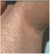
  • After age two years, post-inflammatory poikiloderma (areas of hyper- and hypopigmentation, atrophy, and telangiectasias) (See Figure 1.)
    Note: The telangiectasia may be subclinical and seen only on skin biopsy (which is not necessary for diagnosis).
Figure 1. . Post-inflammatory poikiloderma in a boy age ten years; note hypo- and hyperpigmentation.

Figure 1.

Post-inflammatory poikiloderma in a boy age ten years; note hypo- and hyperpigmentation.

Recurrent infections (as evidence of neutropenia)

  • In the first two years of life, recurrent sinopulmonary infections, often complicated by bronchiectasis
  • Adolescent- and adult-onset non-healing skin ulcers
  • Cellulitis, osteomyelitis

Laboratory Findings

Congenital chronic noncyclic neutropenia that is moderate to severe:

  • Moderate neutropenia: absolute neutrophil count (ANC)* of 500-1,000/µL
  • Severe neutropenia: ANC <500/µL
    * ANC = white blood cell count (WBC) x % neutrophils

Family History

Family history is consistent with autosomal recessive inheritance (e.g., affected sibs and/or parental consanguinity). Absence of a known family history does not preclude the diagnosis.

Establishing the Diagnosis

In many instances, the clinical diagnosis of poikiloderma with neutropenia (PN) can be established in a proband with post-inflammatory poikiloderma and congenital chronic neutropenia, or the molecular diagnosis can be established in a proband with suggestive findings and biallelic pathogenic (or likely pathogenic) variants in USB1 identified by molecular genetic testing (see Table 1).

Note: (1) Per ACMG/AMP variant interpretation guidelines, the terms "pathogenic variant" and "likely pathogenic variant" are synonymous in a clinical setting, meaning that both are considered diagnostic and can be used for clinical decision making [Richards et al 2015]. Reference to "pathogenic variants" in this GeneReview is understood to include likely pathogenic variants. (2) Identification of biallelic USB1 variants of uncertain significance (or of one known USB1 pathogenic variant and one USB1 variant of uncertain significance) does not establish or rule out the diagnosis.

Molecular genetic testing approaches can include a combination of gene-targeted testing (single-gene testing, multigene panel) and comprehensive genomic testing (exome sequencing, genome sequencing). Gene-targeted testing requires that the clinician determine which gene(s) are likely involved (see Option 1), whereas comprehensive genomic testing does not (see Option 2).

Option 1

Single-gene testing. Sequence analysis of USB1 is performed first to detect missense, nonsense, and splice site variants and small intragenic deletions/insertions. Typically, if only one or no variant is detected by the sequencing method used, the next step is to perform gene-targeted deletion/duplication analysis to detect exon and whole-gene deletions or duplications; however, to date such variants have not been identified as a cause of this disorder.

Note: Targeted analysis for pathogenic variants can be performed first in individuals of geographic regions with known founder pathogenic variants (see Table 7).

A multigene panel that includes USB1 and other genes of interest (see Differential Diagnosis) may also be considered to identify the genetic cause of the condition while limiting identification of variants of uncertain significance and pathogenic variants in genes that do not explain the underlying phenotype. Note: (1) The genes included in the panel and the diagnostic sensitivity of the testing used for each gene vary by laboratory and are likely to change over time. (2) In some laboratories, panel options may include a custom laboratory-designed panel and/or custom phenotype-focused exome analysis that includes genes specified by the clinician. (3) Methods used in a panel may include sequence analysis, deletion/duplication analysis, and/or other non-sequencing-based tests.

For an introduction to multigene panels click here. More detailed information for clinicians ordering genetic tests can be found here.

Option 2

Comprehensive genomic testing does not require the clinician to determine which gene is likely involved. Exome sequencing is most commonly used; genome sequencing is also possible.

For an introduction to comprehensive genomic testing click here. More detailed information for clinicians ordering genomic testing can be found here.

Table 1.

Molecular Genetic Testing Used in Poikiloderma with Neutropenia

Gene 1MethodProportion of Pathogenic Variants 2 Detectable by Method
USB1 Sequence analysis 3100% 4
Gene-targeted deletion/duplication analysis 5None reported 4
1.
2.

See Molecular Genetics for information on variants detected in this gene.

3.

Sequence analysis detects variants that are benign, likely benign, of uncertain significance, likely pathogenic, or pathogenic. Variants may include missense, nonsense, and canonical and non-canonical splice site variants and small intragenic deletions/insertions; typically, structural rearrangements involving the gene and/or its flanking regions require long read sequencing techniques. For issues to consider in interpretation of sequence analysis results, click here.

4.

Data derived from LOVD and the subscription-based professional view of Human Gene Mutation Database [Stenson et al 2020]

5.

Gene-targeted deletion/duplication analysis detects intragenic deletions or duplications. Methods used may include a range of techniques such as quantitative PCR, long-range PCR, multiplex ligation-dependent probe amplification (MLPA), and a gene-targeted microarray designed to detect single-exon deletions or duplications. Exome and genome sequencing may be able to detect deletions/duplications using breakpoint detection or read depth; however, sensitivity can be lower than gene-targeted deletion/duplication analysis.

Clinical Characteristics

Clinical Description

Poikiloderma with neutropenia (PN) is characterized by post-inflammatory poikiloderma and chronic noncyclic neutropenia typically associated with recurrent sinopulmonary infections and often bronchiectasis. There is increased risk for myelodysplastic syndrome, which may evolve into acute myelogenous leukemia, and skin squamous cell carcinoma. Other ectodermal findings include thickened nails, nail dystrophy, and palmar/plantar hyperkeratosis. Most affected individuals also have reactive airway disease, and some have short stature, hypogonadotropic hypogonadism, midfacial retrusion, calcinosis cutis, and non-healing skin ulcers [Colombo et al 2012]. Intrafamilial clinical variability has been observed. The clinical information that follows is based on around 100 individuals with a clinical and molecular diagnosis of PN [Larizza 2024].

Table 2.

Poikiloderma with Neutropenia: Frequency of Select Features

Feature% of Persons w/FeatureComment
Poikiloderma >95%Post-inflammatory
Palmar/plantar hyperkeratosis 50%-75%
Nail abnormalities >95%Thick nails, dystrophic nails
Hair abnormalities 50%-75%
Dental abnormalities 20%-40%
Neutropenia >95%Chronic noncyclic, moderate to severe
Recurrent infections 90%Respiratory infections, otitis media, sinusitis, cellulitis
Malignancies 5%-10%Myelodysplasia, hematologic malignancies; rarely, skin cancer
Facial dysmorphism 50%-75%Frontal bossing, midface hypoplasia, depressed nasal bridge, hypertelorism

Ectodermal Features

Skin. Typically, the skin is normal at birth, and at age six to 12 months, a nonpruritic acral eczematous-like rash develops that progresses to the trunk and face. Rarely, the rash can start on the face [Tadros et al 2021]. Over the next year or so the inflammatory rash resolves, the skin becomes dry, and poikiloderma becomes evident as areas of hyper- and hypopigmentation, atrophy, and telangiectasias develop (see Figure 1). Poikiloderma is not photodistributed, persists throughout life, and may be more noticeable in individuals who have constitutionally darker skin.

Palmar/plantar hyperkeratosis is common, can range from mild to severe, and can be debilitating [Concolino et al 2019, Akdogan et al 2020, Bilgic Eltan et al 2021].

Calcinosis cutis – small nodules that may be localized to the elbows, knees, and pinnae or can be more diffuse – may develop in childhood [Clericuzio et al 2011, Bishnoi et al 2021, Kreuter et al 2021, Tadros et al 2021, Parajuli et al 2024].

Children and adults are prone to cellulitis (a manifestation of neutropenia) that may progress to non-healing skin ulcers or even abscesses [Parperis et al 2020, Kreuter et al 2021, Roebke et al 2021, Parajuli et al 2024]. One individual has been described with pyoderma gangrenosum [Al Haddabi et al, unpublished data].

Photosensitivity and blistering have been reported in a few individuals [Kreuter et al 2021, Vahidnezhad et al 2021, Peterson et al 2022].

Squamous cell carcinoma of the skin has been reported in a handful of individuals at young ages (age 13 to 20 years) [Walne et al 2010, Rodgers et al 2013, Colombo et al 2018, Hertel et al 2018, Concolino et al 2019, Kreuter et al 2021].

Nails. Thickened, hyperkeratotic toenails and/or fingernails (pachyonychia) are common; dystrophic nails, which can slough, may also be seen (see Figure 2) [Bilgic Eltan et al 2021, Bishnoi et al 2021, Kreuter et al 2021, Peterson et al 2022, Yan et al 2023]. Anonychia was observed in one individual [Colombo et al 2012].

Figure 2. A.

Figure 2

A. Nail dystrophy in a girl age five years B. Dysplastic toenails and squamous cell carcinoma (blue arrow) in a girl age 14 years

Hair. Eyebrows and eyelashes are often sparse; hair can be dry and thin [Parperis et al 2020, Bilgic Eltan et al 2021, Kreuter et al 2021, Vahidnezhad et al 2021].

Teeth. Delayed dental eruption, abnormally shaped teeth, and dental abscesses have been observed. Gingivitis and dental caries leading to tooth loss are common [Concolino et al 2019, Bishnoi et al 2021, Kreuter et al 2021, Tadros et al 2021].

Hematologic Findings

Neutropenia/infections. Neutropenia is usually identified in early infancy. Chronic recurrent otitis media and sinusitis are common in childhood. After age five to ten years, the frequency of acute sinopulmonary infections decreases, but most individuals continue to have bronchiectasis, chronic non-productive cough, and reactive airway disease. Almost 90% of individuals with PN have recurrent pulmonary infections, including lung abscesses and lung granulomas.

Most individuals with PN who are not acutely ill have moderate neutropenia, although some have severe neutropenia [Bilgic Eltan et al 2021, Tadros et al 2021, Peterson et al 2022, Yan et al 2023, Parajuli et al 2024]. While the absolute neutrophil count (ANC) may rise to the low-normal range during acute infection, it is always inappropriately low, and with resolution of the infection reverts to baseline neutropenia.

Quantitative immunoglobulins and lymphocyte subset panels are normal.

Transient thrombocytopenia and variable anemia have been reported [Patiroglu & Akar 2015, Walne et al 2016, Colombo et al 2018, Bilgic Eltan et al 2021, Piccolo et al 2021].

Myelodysplasia and Hematologic Malignancies

Bone marrow studies have been described in a portion of reported individuals and have shown hypocellularity and premyelodysplastic changes (often defined as <10% abnormal cells), including increased number of immature cells and myeloid maturation defects. Some individuals with PN initially have normal bone marrow studies and subsequently develop bone marrow changes. Global impaired bone marrow production has been reported, affecting the development of all three major lineages [Concolino et al 2019, Bilgic Eltan et al 2021, Kreuter et al 2021]. Persons with PN can go on to develop myelodysplastic syndrome as well as acute myelogenous leukemia [Colombo et al 2018].

Facial Features

Facial features are usually normal at birth; however, over time characteristic craniofacial features of prominent forehead, frontal bossing, depressed nasal bridge, and midface retrusion usually develop (see Figure 3) [Concolino et al 2019, Akdogan et al 2020, Bishnoi et al 2021, Parperis et al 2020, Kreuter et al 2021, Vahidnezhad et al 2021].

Figure 3.

Figure 3.

Three Italian sibs with typical midfacial hypoplasia

Endocrine Manifestations

Growth deficiency. Birth length and weight are usually normal; however, intrauterine growth restriction (IUGR) can be seen. Postnatal-onset short stature not associated with growth hormone deficiency is common. One individual was reported to be unresponsive to growth hormone therapy [Koparir et al 2014].

Hypogonadotropic hypogonadism causing delayed puberty is not uncommon [Akdogan et al 2020, Kreuter et al 2021].

Abnormal bone density. Osteopenia, osteoporosis, and increased bone fragility have been reported. Increased bone density has also been reported [Akdogan et al 2020, Bilgic Eltan et al 2021, Kreuter et al 2021, Vahidnezhad et al 2021].

Other Findings

Additional abnormal laboratory findings. Virtually all individuals have elevated serum lactate dehydrogenase of unknown etiology; some have nonspecific mild elevation of aminotransferases, aspartate aminotransferase, ferritin, and creatine phosphokinase [Concolino et al 2019, Bilgic Eltan et al 2021, Kreuter et al 2021, Piccolo et al 2021, Peterson et al 2022].

Hepatosplenomegaly has been described in several individuals [Concolino et al 2019, Bilgic Eltan et al 2021, Piccolo et al 2021, Tadros et al 2021].

Development. Although early developmental delays (possibly related to chronic illness) have been reported, intellectual disability has not been reported.

Muscle weakness. Several individuals with muscle weakness had normal muscle biopsies.

Other rare features

Genotype-Phenotype Correlations

Genotype-phenotype correlations have not been established to date. Individuals homozygous for USB1 pathogenic variants c.531delA, c.243G>A, or c.541C>T may have a higher risk of cancer development [Colombo et al 2018].

Nomenclature

Poikiloderma with neutropenia was termed "immune-deficient poikiloderma" in the publication by Clericuzio et al [1991], who first described the condition in the Navajo population. When it was subsequently identified in individuals of other ethnicities, the condition was renamed poikiloderma with neutropenia [Wang et al 2003].

In 2005 Van Hove proposed renaming the disorder Clericuzio-type poikiloderma with neutropenia [Van Hove et al 2005].

Prevalence

First described in the Navajo Native American population [Clericuzio et al 1991], PN has since been identified in individuals of other ethnicities.

Pathogenic variant c.531delA, identified in 22 individuals from 16 families of Turkish ancestry, is the most prevalent USB1 pathogenic variant identified to date.

Differential Diagnosis

Table 3 summarizes inherited disorders with either poikiloderma or neutropenia in the differential diagnosis of poikiloderma with neutropenia (PN). These disorders can be distinguished from PN by the distinctive characteristics of poikiloderma and neutropenia in PN and by additional clinical features (see Table 3).

Of note, prior to USB1 molecular genetic testing, individuals with PN have been clinically misdiagnosed as having Rothmund-Thomson syndrome, dyskeratosis congenita, and Kindler syndrome (a rare subtype of epidermolysis bullosa) [Walne et al 2016, Akdogan et al 2020,Tadros et al 2021,Vahidnezhad et al 2021].

Additional considerations in the differential diagnosis of PN include severe congenital ELANE-related neutropenia and hereditary fibrosing poikiloderma (with which PN resemblance is evidenced by computational facial analysis).

Table 3.

Disorders to Consider in the Differential Diagnosis of Poikiloderma with Neutropenia

Gene(s)DisorderMOIFeatures of Disorder
Overlapping w/PNDistinguishing From PN
ACD
CTC1
DKC1
NAF1
NHP2
NOP10
PARN
POT1
RPA1
RTEL1
STN1
TERC
TERT
TINF2
WRAP53
ZCCHC8
Dyskeratosis congenita & related telomere biology disorders XL
AD
AR 1
  • Poikiloderma
  • Acute myelogenous leukemia
  • Myelodysplasia
  • Nail dystrophy
  • Poikiloderma on upper chest/neck
  • Oral leukoplakia, pulmonary fibrosis. & short telomeres (on lab testing)
ANAPC1
RECQL4
Rothmund-Thomson syndrome AR
  • Early-onset poikiloderma
  • Dental abnormalities
  • Nail dystrophy
  • Palmar/plantar hyperkeratosis
  • Skin SCC
  • Short stature
  • Sparse hair & eyebrows/lashes
  • Rash typically starts on face & spreads to extremities.
  • Skeletal defects (incl radial ray defects), GI disturbance, & cataracts
  • Osteosarcoma is the predominant cancer.
ELANE ELANE-related neutropenia ADCongenital neutropenia
  • Neutropenia is cyclic & severe.
  • Not assoc w/poikiloderma or nail dystrophy.
FAM111B Hereditary fibrosing poikiloderma w/tendon contractures, myopathy, & pulmonary fibrosis (POIKTMP) AD
  • Poikiloderma
  • Nail dysplasia
  • Palmar/plantar hyperkeratosis
  • Recurrent bronchitis
  • Short stature
  • Sparse/absent eyelashes &/or eyebrows
  • Thrombocytopenia, marrow hypocellularity
  • Poikiloderma is localized to face.
  • Hypohidrosis, muscle contractures, lymphedema of the extremities, myopathy, exocrine pancreatic insufficiency, & pulmonary fibrosis
FERMT1 Kindler syndrome epidermolysis bullosa 2AR
  • Poikiloderma
  • Blistering
  • Photosensitivity
  • Skin atrophy
  • Hyperkeratosis
  • Nail dystrophy
  • Loss of dermatoglyphics
  • Mucocutaneous SCC
  • Blistering is early onset & severe, while poikiloderma is later onset.
  • Generalized atrophy & oral mucosal fragility is prominent & may lead to strictures.

AD = autosomal dominant; AR = autosomal recessive; GI = gastrointestinal; MOI = mode of inheritance; PN = poikiloderma with neutropenia; SCC = squamous cell carcinoma; XL = X-linked

1.

The mode of inheritance of dyskeratosis congenita & related telomere biology disorders varies by gene (see Genetic Counseling in the Dyskeratosis Congenita and Related Telomere Biology Disorders GeneReview).

2.

Management

Evaluations Following Initial Diagnosis

To establish the extent of disease and needs in an individual diagnosed with poikiloderma with neutropenia (PN), the evaluations summarized in Table 4 (if not performed as part of the evaluation that led to the diagnosis) are recommended,

Table 4.

Poikiloderma with Neutropenia: Recommended Evaluations Following Initial Diagnosis

System/ConcernEvaluationComment
Skin Dermatologic eval to assess for calcinosis cutis, cellulitis, palmar/plantar hyperkeratosis, & examine for evidence of non-healing ulcers & skin cancer
Dental Dental eval to assess for gingivitis &/or caries
Otolaryngology Otolaryngology eval for complications of chronic otitis media
Pulmonary Pulmonary consult to incl eval for bronchiectasis & granulomas & to assess for reactive airway disease
Hematology
  • CBC w/differential & platelet count
  • Hematology/oncology consultation to eval for need for bone marrow exam (e.g., if >1 cell line is abnormal on CBC)
Endocrine Assess growth & pubertal development in children,
Gastrointestinal
  • AST & ALT
  • GI eval if hepatosplenomegaly &/or ↑ liver transaminases are present
Development Developmental evalIn children age <5 yrs
Genetic counseling By genetics professionals 1To inform affected persons & their families re nature, MOI, & implications of PN to facilitate medical & personal decision making

ALT = alanine aminotransferase; AST = aspartate aminotransferase; CBC = complete blood count; MOI = mode of inheritance; PN = poikiloderma with neutropenia

1.

Medical geneticist, certified genetic counselor, certified advanced genetic nurse

Treatment of Manifestations

Supportive care to improve quality of life, maximize function, and reduce complications is recommended. This ideally involves multidisciplinary care by specialists in relevant fields (see Table 5).

Table 5.

Poikiloderma with Neutropenia: Treatment of Manifestations

Manifestation/ConcernTreatmentConsiderations/Other
Skin manifestations
  • Gentle skin care using bland emollients (creams or ointments)
  • Use of sunscreens w/UVA & UVB protection to ↓ risk of skin cancer
  • Sun-protective clothing
Consider strong topical steroid (e.g., fluocinonide, clobetasol) 2-3x/day for short duration for keratosis-associated discomfort due to inflammation.If hyperkeratosis is very pruritic & there is no evidence of secondary dermatophyte infection
Another treatment option for hyperkeratosis is topical keratolytics:
  • 40% urea
  • Compounded salicylic acid mixed in cream or propylene glycol
Standard mgmt of skin cancers
Gingivitis /
Dental caries
Dental cleaning every 3-6 mos w/treatment per dentist
Neutropenia Based on lack of evidence of definitive benefit, G-CSF should be considered only in persons w/severe infections assoc w/very low neutrophil count.Although administration of G-CSF to persons w/PN ↑ the ANC, to date no reports have indicated definitive benefit. 1
Sinopulmonary, middle ear, & skin infections
  • Aggressive antibiotic treatment until clinical resolution & normalization of inflammatory markers.
  • Annual influenza vaccine
Consider prophylactic antibiotics during winter, which may ↓ frequency of recurrent sinopulmonary infections [Authors, personal observation].
  • There is no rationale for immunoglobulin therapy in the absence of low immunoglobulin levels.
  • Consider hyperbaric oxygen treatment for refractory cellulitis, as reported in 1 person. 2
Reactive airway disease Standard treatment per pulmonologist
Bone marrow dysplasia /
Malignancy
  • Mgmt of premyelodysplastic changes per hematologist/oncologist
  • Standard mgmt of myelodysplastic syndrome & acute myelogenous leukemia
Growth deficiency No known treatmentThere is no evidence that growth hormone therapy ↑ linear growth.
Delayed puberty Mgmt of hypogonadotropic hypogonadism per endocrinologist may incl hormone replacement therapy.
Osteoporosis Treatment per orthopedist &/or endocrinologist
Developmental delay Developmental support as needed

ANC = absolute neutrophil count; G-CSF = granulocyte-colony stimulating factor; PN = poikiloderma with neutropenia

1.
2.

Surveillance

To monitor existing manifestations, the individual's response to supportive care, and the emergence of new manifestations, the evaluations summarized in Table 6 are recommended.

Table 6.

Poikiloderma with Neutropenia: Recommended Surveillance

System/ConcernEvaluationFrequency
General Exam by physician familiar w/PNAnnually
Skin Assessment for calcinosis cutis, cellulitis, palmar/plantar hyperkeratosis & evidence of non-healing ulcersAnnually or as needed
Exam by dermatologist to assess for skin cancerAnnually beginning at age 10 yrs
Dental Dental cleaning & eval for gingivitis/cariesEvery 3-6 mos
Pulmonology Eval by pulmonologistAnnually in persons w/bronchiectasis, chronic cough, &/or reactive airway disease
Hematology CBC w/differential & platelet count for evidence of anemia &/or thrombocytopenia (signs of possible myelodysplastic syndrome)Annually
Eval by hematologist/oncologist for consideration of bone marrow examAs needed
Endocrine Assessment of growth & pubertal development in childrenAt each visit throughout childhood
Consider DXA scan to eval for low bone density.As needed in adults
Development Assessment for developmental & educational progressAt each visit throughout childhood

CBC = complete blood count; DXA = dual-energy x-ray absorptiometry; PN = poikiloderma with neutropenia

Agents/Circumstances to Avoid

Avoid excessive sun exposure due to the increased risk of skin cancer.

Avoid exposure to secondhand cigarette or wood smoke and persons with respiratory illnesses due to increased risk of respiratory infections.

Evaluation of Relatives at Risk

It is appropriate to evaluate apparently asymptomatic older and younger sibs of a proband in order to identify as early as possible those who would benefit from prompt initiation of treatment and surveillance for potential complications. Evaluations can include the following:

  • If the USB1 pathogenic variants in the family are known: molecular genetic testing
  • If the pathogenic variants in the family are not known:
    • Examination by a clinician familiar with PN to evaluate for the characteristic skin changes
    • Complete blood count with differential and platelet count, especially in newborn sibs who have not manifested a skin rash

See Genetic Counseling for issues related to testing of at-risk relatives for genetic counseling purposes.

Therapies Under Investigation

Search ClinicalTrials.gov in the US and EU Clinical Trials Register in Europe for access to information on clinical studies for a wide range of diseases and conditions. Note: There may not be clinical trials for this disorder.

Genetic Counseling

Genetic counseling is the process of providing individuals and families with information on the nature, mode(s) of inheritance, and implications of genetic disorders to help them make informed medical and personal decisions. The following section deals with genetic risk assessment and the use of family history and genetic testing to clarify genetic status for family members; it is not meant to address all personal, cultural, or ethical issues that may arise or to substitute for consultation with a genetics professional. —ED.

Mode of Inheritance

Poikiloderma with neutropenia (PN) is inherited in an autosomal recessive manner.

Risk to Family Members

Parents of a proband

Sibs of a proband

  • If both parents are known to be heterozygous for a USB1 pathogenic variant, each sib of an affected individual has at conception a 25% chance of being affected, a 50% chance of being an asymptomatic carrier, and a 25% chance of being unaffected and not a carrier.
  • Variability in midfacial hypoplasia, poikiloderma, neutropenia [Concolino et al 2019], and pulmonary involvement [Patiroglu & Akar 2015] has been observed among affected sibs; the most significant instance of intrafamilial clinical variability was acute myelogenous leukemia observed in one of two sibs with PN [Porter et al 1999].
  • Heterozygotes (carriers) are asymptomatic and are not at risk of developing the disorder.

Offspring of a proband. The offspring of an individual with PN are obligate heterozygotes (carriers) for a pathogenic variant in USB1.

Other family members. Each sib of the proband’s parents is at a 50% risk of being a carrier of a USB1 pathogenic variant.

Carrier Detection

Carrier testing for at-risk relatives requires prior identification of the USB1 pathogenic variants in the family.

Related Genetic Counseling Issues

See Management, Evaluation of Relatives at Risk for information on evaluating at-risk relatives for the purpose of early diagnosis and treatment.

Family planning

  • The optimal time for determination of genetic risk and discussion of the availability of prenatal/preimplantation genetic testing is before pregnancy.
  • It is appropriate to offer genetic counseling (including discussion of potential risks to offspring and reproductive options) to young adults who are affected, are carriers, or are at risk of being carriers.
  • Carrier testing for the reproductive partners of known carriers and for the reproductive partners individuals affected with PN should be considered, particularly if consanguinity is likely. Founder variants have been identified in several population groups (see Table 7).

Prenatal Testing and Preimplantation Genetic Testing

Once the USB1 pathogenic variants have been identified in an affected family member, prenatal and preimplantation genetic testing are possible.

Differences in perspective may exist among medical professionals and within families regarding the use of prenatal testing. While most centers would consider use of prenatal testing to be a personal decision, discussion of these issues may be helpful.

Resources

GeneReviews staff has selected the following disease-specific and/or umbrella support organizations and/or registries for the benefit of individuals with this disorder and their families. GeneReviews is not responsible for the information provided by other organizations. For information on selection criteria, click here.

  • National Neutropenia Network
    Phone: 866-600-0799
    Email: stephanie@neutropenianet.org
  • Neutropenia Support Association Inc.
    Canada
    Phone: 800-6-NEUTRO (638876); 204-781-7240
    Email: stevensl@neutropenia.ca
  • European Society for Immunodeficiencies (ESID) Registry
    Email: esid-registry@uniklinik-freiburg.de
  • Severe Chronic Neutropenia International Registry
    Phone: 49-511-557105
    Fax: 49-511-557106
    Email: SCNIR@mh-hannover.de

Molecular Genetics

Information in the Molecular Genetics and OMIM tables may differ from that elsewhere in the GeneReview: tables may contain more recent information. —ED.

Table A.

Poikiloderma with Neutropenia: Genes and Databases

GeneChromosome LocusProteinLocus-Specific DatabasesHGMDClinVar
USB1 16q21 U6 snRNA phosphodiesterase 1 USB1 database USB1 USB1

Data are compiled from the following standard references: gene from HGNC; chromosome locus from OMIM; protein from UniProt. For a description of databases (Locus Specific, HGMD, ClinVar) to which links are provided, click here.

Table B.

OMIM Entries for Poikiloderma with Neutropenia (View All in OMIM)

604173POIKILODERMA WITH NEUTROPENIA; PN
613276U6 SMALL NUCLEAR RNA BIOGENESIS PHOSPHODIESTERASE 1; USB1

Molecular Pathogenesis

USB1 encodes a member of the 2H phosphodiesterase family, U6 snRNA phosphodiesterase 1 (USB1), recognized by its characteristic fold with conserved terminal and transit lobes and the two H x S tetrapeptid motifs that frame the catalytic site [Huynh & Parker 2023]. Based on the bioinformatic prediction, all reported pathogenic variants directly or indirectly affect the enzymatic domain of the protein by either deleting one or both key histidine-serine motifs (His120/208-Ser122/210) – or destroying the native fold and the pseudosymmetric structure of the protein [Colombo et al 2012, Huynh & Parker 2023].

USB1 regulates hematopoietic development by primarily acting as microRNA (miRNA) deadenylase [Jeong et al 2023]. Embryonic human stem cells with USB1 pathogenic variant c.531delA have been show to, upon differentiation, lead to reduced formation of myeloid precursors and mature blood population without splicing impairment. The transcriptome and miRNome of USB1 mutated cells during blood development revealed dysregulated miRNA levels due to the failure to remove their 3' adenylated tails, favoring premature degradation. These findings identify USB1 as a de-tailing enzyme and suggest inhibitors of the adenylating PAPD5/7 enzyme as potential therapy for poikiloderma with neutropenia (PN) [Jeong et al 2023].

A study initiated by the management of two affected sibs (one with a non-healing fistula) demonstrated, by multiparametric analyses of their mononuclear cells, the virtual absence of nonclassic CD16+ monocytes, suggesting a block of differentiation from the classic CD14+ progenitors. The defective monocyte functions – impaired monocyte-macrophage plasticity and lack of interstitial repair – are mirrored by the PN sibs' monocyte transcriptome profiling, which reveal a signature consistent with the hematologic findings and the clinical presentation [Parajuli et al 2024]. These insights confirm the intrinsic requirement of USB1 in human hematopoiesis and highlight monocytes as the linchpin of the intertwined USB1 functions in the hematopoietic and skin tissues, so far only inferred by the clinical signs of PN and the increased risk of developing myelodysplasia, acute myeloid leukemia, and skin cancer.

Mechanism of disease causation. Loss of function

USB1-specific laboratory technical considerations. In individuals with a clinical diagnosis of PN and only one or no USB1 pathogenic variant detected, long-read sequencing should be considered to identify complex structural variants (deletions/duplications >50 bp), missed by the current short-read sequencing testing methods.

Table 7.

USB1 Pathogenic Variants Referenced in This GeneReview

Reference SequencesDNA Nucleotide Change
(Alias 1)
Predicted Protein ChangeComment [Reference]
NM_024598​.4
NP_078874​.2
c.179delCp.Pro60LeufsTer55Founder variant in northwest Africa [Tanaka et al 2010]
c.243G>Ap.Trp81TerCommon variant in European persons [Piard et al 2012]
c.499delA
(496delA)
p.Thr167ProfsTer98Founder variant in persons of Navajo & Apache ancestry [Clericuzio et al 2011]
c.531delAp.His179MetfsTer86Founder variant in persons of Turkish ancestry [Kilic & Cekic 2016]
c.541C>Tp.Gln181TerSee Genotype-Phenotype Correlations.

Variants listed in the table have been provided by the authors. GeneReviews staff have not independently verified the classification of variants.

GeneReviews follows the standard naming conventions of the Human Genome Variation Society (varnomen​.hgvs.org). See Quick Reference for an explanation of nomenclature.

1.

Variant designation that does not conform to current naming conventions.

Chapter Notes

Author Notes

Dr Wang is a pediatric oncologist at Texas Children's Cancer Center with particular interest in treating children with solid tumors, particularly osteosarcoma. She has a long-standing interest in Rothmund-Thomson syndrome and related disorders.

Dr Clericuzio is a clinical geneticist and dysmorphologist at University of New Mexico Health Sciences Center with an interest in cancer syndromes. She was the first to identify poikiloderma with neutropenia as a unique disorder in 1991.

Dr Larizza is a former professor of Medical Genetics at the University of Milan with a long-lasting interest in rare diseases, particularly cancer-predisposing syndromes. In 2010 her group identified USB1 as the gene involved in Clericuzio-type poikiloderma with neutropenia.

Acknowledgments

We thank Dr D Concolino for providing the photographs in Figure 3 and the families who have generously shared their information.

L Larizza thanks Dr EA Colombo (curator of the USB1 LOVD) for updating the USB1 pathogenic variants.

Revision History

  • 22 February 2024 (sw) Comprehensive update posted live
  • 26 October 2017 (bp) Review posted live
  • 19 December 2016 (lw) Original submission

References

Literature Cited

  • Akdogan N, Kindis E, Bostan E, Utine E, Alikasifoglu M, Ersoy-Evans S. Poikiloderma with neutropenia, Clericuzio-type accompanied by loss of digits due to severe osteomyelitis. J Clin Immunol. 2020;40:934-9. [PubMed: 32620997]
  • Bilgic Eltan S, Sefer AP, Karakus İS, Ozen A, Karakoc-Aydiner E, Baris S. Lymphopenia with low T and NK cells in a patient with USB1 mutation, rare findings in Clericuzio-type poikiloderma with neutropenia. J Clin Immunol. 2021;41:1106-11. [PubMed: 33624217]
  • Bishnoi A, Jamwal M, Das R, Scaria V, Vishwajeet V, De D, Saikia UN, Mahajan R. Clericuzio-type poikiloderma with neutropenia in a patient from India. Am J Med Genet A. 2021;185:278-81. [PubMed: 33111394]
  • Clericuzio C, Harutyunyan K, Jin W, Erickson RP, Irvine AD, McLean WH, Wen Y, Bagatell R, Griffin TA, Shwayder TA, Plon SE, Wang LL. Identification of a novel C16orf57 mutation in Athabaskan patients with poikiloderma with neutropenia. Am J Med Genet A. 2011;155A:337-42. [PMC free article: PMC3069503] [PubMed: 21271650]
  • Clericuzio C, Hoyme HE, Asse JM. Immune deficient poikiloderma: a new genodermatosis. Am J Hum Genet. 1991;49:A661.
  • Colombo EA, Bazan JF, Negri G, Gervasini C, Elcioglu NH, Yucelten D, Altunay I, Cetincelik U, Teti A, Del Fattore A, Luciani M, Sullivan SK, Yan AC, Volpi L, Larizza L. Novel C16orf57 mutations in patients with poikiloderma with neutropenia: bioinformatic analysis of the protein and predicted effects of all reported mutations. Orphanet J Rare Dis. 2012;7:7. [PMC free article: PMC3315733] [PubMed: 22269211]
  • Colombo EA, Elcioglu NH, Graziano C, Farinelli P, Di Fede E, Neri I, Facchini E, Greco M, Gervasini C, Larizza L. Insights into mutation effect in three poikiloderma with neutropenia patients by transcript analysis and disease evolution of reported patients with the same pathogenic variants. J Clin Immunol. 2018;38:494-502. [PubMed: 29770900]
  • Concolino D, Roversi G, Muzzi GL, Sestito S, Colombo EA, Volpi L, Larizza L, Strisciuglio P. Clericuzio-type poikiloderma with neutropenia syndrome in three sibs with mutations in the C16orf57 gene: delineation of the phenotype. Am J Med Genet A. 2010;152A:2588-94. [PubMed: 20734427]
  • Concolino D, Sestito S, Falvo F, Romano G, Ceravolo M, Anastasio E, Pensabene L, Colombo EA, Larizza L. Assessment of intrafamilial clinical variability of poikiloderma with neutropenia by a 10-year follow-up of three affected siblings. Eur J Med Genet. 2019;62:73-6. [PubMed: 29753917]
  • Hertel DP, Mohammad TF, Shwayder TA. Poikiloderma with neutropenia and associated squamous cell carcinoma: a case report. Pediatr Dermatol. 2018;35:e366-e367. [PubMed: 30152552]
  • Huynh TN, Parker R. The PARN, TOE1, and USB1 RNA deadenylases and their roles in non-coding RNA regulation. J Biol Chem. 2023;299:105139. [PMC free article: PMC10493513] [PubMed: 37544646]
  • Jeong HC, Shukla S, Fok WC, Huynh TN, Batista LFZ, Parker R. USB1 is a miRNA deadenylase that regulates hematopoietic development. Science. 2023;379:901-7. [PMC free article: PMC10827040] [PubMed: 36862787]
  • Jónsson H, Sulem P, Kehr B, Kristmundsdottir S, Zink F, Hjartarson E, Hardarson MT, Hjorleifsson KE, Eggertsson HP, Gudjonsson SA, Ward LD, Arnadottir GA, Helgason EA, Helgason H, Gylfason A, Jonasdottir A, Jonasdottir A, Rafnar T, Frigge M, Stacey SN, Th Magnusson O, Thorsteinsdottir U, Masson G, Kong A, Halldorsson BV, Helgason A, Gudbjartsson DF, Stefansson K. Parental influence on human germline de novo mutations in 1,548 trios from Iceland. Nature. 2017;549:519-22. [PubMed: 28959963]
  • Kilic SS, Cekic S. Juvenile idiopathic inflammatory myopathy in a patient with dyskeratosis congenita due to C16orf57 mutation. J Pediatr Hematol Oncol. 2016;38:e75-7. [PubMed: 26535771]
  • Koparir A, Gezdirici A, Koparir E, Ulucan H, Yilmaz M, Erdemir A, Yuksel A, Ozen M. Poikiloderma with neutropenia: genotype-ethnic origin correlation, expanding phenotype and literature review. Am J Med Genet A. 2014;164A:2535-40. [PubMed: 25044170]
  • Kreuter A, Koushk-Jalali B, Akgül B, Silling S, Oellig F, Has C, Wieland U. HPV-5-associated cutaneous squamous cell carcinoma in situ in poikiloderma with neutropenia. Clin Exp Dermatol. 2021;46:1619-21. [PubMed: 34155685]
  • Larizza L. From clinical findings to the pathomechanism of poikiloderma with neutropenia. Br J Haematol. 2024;204:395-6. [PubMed: 37932156]
  • Parajuli P, Craig DB, Gadgeel M, Bagla S, Wright RE 3rd, Chu R, Shanti CM, Thirunagari R, Grover SK, Ravindranath Y. Defective monocyte plasticity and altered cAMP pathway characterize USB1-mutated poikiloderma with neutropenia Clericuzio type. Br J Haematol. 2024;204:683-93. [PubMed: 37779259]
  • Parperis K, Letsos K, Loizidou MA, Hadjisavvas A, Tanteles GA. Jaccoud's arthropathy in Clericuzio-type poikiloderma with neutropenia. Rheumatology (Oxford). 2020. Epub ahead of print. [PubMed: 32415769]
  • Patiroglu T, Akar HH. Clericuzio-type poikiloderma with neutropenia syndrome in a Turkish family: a three report of siblings with mutation in the C16orf57 gene. Iran J Allergy Asthma Immunol. 2015;14:331-7. [PubMed: 26546903]
  • Peterson MY, Hanson B, Polcari I. Poikiloderma with neutropenia: An alternate presentation with dyspigmentation and novel USB1 mutation. Pediatr Dermatol. 2022;39:609-12. [PMC free article: PMC9543344] [PubMed: 35522049]
  • Piard J, Holder-Espinasse M, Aral B, Gigot N, Rio M, Tardieu M, Puzenat E, Goldenberg A, Toutain A, Franques J, MacDermot K, Bessis D, Boute O, Callier P, Gueneau L, Huet F, Vabres P, Catteau B, Faivre L, Thauvin-Robinet C. Systematic search for neutropenia should be part of the first screening in patients with poikiloderma. Eur J Med Genet. 2012;55:8-11. [PubMed: 21872685]
  • Piccolo V, Russo T, Di Pinto D, Pota E, Di Martino M, Piluso G, Ronchi A, Argenziano G, Di Brizzi EV, Santoro C. Poikiloderma with neutropenia and mastocytosis: a case report and a review of dermatological signs. Front Med (Lausanne). 2021;8:680363. [PMC free article: PMC8222900] [PubMed: 34179048]
  • Porter WM, Hardman CM, Abdalla SH, Powles AV. Haematological disease in siblings with Rothmund-Thomson syndrome. Clin Exp Dermatol. 1999;24:452-4. [PubMed: 10606946]
  • Richards S, Aziz N, Bale S, Bick D, Das S, Gastier-Foster J, Grody WW, Hegde M, Lyon E, Spector E, Voelkerding K, Rehm HL; ACMG Laboratory Quality Assurance Committee. Standards and guidelines for the interpretation of sequence variants: a joint consensus recommendation of the American College of Medical Genetics and Genomics and the Association for Molecular Pathology. Genet Med. 2015;17:405-24. [PMC free article: PMC4544753] [PubMed: 25741868]
  • Rodgers W, Ancliff P, Ponting CP, Sanchez-Pulido L, Burns S, Hayman M, Kimonis V, Sebire N, Bulstrode N, Harper JI. Squamous cell carcinoma in a child with Clericuzio-type poikiloderma with neutropenia. Br J Dermatol. 2013;168:665-7. [PubMed: 22924337]
  • Roebke LJ, Vander Maten JW, Alkhoury G. Hyperbaric oxygen management of recurrent cellulitis in poikiloderma with neutropenia. Am J Med Genet A. 2021;185:2150-2. [PubMed: 33836117]
  • Stenson PD, Mort M, Ball EV, Chapman M, Evans K, Azevedo L, Hayden M, Heywood S, Millar DS, Phillips AD, Cooper DN. The Human Gene Mutation Database (HGMD®): optimizing its use in a clinical diagnostic or research setting. Hum Genet. 2020;139:1197-207. [PMC free article: PMC7497289] [PubMed: 32596782]
  • Tadros S, Forde K, Syed S, Gholam K, Hurst J. When Rothmund-Thomson syndrome is not: two new cases of Clericuzio-type poikiloderma with neutropenia. Clin Dysmorphol. 2021;30:50-3. [PubMed: 32897901]
  • Tanaka A, Morice-Picard F, Lacombe D, Nagy N, Hide M, Taïeb A, McGrath J. Identification of a homozygous deletion mutation in C16orf57 in a family with Clericuzio-type poikiloderma with neutropenia. Am J Med Genet A. 2010;152A:1347-8. [PubMed: 20503306]
  • Vahidnezhad H, Youssefian L, Saeidian AH, Boyden LM, Touati A, Harvey N, Naji M, Zabihi M, Barzegar M, Sotoudeh S, Liu L, Guy A, Kariminejad A, Zeinali S, Choate KA, McGrath JA, Uitto Kindler epidermolysis bullosa-like skin phenotype and downregulated basement membrane zone gene expression in poikiloderma with neutropenia and a homozygous USB1 mutation. Matrix Biol. 2021;99:43-57. [PubMed: 34004352]
  • Van Hove JL, Jaeken J, Proesmans M, Boeck KD, Minner K, Matthijs G, Verbeken E, Demunter A, Boogaerts M. Clericuzio type poikiloderma with neutropenia is distinct from Rothmund-Thomson syndrome. Am J Med Genet A. 2005;132A:152-8. [PubMed: 15558713]
  • Walne AJ, Collopy L, Cardoso S, Ellison A, Plagnol V, Albayrak C, Albayrak D, Kilic SS, Patıroglu T, Akar H, Godfrey K, Carter T, Marafie M, Vora A, Sundin M, Vulliamy T, Tummala H, Dokal I. Marked overlap of four genetic syndromes with dyskeratosis congenita confounds clinical diagnosis. Haematologica. 2016;101:1180-9. [PMC free article: PMC5046647] [PubMed: 27612988]
  • Walne AJ, Vulliamy T, Beswick R, Kirwan M, Dokal I. Mutations in C16orf57 and normal-length telomeres unify a subset of patients with dyskeratosis congenita, poikiloderma with neutropenia and Rothmund-Thomson syndrome. Hum Mol Genet. 2010;19:4453-61. [PMC free article: PMC2957322] [PubMed: 20817924]
  • Wang LL, Gannavarapu A, Clericuzio CL, Erickson RP, Irvine AD, Plon SE. Absence of RECQL4 mutations in poikiloderma with neutropenia in Navajo and non-Navajo patients. Am J Med Genet A. 2003;118A:299-301. [PubMed: 12673665]
  • Yan C, Zhou T, Geng S. Chinese nontwin sisters suffer from poikiloderma with neutropenia harboring novel compound heterozygous USB1 gene mutations. J Dermatol. 2023;50:e278-e279. [PubMed: 36938655]
Copyright © 1993-2024, University of Washington, Seattle. GeneReviews is a registered trademark of the University of Washington, Seattle. All rights reserved.

GeneReviews® chapters are owned by the University of Washington. Permission is hereby granted to reproduce, distribute, and translate copies of content materials for noncommercial research purposes only, provided that (i) credit for source (http://www.genereviews.org/) and copyright (© 1993-2024 University of Washington) are included with each copy; (ii) a link to the original material is provided whenever the material is published elsewhere on the Web; and (iii) reproducers, distributors, and/or translators comply with the GeneReviews® Copyright Notice and Usage Disclaimer. No further modifications are allowed. For clarity, excerpts of GeneReviews chapters for use in lab reports and clinic notes are a permitted use.

For more information, see the GeneReviews® Copyright Notice and Usage Disclaimer.

For questions regarding permissions or whether a specified use is allowed, contact: ude.wu@tssamda.

Bookshelf ID: NBK459118PMID: 29072891

Views

Tests in GTR by Gene

Related information

  • OMIM
    Related OMIM records
  • PMC
    PubMed Central citations
  • PubMed
    Links to PubMed
  • Gene
    Locus Links

Similar articles in PubMed

See reviews...See all...

Recent Activity

Your browsing activity is empty.

Activity recording is turned off.

Turn recording back on

See more...