U.S. flag

An official website of the United States government

NCBI Bookshelf. A service of the National Library of Medicine, National Institutes of Health.

Adam MP, Feldman J, Mirzaa GM, et al., editors. GeneReviews® [Internet]. Seattle (WA): University of Washington, Seattle; 1993-2024.

Cover of GeneReviews®

GeneReviews® [Internet].

Show details

Kindler Syndrome

Synonym: Congenital Bullous Poikiloderma

, PhD, , PhD, and , MD, PhD.

Author Information and Affiliations

Initial Posting: ; Last Update: January 6, 2022.

Estimated reading time: 23 minutes

Summary

Clinical characteristics.

Kindler syndrome (KS), a rare subtype of inherited epidermolysis bullosa, is characterized by skin fragility and acral blister formation beginning at birth, diffuse cutaneous atrophy, photosensitivity (most prominent during childhood and usually decreasing after adolescence), poikiloderma, diffuse palmoplantar hyperkeratosis, and pseudosyndactyly. Mucosal manifestations are also common and include hemorrhagic mucositis and gingivitis, periodontal disease, premature loss of teeth, and labial leukokeratosis. Other mucosal findings can include ectropion, urethral stenosis, and severe phimosis. Severe long-term complications of KS include periodontitis, mucosal strictures, and aggressive squamous cell carcinomas. Manifestations can range from mild to severe.

Diagnosis/testing.

The diagnosis of KS is established in a proband with suggestive clinical findings and biallelic pathogenic variants in FERMT1 identified by molecular genetic testing.

Management.

Treatment of manifestations: When possible, children with KS should be managed by a multidisciplinary team (dermatologist, pediatrician, ophthalmologist, dentist, gastroenterologist, urologist, nurse specialist, and dietitian) in a center experienced in caring for children with skin fragility. Skin care includes standard blister care, use of moisturizers, and protection from trauma and the sun. Mucosal involvement can require lubrication of the cornea, regular dental care to ensure optimal oral hygiene to reduce periodontal disease, and management of gastrointestinal and urethral complications.

Surveillance: Screening for premalignant keratoses and early squamous cell carcinomas starting in adolescence and repeated annually. Screening for oral ulcerations, gingivitis, and periodontitis starting in adolescence with regular dental checkups.

Agents/circumstances to avoid: Sun exposure.

Pregnancy management: Planning for potential complications at delivery (e.g., vaginal stenosis, labial synechiae). Breast-feeding is not advised due to risk of blistering breasts.

Genetic counseling.

KS is inherited in an autosomal recessive manner. If both parents are known to be heterozygous for an FERMT1 pathogenic variant, each sib of an affected individual has at conception a 25% chance of being affected, a 50% chance of being an asymptomatic carrier, and a 25% chance of being unaffected and not a carrier. Once the FERMT1 pathogenic variants have been identified in an affected family member, carrier testing for at-risk relatives and prenatal and preimplantation genetic testing are possible.

Diagnosis

No consensus clinical diagnostic criteria for Kindler syndrome (KS) have been published.

Suggestive Findings

KS should be suspected in individuals with the following clinical findings, skin biopsy results, and family history.

Clinical findings

  • Skin fragility with trauma-induced blistering. Blisters are present at birth and result from cutaneous trauma and/or exposure to sunlight.
  • Skin atrophy. Thin, wrinkled (cigarette-paper) skin, developing early in life, particularly present on the dorsa of the hands and feet
  • Photosensitivity. Erythema and burning within minutes of sun exposure; tends to improve with age.
  • Poikiloderma. Reticular telangiectasia and mottled hypo- and hyperpigmentation of the skin appear between ages two and three years.
  • Hyperkeratosis of the palms and soles with fissuring; a waxy appearance on the palms may be associated with the loss of the dermal ridges (i.e., fingerprints).
  • Pseudosyndactyly (i.e., partial fusion of the third and fourth and fourth and fifth toes) is associated with repeated blistering and scarring in infancy.
  • Fragility of mucosal surfaces including the conjunctiva, periodontal tissues, gastrointestinal tract, and/or external genitourinary structures

Skin biopsy results

  • Histopathologic examination shows hyperkeratosis, nonspecific epidermal atrophy, dermal edema, incontinence of pigment with or without cytoid bodies, loss of rete ridges, and focal vacuolization of the basal layer of the epidermis and pigmentary incontinence in the upper dermis, consistent with poikiloderma [Shimizu et al 1997]. A characteristic feature is a split within the basement membrane zone at different levels. However, the most frequently encountered cleavage plane is through the lowermost portion of the basal layer of the epidermis. Extensive reduplication of the basement membrane and associated collagen deposition within the clefts are unique ultrastructural and histopathologic findings suggestive of KS.
  • Electron microscopy. The level of cleavage of blisters can be variable; intradermal, junctional, and dermal cleavage planes have been reported in a single biopsy from one individual.
  • Transmission electron microscopy of nonblistered skin demonstrates marked disorganization of the dermoepidermal basement membrane with reduplication of the basal lamina, focal interruptions of the lamina densa, and cleavage at or close to the dermoepidermal junction. Desmosomes, hemidesosomes, tonofilaments, anchoring filaments, and anchoring fibrils appear normal [Shimizu et al 1997, Lanschuetzer et al 2003].
  • Immunofluorescence staining. Antigen mapping analysis revealed that the major diagnostic criterion for KS is intense, broad, reticulate, and branching staining pattern with antibodies directed against laminin-332 and type IV and type VII collagen. Although anti-kindlin-1 immunostaining is also applied as a diagnostic test for KS, the final confirmation is made by FERMT1 molecular genetic testing [Lanschuetzer et al 2003, Barzegar et al 2015].

Family history is consistent with autosomal recessive inheritance (e.g., affected sibs and/or parental consanguinity). Absence of a known family history does not preclude the diagnosis.

Establishing the Diagnosis

The diagnosis of KS is established in a proband with suggestive clinical findings and biallelic pathogenic (or likely pathogenic) variants in FERMT1 identified by molecular genetic testing (see Table 1).

Note: (1) Per ACMG/AMP variant interpretation guidelines, the terms "pathogenic variants" and "likely pathogenic variants" are synonymous in a clinical setting, meaning that both are considered diagnostic and both can be used for clinical decision making [Richards et al 2015]. Reference to "pathogenic variants" in this section is understood to include any likely pathogenic variants. (2) Identification of biallelic FERMT1 variants of uncertain significance (or of one known FERMT1 pathogenic variant and one FERMT1 variant of uncertain significance) does not establish or rule out the diagnosis.

Because the phenotype of KS may be indistinguishable from many other inherited disorders with cutaneous fragility, recommended molecular genetic testing approaches include use of a multigene panel or comprehensive genomic testing:

  • A skin fragility multigene panel that includes FERMT1 and other genes of interest (see Differential Diagnosis) is most likely to identify the genetic cause of the condition while limiting identification of variants of uncertain significance and pathogenic variants in genes that do not explain the underlying phenotype. Note: (1) The genes included in the panel and the diagnostic sensitivity of the testing used for each gene vary by laboratory and are likely to change over time. (2) Some multigene panels may include genes not associated with the condition discussed in this GeneReview. (3) In some laboratories, panel options may include a custom laboratory-designed panel and/or custom phenotype-focused exome analysis that includes genes specified by the clinician. (4) Methods used in a panel may include sequence analysis, deletion/duplication analysis, and/or other non-sequencing-based tests.
    For an introduction to multigene panels click here. More detailed information for clinicians ordering genetic tests can be found here.
  • Comprehensive genomic testing does not require the clinician to determine which gene is likely involved. Exome sequencing is most commonly used; genome sequencing is also possible.
    For an introduction to comprehensive genomic testing click here. More detailed information for clinicians ordering genomic testing can be found here.

Table 1.

Molecular Genetic Testing Used in Kindler Syndrome

Gene 1MethodProportion of Pathogenic Variants 2 Detectable by Method
FERMT1 Sequence analysis 3~95% 4
Gene-targeted deletion/duplication analysis 5~3% 4
1.
2.

See Molecular Genetics for information on variants detected in this gene.

3.

Sequence analysis detects variants that are benign, likely benign, of uncertain significance, likely pathogenic, or pathogenic. Variants may include small intragenic deletions/insertions and missense, nonsense, and splice site variants; typically, exon or whole-gene deletions/duplications are not detected. For issues to consider in interpretation of sequence analysis results, click here.

4.

Data derived from the subscription-based professional view of Human Gene Mutation Database [Stenson et al 2020]

5.

Gene-targeted deletion/duplication analysis detects intragenic deletions or duplications. Methods used may include a range of techniques such as quantitative PCR, long-range PCR, multiplex ligation-dependent probe amplification (MLPA), and a gene-targeted microarray designed to detect single-exon deletions or duplications.

Clinical Characteristics

Clinical Description

Kindler syndrome (KS), a rare subtype of epidermolysis bullosa, is characterized by skin fragility and acral blister formation beginning at birth or in early infancy, diffuse cutaneous atrophy, photosensitivity (most prominent during childhood and usually decreasing after adolescence), poikiloderma, palmoplantar hyperkeratosis, and pseudosyndactyly. Mucosal manifestations are also common and include hemorrhagic mucositis and gingivitis, periodontal disease, premature loss of teeth, and labial leukokeratosis. Other mucosal findings include ectropion, urethral stenosis, and severe phimosis. Severe long-term complications of KS include periodontitis, mucosal strictures, and aggressive squamous cell carcinomas.

To date, about 400 individuals have been identified with biallelic pathogenic variants in FERMT1 [Guerrero-Aspizua et al 2019]. The following description of the phenotypic features associated with this condition is based on these reports.

Table 2.

Kindler Syndrome: Frequency of Select Features

FeatureFrequency
Nearly allCommonInfrequent
Blisters
Skin atrophy
Photosensitivity
Poikiloderma
Axillary freckling
Hyperkeratosis of palms & soles
Pseudosyndactyly
Mucosal fragility
Malignancy

The phenotypic spectrum ranges from mild to severe based on age of onset, organs involved, and severity of manifestations. The mild end of the spectrum is characterized by minimal skin involvement (such as that observed in adults with KS), with or without other mild manifestations. Some individuals with mild manifestations are not diagnosed until late in life; for example, two individuals were diagnosed by molecular genetic testing in their 60s and 70s after early-stage cutaneous precancerous lesions and epithelial skin cancer were identified and treated in their 50s [Has et al 2010]. In contrast, the severe end of the spectrum is characterized by such findings as severe mucosal involvement, severe esophageal stenosis, pseudoainhum, anemia, and/or malignancies.

Blisters are present at birth; those observed in childhood are mainly localized to extremities.

  • Blisters result from trauma and/or exposure to sunlight.
  • The number of blisters decreases by age ten to 12 years [Siegel et al 2003, Penagos et al 2004].
  • Pyogenic skin infections can be a complication of blistering.

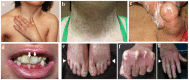
Skin atrophy, characterized by thin, wrinkled (cigarette-paper) skin is initially primarily localized to hands and feet; atrophy becomes generalized by adolescence and is often present on the abdomen, thighs, knees, and elbows (Figure 1a, 1f, 1g) [Jobard et al 2003].

Figure 1.

Figure 1.

Characteristic clinical features of Kindler syndrome a. Child age nine years: atrophy of skin on the dorsum of the hands and poikiloderma on the neck and axillary area

Photosensitivity, characterized by erythema and burning after sun exposure, tends to improve with age; however, some degree of photosensitivity usually persists (e.g., facial erythema after minimal sun exposure) [Ashton et al 2004]. Note: Affected individuals can develop redness within minutes of sun exposure.

Poikiloderma, which is not present at birth, appears first on sun-exposed areas, progressing with age to non-sun-exposed areas (Figure 1b). This is characterized by reticular telangiectasia and mottled hypo- and hyperpigmentation of the skin, frequently appearing between ages two and three years. Generalized poikiloderma (in both sun-exposed and non-sun-exposed areas) eventually develops and persists throughout adult life in most affected individuals.

Axillary freckling may be observed in some (Figure 1a) [Jobard et al 2003, Siegel et al 2003, Rayinda et al 2021].

Hyperkeratosis of the palms and soles has been reported as fissured or punctate [Penagos et al 2004] and is observed in about 65% of affected individuals. Palmar hyperkeratosis often has a waxy appearance, occasionally leading to the loss of the dermal ridges (i.e., fingerprints). Dermatoglyphics can be flattened or lost. Some individuals may have ridged, ribbed hyperkeratosis of the lateral and anterior ankles reminiscent of epidermolytic hyperkeratosis.

Pseudosyndactyly. Typically, interdigital webbing develops (Figure 1f), but without the scarring or milia noted in other forms of epidermolysis bullosa (EB) [Jobard et al 2003, Siegel et al 2003].

Constricting bands of the pseudoainhum type have also been reported (Figure 1e, 1g) [Penagos et al 2004].

Mucosal involvement can include the following [Jobard et al 2003, Penagos et al 2004]:

  • Eyes. Conjunctivitis, conjunctival scarring, corneal erosion, and ectropion of the lower eyelids [Lelli 2010, Martinez & Siegel 2011, Signes-Soler et al 2013]
  • Mouth and periodontium. Severe periodontal disease (e.g., hemorrhagic mucositis, gingivitis, periodontitis, premature loss of teeth, and labial leukokeratosis), usually beginning in early adolescence [Lai-Cheong & McGrath 2010]
  • Gastrointestinal tract. Esophageal stenosis, severe colitis, bloody diarrhea, constipation, and rectal mucosal fissures and stenosis. Affected children born with an imperforate anus that required surgical repair have also been reported [Lai-Cheong & McGrath 2010].
  • Genitourinary tract
    • Vaginal stenosis and labial synechiae
    • Urethral meatal stenosis and urethral strictures, phimosis

Malignancy. About half of individuals with KS will develop squamous cell carcinoma in their lifetime. An increased risk for malignancies has been reported:

Other findings that may be present:

  • Xerosis, eczema, and dermatitis
  • Variable hypermobility of the thumb, fingers, knees, and elbows without skin hyperextensibility [Penagos et al 2004]
  • Nail dystrophy (Figure 1e)

Morbidity and mortality mostly result from mucosal strictures and associated complications, secondary infections or cutaneous bullae, and cancer.

Genotype-Phenotype Correlations

No genotype-phenotype correlations have been identified.

Most FERMT1 variants associated with Kindler syndrome are null variants. It has been proposed that FERMT1 pathogenic missense variants and in-frame deletions are associated with milder disease manifestations and later onset of complications [Maier et al 2016].

Nomenclature

KS was first described by Theresa Kindler [Kindler 1954].

In May 2007, 18 leading authorities on epidermolysis bullosa (EB) revised the EB classification to include KS based on its biologic and clinical findings [Fine et al 2008, Fine et al 2014].

Prevalence

Since the first description of KS in 1954 [Kindler 1954], about 400 affected individuals have been reported worldwide.

Persons of any geographic origin can be affected and there is no sex predilection [Siegel et al 2003, Has et al 2011]. A cluster of 26 Panamanian affected individuals and 24 Iranian affected individuals with the syndrome have been identified [Penagos et al 2004, Youssefian et al 2015].

Differential Diagnosis

Before the onset of the photosensitivity and poikiloderma in the first few years of life, Kindler syndrome (KS) is frequently confused with other variants of epidermolysis bullosa (e.g., dystrophic, junctional, and simplex epidermolysis bullosa); however, acral skin atrophy is indicative of KS. Furthermore, in contrast to blistering in other types of epidermolysis bullosa, the blistering in KS significantly improves with age.

Disorders of known genetic cause that can exhibit features of poikiloderma but are distinguishable by other clinical features are summarized Table 3.

Table 3.

Disorders with Features of Poikiloderma in the Differential Diagnosis of Kindler Syndrome

Gene(s)DisorderMOIClinical Characteristics
Ectodermal FindingsOther Findings
ACD
CTC1
DKC1
NHP2
NOP10
PARN
RTEL1
TERC
TERT
TINF2
WRAP53
Dyskeratosis congenita XL
AD
AR
Dysplastic nails, lacy reticular pigmentation of upper chest &/or neck. Poikiloderma & nail dystrophy occur in late childhood.Oral leukoplakia. ↑ risk for progressive bone marrow failure, myelodysplastic syndrome or acute myelogenous leukemia, solid tumors, & pulmonary fibrosis.
ANAPC1
RECQL4
Rothmund-Thomson syndrome ARRash that typically develops ages 3-6 mos (occasionally as late as age 2 yrs) as erythema, swelling, & blistering on face, then spreading to buttocks & extremities. Over mos to yrs, rash evolves into poikiloderma. Sparse hair, eyelashes, &/or eyebrows.Small size; skeletal & dental abnormalities; juvenile cataracts; ↑ risk for cancer, esp osteosarcoma
BLM Bloom syndrome ARSkin at birth & during early infancy appears normal. During 1st or 2nd yr of life, typically following sun exposure, red, sun-sensitive rash appears on nose & cheeks (sometimes also on dorsa of hands & forearms). Rash varies in severity & extent & is usually characterized by telangiectasia but in others is described as poikiloderma.Severe pre- & postnatal growth deficiency, highly characteristic sparseness of subcutaneous fat tissue throughout infancy & early childhood, & short stature throughout postnatal life. Gastroesophageal reflux is common.
CD151 KS-like epidermolysis bullosa 1ARFacial freckling, poikiloderma, atrophy of skin, & acrogeria of backs of hands on sun-exposed areas. Unlike KS, which presents w/blistering during childhood, 1 person described 1 presented widespread blistering & erosions primarily in pretibial area in adulthood. Dystrophic nails in all fingers & toes & early-onset alopecia were also observed.The person described 1 had history of nephropathy manifesting w/proteinuria.
DDB2
ERCC1
ERCC2
ERCC3
ERCC4
ERCC5
POLH
XPA
XPC
Xeroderma pigmentosum (XP)ARSun sensitivity w/marked freckle-like pigmentation of face before age 2 yrs. Sunlight-induced ocular involvement. Greatly ↑ risk of sunlight-induced cutaneous neoplasms. XP typically does not have acral bullae whereas in KS acral bullae are observed in childhood.~25% of affected persons have neurologic manifestations.
FAM111B Hereditary fibrosing poikiloderma w/tendon contractures, myopathy, & pulmonary fibrosis ADPoikiloderma (typically beginning in 1st 6 mos & mainly localized to face), hypohidrosis w/heat intolerance, mild lymphedema of extremities, chronic erythematous & scaly skin lesions on extremities, sclerosis of digits, & mild palmoplantar keratoderma. Typically scalp hair, eyelashes, &/or eyebrows are sparse; nail dysplasia may be assoc.Muscle contractures usually seen in childhood; progressive weakness of proximal & distal muscles of all 4 limbs
USB1 Poikiloderma w/neutropenia (Clericuzio-type poikiloderma w/neutropenia) 2ARInflammatory eczematous rash (ages 6-12 mos) followed by post-inflammatory poikiloderma (age >2 yrs); nail dystrophy & palmar/plantar hyperkeratosisChronic noncyclic neutropenia typically assoc w/recurrent sinopulmonary infections in 1st 2 yrs of life & (often) bronchiectasis. ↑ risk for myelodysplastic syndrome. Reactive airway disease.

Disorders of unknown genetic cause with features of poikiloderma

  • The designation Weary-Kindler syndrome (WKS) has been used for a disorder with features that overlap with KS including vesicopustules, eczema, poikiloderma, and acral keratotic papules in infancy. In WKS the bullae are not congenital and photosensitivity and mucosal involvement are not observed [Weary et al 1971, Larrègue et al 1981, Lai-Cheong et al 2009]. It has been suggested that Weary and Kindler syndromes are aspects of the same disorder [Lee et al 2012].
  • Mendes da Costa syndrome, also referred to as hereditary bullous dystrophy, macular type (OMIM 302000), is characterized by microcephaly, short stature, mild intellectual disability, cone-shaped fingers, and poikiloderma. The disorder has been described in Dutch and Italian families [Sybert 2010].
  • Hereditary sclerosing poikiloderma (OMIM 173700) is characterized by progressive poikiloderma in flexural areas (manifest as hyper- and hypopigmentation without telangiectasia or atrophy), sclerotic bands, poor dentition, and occasionally calcinosis cutis [Weary et al 1969]. A later-onset complication is stenosis of cardiac valves. Absence of bullae and photosensitivity distinguish hereditary sclerosing poikiloderma from KS.

Management

No clinical practice guidelines for Kindler syndrome (KS) have been published.

Evaluation Following Initial Diagnosis

To establish the extent of disease and needs in an individual diagnosed with KS, the evaluations summarized in Table 4 (if not performed as part of the evaluation that led to the diagnosis) are recommended.

Table 4.

Recommended Evaluations Following Initial Diagnosis in Individuals with Kindler Syndrome

System/ConcernEvaluationComment
Skin issues Comprehensive dermatologic eval for skin fragility, blistering, photosensitivity, & risk for squamous cell carcinoma
Eyes Ophthalmologic consult for conjunctival involvement
Mouth Dental consult for periodontal involvement
GI tract
  • Gastroenterology eval for strictures affecting GI tract
  • Incl eval of nutritional status, diet, & oral intake
Genitourinary
tract
  • Males: urology consult for urethra or foreskin stricture/stenosis
  • Females: gynecology eval for vaginal or labial stricture/stenosis
Genetic
counseling
By genetics professionals 1To inform affected persons & families re nature, MOI, & implications of KS to facilitate medical & personal decision making
Family support
& resources
Assess need for:

GI = gastrointestinal; KS = Kindler syndrome; MOI = mode of inheritance

1.

Medical geneticist, certified genetic counselor, certified advanced genetic nurse

Treatment of Manifestations

No established treatment for KS exists. The goals of care are to treat manifestations and prevent complications.

When possible, children with KS should be managed in a center experienced in caring for children with skin fragility by a multidisciplinary team that includes a dermatologist, pediatrician, ophthalmologist, dentist, gastroenterologist, urologist, nurse specialist, and dietitian.

Table 5.

Treatment of Manifestations in Individuals with Kindler Syndrome

Manifestation/ConcernTreatment
Skin blistering
  • If blister is not painful, it should be kept intact.
  • Otherwise alleviate blister-related pain by draining fluid using a sterile needle, leaving overlying skin in place.
  • Apply ointment to blister & cover w/nonstick gauze bandage.
  • Antibiotics may be used to treat infected blister.
Skin fragility Protection from trauma, e.g., by use of soft & protective clothing & avoiding contact sports to prevent physical trauma
Skin atrophy Use of moisturizers for dry, pruritic skin
Photosensitivity Sun safety education, incl use of high sun-protective factor (>30 SPF) sunscreens, use of sun-protective clothing (hats & long-sleeve shirts), & avoidance of sun exposure as much as possible
Eyes
  • Lubrication of cornea by artificial tears & eye drops
  • Prevention of infections by use of local antibiotics
  • Surgical correction of corneal scarring by ophthalmologist as needed
Mouth &
periodontium
Regular dental care to ensure optimal oral hygiene to reduce periodontal disease
GI tract
  • Esophageal dilation may be indicated for those w/dysphagia.
  • Esophageal strictures & stenosis may require fluoroscopically guided balloon dilations [Sadler et al 2006].
  • Temporary parenteral nutrition may be necessary when esophageal dysfunction is severe.
  • Anal stenosis & bleeding requires regular laxatives.
  • Severe colitis may require surgical bowel resection in some cases.
Genitourinary
tract
  • Urethral meatal stenosis may require dilation. Strictures may require stenting &/or surgical intervention.
  • Most males require circumcision for phimosis.
  • Vaginal or labial stricture/stenosis may require surgical intervention. In case of pregnancy, delivery should be considered by an elective cesarean section.

GI = gastrointestinal

Surveillance

Table 6.

Recommended Surveillance for Individuals with Kindler Syndrome

System/ConcernEvaluationFrequency
Risk for skin cancer Screen for premalignant keratoses & early squamous cell carcinomas.Starting in adolescence; annually thereafter
Risk for oral mucosa tumors Screen for oral ulcerations, gingivitis, & periodontitis.Starting in adolescence & w/regular dental checkups

Agents/Circumstances to Avoid

Avoid sun exposure by using sunscreen (SFP >30) and sun-protective clothing.

Evaluation of Relatives at Risk

See Genetic Counseling for issues related to testing of at-risk relatives for genetic counseling purposes.

Pregnancy Management

Although the cutaneous manifestations of KS are not exacerbated by pregnancy, vaginal stenosis and labial synechiae have been reported; thus, obstetric planning, such as consideration of delivery by elective cesarean section, warrants consideration [Mansur et al 2007]. Of note, specialized perioperative cesarean section management is needed to protect vulnerable skin and mucosa.

Breastfeeding is not advised because of the risk of blistering the breasts [Hayashi et al 2007].

Therapies Under Investigation

Search ClinicalTrials.gov in the US and EU Clinical Trials Register in Europe for access to information on clinical studies for a wide range of diseases and conditions. Note: There may not be clinical trials for this disorder.

Genetic Counseling

Genetic counseling is the process of providing individuals and families with information on the nature, mode(s) of inheritance, and implications of genetic disorders to help them make informed medical and personal decisions. The following section deals with genetic risk assessment and the use of family history and genetic testing to clarify genetic status for family members; it is not meant to address all personal, cultural, or ethical issues that may arise or to substitute for consultation with a genetics professional. —ED.

Mode of Inheritance

Kindler syndrome (KS) is inherited in an autosomal recessive manner.

Risk to Family Members

Parents of a proband

  • The parents of an affected individual are obligate heterozygotes (i.e., presumed to be carriers of one FERMT1 pathogenic variant based on family history).
  • Molecular genetic testing is recommended for the parents of the proband to confirm that both parents are heterozygous for an FERMT1 pathogenic variant and to allow reliable recurrence risk assessment. If a pathogenic variant is detected in only one parent and parental identity testing has confirmed biological maternity and paternity, the following possibilities should be considered:
  • Heterozygotes (carriers) are asymptomatic and are not at risk of developing the disorder.

Sibs of a proband

  • If both parents are known to be heterozygous for an FERMT1 pathogenic variant, each sib of an affected individual has at conception a 25% chance of being affected, a 50% chance of being an asymptomatic carrier, and a 25% chance of being unaffected and not a carrier.
  • Heterozygotes (carriers) are asymptomatic and are not at risk of developing the disorder.

Offspring of a proband

Other family members. Each sib of the proband's parents is at a 50% risk of being a carrier of an FERMT1 pathogenic variant.

Carrier Detection

Carrier testing for at-risk relatives requires prior identification of the FERMT1 pathogenic variants in the family.

Related Genetic Counseling Issues

Family planning

  • The optimal time for determination of genetic risk and discussion of the availability of prenatal/preimplantation genetic testing is before pregnancy.
  • It is appropriate to offer genetic counseling (including discussion of potential risks to offspring and reproductive options) to young adults who are affected, are carriers, or are at risk of being carriers.
  • FERMT1 molecular genetic testing for reproductive partners of known carriers is appropriate, particularly if consanguinity is likely.

Prenatal Testing and Preimplantation Genetic Testing

Once the FERMT1 pathogenic variants have been identified in an affected family member, prenatal and preimplantation genetic testing for KS are possible.

Differences in perspective may exist among medical professionals and within families regarding the use of prenatal testing. While most centers would consider use of prenatal testing to be a personal decision, discussion of these issues may be helpful.

Resources

GeneReviews staff has selected the following disease-specific and/or umbrella support organizations and/or registries for the benefit of individuals with this disorder and their families. GeneReviews is not responsible for the information provided by other organizations. For information on selection criteria, click here.

Molecular Genetics

Information in the Molecular Genetics and OMIM tables may differ from that elsewhere in the GeneReview: tables may contain more recent information. —ED.

Table A.

Kindler Syndrome: Genes and Databases

GeneChromosome LocusProteinLocus-Specific DatabasesHGMDClinVar
FERMT1 20p12​.3 Fermitin family homolog 1 FERMT1 database FERMT1 FERMT1

Data are compiled from the following standard references: gene from HGNC; chromosome locus from OMIM; protein from UniProt. For a description of databases (Locus Specific, HGMD, ClinVar) to which links are provided, click here.

Table B.

OMIM Entries for Kindler Syndrome (View All in OMIM)

173650KINDLER SYNDROME; KNDLRS
607900FERM DOMAIN-CONTAINING KINDLIN 1; FERMT1

Molecular Pathogenesis

FERMT1 is located on chromosome 20; it consists of 15 exons and encodes kindlin-1 protein with 677 amino acids (~77.4 kd). The gene is expressed in different tissues including skin keratinocytes and epithelial cells of colon, lung, placenta, adrenal, and prostate [Jobard et al 2003]. Kindlin-1 is a focal adhesion protein that like other cytoskeletal proteins has "the four point one ezrin radixin and moesin" (FERM) and "pleckstrin" homology (PH) domains [Larjava et al 2008]. Kindlin-1 is an adaptor protein that links filamentous actin in the cell to membrane proteins and is involved in integrin-associated signaling pathways [Has et al 2011].

To date, a total of 91 loss-of-function pathogenic variants have been reported in FERMT1, including: missense, nonsense, and splicing variants; indels; and Alu-mediated gene rearrangements. The absence of kindlin-1 leads to disorganized keratinocytes as a result of aberrant integrin-mediated cell adhesion and migration [Larjava et al 2008, Rognoni et al 2016].

Mechanism of disease causation. Kindler syndrome is thought to occur via a loss-of-function mechanism; pathogenic variants may lead to absent kindlin-1 protein or production of nonfunctional protein.

Table 7.

Notable FERMT1 Pathogenic Variants

Variants listed in the table have been provided by the authors. GeneReviews staff have not independently verified the classification of variants.

GeneReviews follows the standard naming conventions of the Human Genome Variation Society (varnomen​.hgvs.org). See Quick Reference for an explanation of nomenclature.

Chapter Notes

Author Notes

Jouni Uitto, MD, PhD has been Professor of Dermatology and Cutaneous Biology, and Biochemistry and Molecular Biology, and Chair of the Department of Dermatology and Cutaneous Biology at The Sidney Kimmel Medical College at Thomas Jefferson University, in Philadelphia, Pennsylvania, since 1986. He is also Director of the Jefferson Institute of Molecular Medicine at Thomas Jefferson University. He received his MD and PhD degrees from the University of Helsinki, Finland, and completed his residency training in dermatology at Washington University School of Medicine, St. Louis, Missouri. Dr Uitto is internationally recognized for his research on connective tissue biology and molecular genetics in relation to cutaneous diseases. Dr Uitto's publications (as of November 2021) include 756 original articles in peer-reviewed journals, 364 text book chapters and review articles, and 1,057 abstracts on presentations in national and international meetings. Dr Uitto has been the recipient of numerous national and international awards, including honorary doctorate degrees from the University of Kuopio, University of Oulu, and University of Turku, all in Finland, as well as University of Buenos Aires, Argentina. He also has honorary professorships at China Medical University, Shenyang; Hebei United University, Tangshan; and The Fourth Military Medical University, Xi'an, all in China. Dr Uitto has held office in several scientific and professional societies, including as President of the Society for Investigative Dermatology, and President and Chair of the Board of Trustees of Dermatology Foundation. Dr Uitto is also Section Editor of the Journal of Investigative Dermatology, Associate Editor of the American Journal of Pathology, and he is on the editorial boards of numerous peer-reviewed journals.

Leila Youssefian, PhD and Hassan Vahidnezhad, PhD are researchers in medical genetics working on genodermatoses at Jefferson Institute of Molecular Medicine of Thomas Jefferson University in Philadelphia, Pennsylvania.

Leila Youssefian and Hassan Vahidnezhad contributed equally to this work.

DEBRA Molecular Diagnostics Laboratory

Department of Dermatology and Cutaneous Biology
Thomas Jefferson University
233 South 10th Street
Bluemle Life Sciences Building, Suite 431
Philadelphia, PA, USA 19107
Phone: 215-503-5785

Individuals with Kindler syndrome, Kindler epidermolysis bullosa, or Kindler-like syndrome interested in receiving information and free genetic testing can contact Leila Youssefian (email: leila.youssefian@jefferson.edu or phone: 610-999-9402).

Acknowledgments

Carol Kelly assisted in manuscript preparation.

Revision History

  • 6 January 2022 (ha) Comprehensive update posted live
  • 1 December 2016 (aa) Revision: addition to Differential Diagnosis
  • 3 March 2016 (bp) Review posted live
  • 3 August 2015 (ly) Original submission

References

Literature Cited

  • Alper JC, Baden HP, Goldsmith LA. Kindler's syndrome. Arch Dermatol. 1978;114:457. [PubMed: 629586]
  • Ashton GH, McLean WH, South AP, Oyama N, Smith FJ, Al-Suwaid R, Al-Ismaily A, Atherton DJ, Harwood CA, Leigh IM, Moss C, Didona B, Zambruno G, Patrizi A, Eady RA, McGrath JA. Recurrent mutations in kindlin-1, a novel keratinocyte focal contact protein, in the autosomal recessive skin fragility and photosensitivity disorder, Kindler syndrome. J Invest Dermatol. 2004;122:78–83. [PubMed: 14962093]
  • Barzegar M, Asadi-Kani Z, Mozafari N, Vahidnezhad H, Kariminejad A, Toossi P. Using immunofluorescence (antigen) mapping in the diagnosis and classification of epidermolysis bullosa: a first report from Iran. Int J Dermatol. 2015;54:e416–23. [PubMed: 26220012]
  • Burch JM, Fassihi H, Jones CA, Mengshol SC, Fitzpatrick JE, McGrath JA. Kindler syndrome: a new mutation and new diagnostic possibilities. Arch Dermatol. 2006;142:620–4. [PubMed: 16702500]
  • Almeida HL Jr, Heckler GT, Fong K, Lai-Cheong J, McGrath J. Sporadic Kindler syndrome with a novel mutation. An Bras Dermatol. 2013;88:212–5. [PMC free article: PMC3875998] [PubMed: 24346923]
  • Emanuel PO, Rudikoff D, Phelps RG. Aggressive squamous cell carcinoma in Kindler syndrome. Skinmed. 2006;5:305–7. [PubMed: 17086002]
  • Fine JD, Bruckner-Tuderman L, Eady RA, Bauer EA, Bauer JW, Has C, Heagerty A, Hintner H, Hovnanian A, Jonkman MF, Leigh I, Marinkovich MP, Martinez AE, McGrath JA, Mellerio JE, Moss C, Murrell DF, Shimizu H, Uitto J, Woodley D, Zambruno G. Inherited epidermolysis bullosa: updated recommendations on diagnosis and classification. J Am Acad Dermatol. 2014;70:1103–26. [PubMed: 24690439]
  • Fine JD, Eady RA, Bauer EA, Bauer JW, Bruckner-Tuderman L, Heagerty A, Hintner H, Hovnanian A, Jonkman MF, Leigh I, McGrath JA, Mellerio JE, Murrell DF, Shimizu H, Uitto J, Vahlquist A, Woodley D, Zambruno G. The classification of inherited epidermolysis bullosa (EB): report of the Third International Consensus Meeting on Diagnosis and Classification of EB. J Am Acad Dermatol. 2008;58:931–50. [PubMed: 18374450]
  • Guerrero-Aspizua S, Conti CJ, Escamez MJ, Castiglia D, Zambruno G, Youssefian L, Vahidnezhad H, Requena L, Itin P, Tadini G, Yordanova I, Martin L, Uitto J, Has C, Del Rio M. Assessment of the risk and characterization of non-melanoma skin cancer in Kindler syndrome: study of a series of 91 patients. Orphanet J Rare Dis. 2019;14:183. [PMC free article: PMC6657209] [PubMed: 31340837]
  • Has C, Burger B, Volz A, Kohlhase J, Bruckner-Tuderman L, Itin P. Mild clinical phenotype of Kindler syndrome associated with late diagnosis and skin cancer. Dermatology. 2010;221:309–12. [PubMed: 20938162]
  • Has C, Castiglia D, del Rio M, Diez MG, Piccinni E, Kiritsi D, Kohlhase J, Itin P, Martin L, Fischer J, Zambruno G, Bruckner-Tuderman L. Kindler syndrome: extension of FERMT1 mutational spectrum and natural history. Hum Mutat. 2011;32:1204–12. [PubMed: 21936020]
  • Hayashi S, Shimoya K, Itami S, Murata Y. Pregnancy and delivery with Kindler syndrome. Gynecol Obstet Invest. 2007;64:72–4. [PubMed: 17284921]
  • Heidari MM, Khatami M, Kargar S, Azari M, Hoseinzadeh H, Fallah H. A novel nonsense mutation in exon 5 of KIND1 gene in an Iranian family with Kindler syndrome. Arch Iran Med. 2016;19:403–8. [PubMed: 27293055]
  • Jobard F, Bouadjar B, Caux F, Hadj-Rabia S, Has C, Matsuda F, Weissenbach J, Lathrop M, Prud'homme JF, Fischer J. Identification of mutations in a new gene encoding a FERM family protein with a pleckstrin homology domain in Kindler syndrome. Hum Mol Genet. 2003;12:925–35. [PubMed: 12668616]
  • Jónsson H, Sulem P, Kehr B, Kristmundsdottir S, Zink F, Hjartarson E, Hardarson MT, Hjorleifsson KE, Eggertsson HP, Gudjonsson SA, Ward LD, Arnadottir GA, Helgason EA, Helgason H, Gylfason A, Jonasdottir A, Jonasdottir A, Rafnar T, Frigge M, Stacey SN, Th Magnusson O, Thorsteinsdottir U, Masson G, Kong A, Halldorsson BV, Helgason A, Gudbjartsson DF, Stefansson K. Parental influence on human germline de novo mutations in 1,548 trios from Iceland. Nature. 2017;549:519–22. [PubMed: 28959963]
  • Kindler T. Congenital poikiloderma with traumatic bulla formation and progressive cutaneous atrophy. Br J Dermatol. 1954;66:104–11. [PubMed: 13149722]
  • Kiritsi D, He Y, Pasmooij AM, Onder M, Happle R, Jonkman MF, Bruckner-Tuderman L, Has C. Revertant mosaicism in a human skin fragility disorder results from slipped mispairing and mitotic recombination. J Clin Invest. 2012;122:1742–6. [PMC free article: PMC3336993] [PubMed: 22466645]
  • Krishna CV, Parmar NV, Has C. Kindler syndrome with severe mucosal involvement in childhood. Clin Exp Dermatol. 2014;39:340–3. [PubMed: 24635075]
  • Lai-Cheong JE, McGrath JA. Kindler syndrome. Dermatol Clin. 2010;28:119–24. [PubMed: 19945624]
  • Lai-Cheong JE, Tanaka A, Hawche G, Emanuel P, Maari C, Taskesen M, Akdeniz S, Liu L, McGrath JA. Kindler syndrome: a focal adhesion genodermatosis. Br J Dermatol. 2009;160:233–42. [PubMed: 19120339]
  • Lanschuetzer CM, Muss WH, Emberger M, Pohla-Gubo G, Klausegger A, Bauer JW, Hintner H. Characteristic immunohistochemical and ultrastructural findings indicate that Kindler's syndrome is an apoptotic skin disorder. J Cutan Pathol. 2003;30:553–60. [PubMed: 14507403]
  • Larjava H, Plow EF, Wu C. Kindlins: essential regulators of integrin signalling and cell-matrix adhesion. EMBO Rep. 2008;9:1203–8. [PMC free article: PMC2603460] [PubMed: 18997731]
  • Larrègue M, Prigent F, Lorette G, Canuel C, Ramdenee P. Bullous and hereditary Weary-Kindler's acrokeratotic poikiloderma (author's transl). Ann Dermatol Venereol. 1981;108:69–76. [PubMed: 7015974]
  • Lee HJ, Shin DH, Choi JS, Kim KH. Hereditary sclerosing poikiloderma. J Korean Med Sci. 2012;27:225–7. [PMC free article: PMC3271301] [PubMed: 22323875]
  • Lelli GJ Jr. Kindler syndrome causing severe cicatricial ectropion. Ophthal Plast Reconstr Surg. 2010;26:368–9. [PubMed: 20592640]
  • Lotem M, Raben M, Zeltser R, Landau M, Sela M, Wygoda M, Tochner ZA. Kindler syndrome complicated by squamous cell carcinoma of the hard palate: successful treatment with high-dose radiation therapy and granulocyte-macrophage colony-stimulating factor. Br J Dermatol. 2001;144:1284–6. [PubMed: 11422071]
  • Maier K, He Y, Esser PR, Thriene K, Sarca D, Kohlhase J, Dengjel J, Martin L, Has C. Single amino acid deletion in kindlin-1 results in partial protein degradation which can be rescued by chaperone treatment. J Invest Dermatol. 2016;136:920–9. [PubMed: 26827766]
  • Mansur AT, Elcioglu NH, Aydingöz IE, Akkaya AD, Serdar ZA, Herz C, Bruckner-Tuderman L, Has C. Novel and recurrent KIND1 mutations in two patients with Kindler syndrome and severe mucosal involvement. Acta Derm Venereol. 2007;87:563–5. [PubMed: 17989907]
  • Martignago BC, Lai-Cheong JE, Liu L, McGrath JA, Cestari TF. Recurrent KIND1 (C20orf42) gene mutation, c.676insC, in a Brazilian pedigree with Kindler syndrome. Br J Dermatol. 2007;157:1281–4. [PubMed: 17916195]
  • Martinez AE, Siegel D. Kindler syndrome. In: Irvine A, Hoeger p, Yan A, eds. Harper's Textbook of Pediatric Dermatology. 3 ed. Hoboken, NJ: Blackwell Publishing Ltd; 2011.
  • Penagos H, Jaen M, Sancho MT, Saborio MR, Fallas VG, Siegel DH, Frieden IJ. Kindler syndrome in native Americans from Panama: report of 26 cases. Arch Dermatol. 2004;140:939–44. [PubMed: 15313809]
  • Rayinda T, van Steensel M, Danarti R. Inherited skin disorders presenting with poikiloderma. Int J Dermatol. 2021;60:1343–53. [PubMed: 33739439]
  • Richards S, Aziz N, Bale S, Bick D, Das S, Gastier-Foster J, Grody WW, Hegde M, Lyon E, Spector E, Voelkerding K, Rehm HL, et al. Standards and guidelines for the interpretation of sequence variants: a joint consensus recommendation of the American College of Medical Genetics and Genomics and the Association for Molecular Pathology. Genet Med. 2015;17:405–24. [PMC free article: PMC4544753] [PubMed: 25741868]
  • Rognoni E, Ruppert R, Fässler R. The kindlin family: functions, signaling properties and implications for human disease. Journal of cell science. 2016;129:17–27. [PubMed: 26729028]
  • Sadler E, Klausegger A, Muss W, Deinsberger U, Pohla-Gubo G, Laimer M, Lanschuetzer C, Bauer JW, Hintner H. Novel KIND1 gene mutation in Kindler syndrome with severe gastrointestinal tract involvement. Arch Dermatol. 2006;142:1619–24. [PubMed: 17178989]
  • Shimizu H, Sato M, Ban M, Kitajima Y, Ishizaki S, Harada T, Bruckner-Tuderman L, Fine JD, Burgeson R, Kon A, McGrath JA, Christiano AM, Uitto J, Nishikawa T. Immunohistochemical, ultrastructural, and molecular features of Kindler syndrome distinguish it from dystrophic epidermolysis bullosa. Arch Dermatol. 1997;133:1111–7. [PubMed: 9301588]
  • Siegel DH, Ashton GH, Penagos HG, Lee JV, Feiler HS, Wilhelmsen KC, South AP, Smith FJ, Prescott AR, Wessagowit V, Oyama N, Akiyama M, Al Aboud D, Al Aboud K, Al Githami A, Al Hawsawi K, Al Ismaily A, Al-Suwaid R, Atherton DJ, Caputo R, Fine JD, Frieden IJ, Fuchs E, Haber RM, Harada T, Kitajima Y, Mallory SB, Ogawa H, Sahin S, Shimizu H, Suga Y, Tadini G, Tsuchiya K, Wiebe CB, Wojnarowska F, Zaghloul AB, Hamada T, Mallipeddi R, Eady RA, McLean WH, McGrath JA, Epstein EH. Loss of kindlin-1, a human homolog of the Caenorhabditis elegans actin-extracellular-matrix linker protein UNC-112, causes Kindler syndrome. Am J Hum Genet. 2003;73:174–87. [PMC free article: PMC1180579] [PubMed: 12789646]
  • Signes-Soler I, Rodriguez-Prats JL, Carbonell S, Tañá-Rivero P. Corneal erosion and Kindler syndrome. Optom Vis Sci. 2013;90:e9–10. [PubMed: 23241824]
  • Stenson PD, Mort M, Ball EV, Chapman M, Evans K, Azevedo L, Hayden M, Heywood S, Millar DS, Phillips AD, Cooper DN. The Human Gene Mutation Database (HGMD®): optimizing its use in a clinical diagnostic or research setting. Hum Genet. 2020;139:1197–207. [PMC free article: PMC7497289] [PubMed: 32596782]
  • Sybert VP. Genetic Skin Disorders. 2 ed. Oxford, UK: Oxford University Press; 2010.
  • Techanukul T, Sethuraman G, Zlotogorski A, Horev L, Macarov M, Trainer A, Fong K, Lens M, Medenica L, Ramesh V, McGrath JA, Lai-Cheong JE. Novel and recurrent FERMT1 gene mutations in Kindler syndrome. Acta Derm Venereol. 2011;91:267–70. [PubMed: 21336475]
  • Vahidnezhad H, Youssefian L, Saeidian AH, Boyden LM, Touati A, Harvey N, Naji M, Zabihi M, Barzegar M, Sotoudeh S, Liu L, Guy A, Kariminejad A, Zeinali S, Choate KA, McGrath JA, Uitto J. Kindler epidermolysis bullosa-like skin phenotype and downregulated basement membrane zone gene expression in poikiloderma with neutropenia and a homozygous USB1 mutation. Matrix Biol. 2021;99:43–57. [PubMed: 34004352]
  • Vahidnezhad H, Youssefian L, Saeidian AH, Mahmoudi H, Touati A, Abiri M, Kajbafzadeh AM, Aristodemou S, Liu L, McGrath JA, Ertel A, Londin E, Kariminejad A, Zeinali S, Fortina P, Uitto J. Recessive mutation in tetraspanin CD151 causes Kindler syndrome-like epidermolysis bullosa with multi-systemic manifestations including nephropathy. Matrix Biol. 2018;66:22–33. [PubMed: 29138120]
  • Valinotto LE, Natale MI, Lusso SB, Cella E, Gutiérrez O, Sebastiani F, Manzur GB. A novel pathogenic FERMT1 variant in four families with Kindler syndrome in Argentina. Pediatr Dermatol. 2020;37:337–341. [PubMed: 31957900]
  • Weary PE, Hsu YT, Richardson DR, Caravati CM, Wood BT. Hereditary sclerosing poikiloderma. Report of two families with an unusual and distinctive genodermatosis. Arch Dermatol. 1969;100:413–22. [PubMed: 5358102]
  • Weary PE, Manley WF Jr, Graham GF. Hereditary acrokeratotic poikiloderma. Arch Dermatol. 1971;103:409–22. [PubMed: 4253719]
  • Youssefian L, Vahidnezhad H, Barzegar M, Li Q, Sotoudeh S, Yazdanfar A, Ehsani AH, Kajbafzadeh AM, Mozafari N, Ebrahimi Daryani N, Agha-Hosseini F, Zeinali S, Uitto J. The Kindler syndrome: a spectrum of FERMT1 mutations in Iranian families. J Invest Dermatol. 2015;135:1447–50. [PubMed: 25599393]
Copyright © 1993-2024, University of Washington, Seattle. GeneReviews is a registered trademark of the University of Washington, Seattle. All rights reserved.

GeneReviews® chapters are owned by the University of Washington. Permission is hereby granted to reproduce, distribute, and translate copies of content materials for noncommercial research purposes only, provided that (i) credit for source (http://www.genereviews.org/) and copyright (© 1993-2024 University of Washington) are included with each copy; (ii) a link to the original material is provided whenever the material is published elsewhere on the Web; and (iii) reproducers, distributors, and/or translators comply with the GeneReviews® Copyright Notice and Usage Disclaimer. No further modifications are allowed. For clarity, excerpts of GeneReviews chapters for use in lab reports and clinic notes are a permitted use.

For more information, see the GeneReviews® Copyright Notice and Usage Disclaimer.

For questions regarding permissions or whether a specified use is allowed, contact: ude.wu@tssamda.

Bookshelf ID: NBK349072PMID: 26937547

Views

Tests in GTR by Gene

Related information

  • OMIM
    Related OMIM records
  • PMC
    PubMed Central citations
  • PubMed
    Links to PubMed
  • Gene
    Locus Links

Similar articles in PubMed

See reviews...See all...

Recent Activity

Your browsing activity is empty.

Activity recording is turned off.

Turn recording back on

See more...