U.S. flag

An official website of the United States government

NCBI Bookshelf. A service of the National Library of Medicine, National Institutes of Health.

Adam MP, Feldman J, Mirzaa GM, et al., editors. GeneReviews® [Internet]. Seattle (WA): University of Washington, Seattle; 1993-2024.

Cover of GeneReviews®

GeneReviews® [Internet].

Show details

Hyperphosphatemic Familial Tumoral Calcinosis

Synonyms: Familial Tumoral Calcinosis/Hyperostosis-Hyperphosphatemia Syndrome (FTC/HHS), HFTC, Hyperostosis-Hyperphosphatemia Syndrome, Primary Hyperphosphatemic Tumoral Calcinosis

, MD, , MD, and , MD.

Author Information and Affiliations

Initial Posting: .

Estimated reading time: 32 minutes

Summary

Clinical characteristics.

Hyperphosphatemic familial tumoral calcinosis (HFTC) is characterized by:

  • Ectopic calcifications (tumoral calcinosis) typically found in periarticular soft tissues exposed to repetitive trauma or prolonged pressure (e.g., hips, elbows, and shoulders); and
  • Painful swellings (referred to as hyperostosis) in the areas overlying the diaphyses of the tibiae (and less often the ulna, metacarpal bones, and radius).

The dental phenotype unique to HFTC includes enamel hypoplasia, short and bulbous roots, obliteration of pulp chambers and canals, and pulp stones. Less common are large and small vessel calcifications that are often asymptomatic incidental findings on radiologic studies but can also cause peripheral vascular insufficiency (e.g., pain, cold extremities, and decreased peripheral pulses). Less frequently reported findings include testicular microlithiasis and angioid streaks of the retina.

Diagnosis/testing.

HFTC results from a relative deficiency of – or resistance to – the phosphate-regulating hormone, fibroblast growth factor 23 (FGF23). The clinical diagnosis of HFTC is established by the presence of tumoral calcinosis and/or characteristic laboratory findings of hyperphosphatemia in the setting of inappropriately increased renal tubular reabsorption of phosphorus (TRP), elevated or inappropriately normal 1,25-dihydroxyvitamin D3 (1,25D) levels, and elevated C-terminal FGF23 fragments. Identification of biallelic pathogenic variants in FGF23, GALNT3, or KL by molecular genetic testing confirms the diagnosis when clinical and laboratory findings are inconclusive.

Management.

Treatment of manifestations: No randomized clinical trials have been performed; studies of the treatment of HFTC consist of case reports or case series. Since FGF23 replacement therapy is not available, treatment relies on lowering blood phosphorus by blocking absorption from the diet (i.e., use of low-phosphate diet and phosphate binders) and/or by increasing renal phosphate excretion (i.e., use of acetazolamide, probenecid, or niacinamide/nicotinamide); however, clinical responses vary. Surgical resection of tumoral calcinosis lesions – generally reserved for those patients with significant pain or functional impairment – has variable success. Treatment of pain associated with hyperostosis is symptomatic. No data on management of vascular calcification or testicular microlithiasis are available.

Surveillance: No specific guidelines address the issue of surveillance in HFTC. The frequency of monitoring depends on the clinical findings and medical treatment, as no data support an appropriate monitoring interval.

Agents/circumstances to avoid: It is prudent to avoid: use of calcium salts as phosphate binders; excessive dietary calcium intake; excessive vitamin D intake/supplements; foods high in phosphorus.

Evaluation of relatives at risk: It is appropriate to evaluate apparently asymptomatic older and younger sibs of a proband in order to identify as early as possible those who would benefit from prompt initiation of treatment and preventive measures to avoid development of ectopic calcifications.

Genetic counseling.

HFTC is inherited in an autosomal recessive manner. At conception, each sib of an affected individual has a 25% chance of being affected, a 50% chance of being an asymptomatic carrier, and a 25% chance of being unaffected and not a carrier. Once the FGF23, GALNT3, or KL pathogenic variants have been identified in an affected family member, carrier testing for at-risk relatives, prenatal testing for a pregnancy at increased risk, and preimplantation genetic testing are possible.

Diagnosis

Suggestive Findings

Hyperphosphatemic familial tumoral calcinosis (HFTC) should be suspected in individuals with the following clinical, laboratory, and/or imaging findings.

Clinical Findings

Tumoral calcinosis or ectopic calcifications are typically seen in periarticular soft tissues exposed to repetitive trauma or prolonged pressure, such as the hips, elbows, and shoulders; they can also be observed in other locations ranging from the eyelid [Ichikawa et al 2006] to the submucosa of the large intestines (Figure 1A&D) [Ramnitz et al 2016].

Figure 1. A.

Figure 1

A. Computed tomography (CT) scan showing calcifications in an adult with HFTC including shoulder and hip soft tissue calcifications (white arrows), aortic calcification (dashed arrow), papillary muscle calcification (black arrow), and submucosal gut calcifications (more...)

Hyperostosis. Some affected individuals experience transient but recurrent episodes of pain in the long bones, typically the tibia, associated with erythema, edema, and warmth of the overlying skin [Ichikawa et al 2007a, Gok et al 2009].

Vascular calcifications occur in both large and small vessels; they are often asymptomatic but can cause symptoms of peripheral vascular insufficiency (e.g., pain, cold extremities, and decreased peripheral pulses). Although rarely the presenting feature, the risk is likely increased over the general population; the true frequency is unknown.

Laboratory Findings

  • Hyperphosphatemia
  • Inappropriately increased renal tubular reabsorption of phosphorus (TRP)
  • Elevated or inappropriately normal 1,25-dihydroxyvitamin D3 (1,25D) for the degree of hyperphosphatemia
  • Renal function and serum calcium levels typically normal
  • Parathyroid hormone levels tend to be at the lower end of the normal range.
  • Elevated plasma levels of the C-terminal portion of the phosphate-regulating hormone, fibroblast growth factor 23 (FGF23): measured (in clinical laboratories) by enzyme-linked immunosorbent assay (ELISA) utilizing antibodies that bind to the C-terminal portion of FGF23; detecting the combination of the biologically active intact hormone and the biologically inactive C-terminal fragments
    Note: Assays to measure intact FGF23 are currently available in research laboratories only. Intact FGF23 is low or inappropriately normal with FGF23-related HFTC and GALNT3-related HFTC, and elevated with KL-related HFTC.

Radiographs

Tumoral calcinosis. Radiographs of affected lesions show heterogeneous calcified masses (Figure 2A&B).

Figure 2. A&B.

Figure 2

A&B. Radiographs in a child with hyperphosphatemic familial tumoral calcinosis showing tumoral calcinosis of the lateral proximal femur (A) and elbow (B) C. Radiograph of tibias/fibulas in a child showing hyperostosis with hypermineralization (more...)

Hyperostosis. When hyperostosis is symptomatic (i.e., causes pain, erythema, edema, and warmth), long-bone radiographs show hypermineralization of cortical bone, patchy sclerosis of the medullary cavities, diaphysitis, and marked periosteal reaction (Figure 2C) [Clarke et al 1984, Ichikawa et al 2007a, Olauson et al 2008, Gok et al 2009, Joseph et al 2010].

Dental radiographs often show the typical findings of short bulbous roots, obliteration of pulp chambers and root canals, abnormal curvature of the dental roots (known as root dilacerations), thistle-shaped dental pulps, and pulp stones (Figure 2D&E) [Foster et al 2014].

Computed Tomography (CT)

Tumoral calcinosis. Although CT is the preferred imaging modality to identify and measure calcifications and changes in size, it carries a risk of radiation exposure and should be used conservatively, especially in children (Figure 2A&B, Figure 3A).

Figure 3. A.

Figure 3

A. CT 3D reconstruction in an adult with HFTC demonstrating bilateral vascular calcifications in superficial femoral arteries (solid arrows) and multifocal sheet-like calcification of the subcutaneous tissues at the level of the distal tibiae and fibulae (more...)

Vascular calcifications. CT can be particularly helpful in identifying vascular calcifications, which appear hyperattenuated when calcium is present (Figure 3A, C, D).

Establishing the Diagnosis

The diagnosis of HFTC is established in a proband with the clinical findings of tumoral calcinosis and/or characteristic laboratory findings of hyperphosphatemia, increased TRP, elevated or inappropriately normal 1,25D levels, and elevated C-terminal FGF23 levels.

Identification of biallelic germline loss-of-function variants in FGF23, GALNT3, or KL by molecular genetic testing (Table 1) confirms the diagnosis when clinical and laboratory findings are inconclusive.

Molecular testing approaches can include serial single-gene testing, use of a multigene panel, or comprehensive genomic testing.

Gene-targeted testing requires that the clinician determine which gene(s) are likely involved, whereas genomic testing does not. Persons with the distinctive findings described in Suggestive Findings are likely to be diagnosed using gene-targeted testing (see Option 1), whereas those in whom a specific diagnosis has been elusive are more likely to be diagnosed using comprehensive genomic testing (see Option 2).

Option 1

When the phenotypic and laboratory findings suggest the diagnosis of HFTC, molecular genetic testing approaches can include serial single-gene testing or use of a multigene panel.

  • Serial single-gene testing is based on the order in which pathogenic variants most commonly occur: GALNT3 followed by FGF23, then KL. Sequence analysis of the gene of interest is performed first, followed by gene-targeted deletion/duplication analysis if only one or no pathogenic variant is found.
  • Use of a multigene panel that includes FGF23, GALNT3, KL, and other genes of interest (see Differential Diagnosis) can be considered. Note: (1) The genes included in the panel and the diagnostic sensitivity of the testing used for each gene vary by laboratory and are likely to change over time. (2) Some multigene panels may include genes not associated with the condition discussed in this GeneReview; thus, clinicians need to determine which multigene panel is most likely to identify the genetic cause of the condition while limiting identification of variants of uncertain significance and pathogenic variants in genes that do not explain the underlying phenotype. (3) In some laboratories, panel options may include a custom laboratory-designed panel and/or custom phenotype-focused exome analysis that includes genes specified by the clinician. (4) Methods used in a panel may include sequence analysis, deletion/duplication analysis, and/or other non-sequencing-based tests.
    For an introduction to multigene panels click here. More detailed information for clinicians ordering genetic tests can be found here.

Option 2

In the event that the diagnosis of HFTC has not been considered, comprehensive genomic testing (which includes exome sequencing and genome sequencing) is likely to be the diagnostic modality selected. For an introduction to comprehensive genomic testing click here. More detailed information for clinicians ordering genomic testing can be found here.

Table 1.

Molecular Genetic Testing Used in Hyperphosphatemic Familial Tumoral Calcinosis

Gene 1Proportion of Probands with Pathogenic Variants in GeneProportion of Pathogenic Variants 2 Detectable by Method
Sequence analysis 3Gene-targeted deletion/duplication analysis 4
FGF23 12/50 5, 68/9 71/9 7, 8
GALNT3 37/50 5, 935/35None reported
KL 1/50 101 reported 7, 11None reported
Unknown3% 12, 13NA
1.
2.

See Molecular Genetics for information on allelic variants detected in this gene.

3.

Sequence analysis detects variants that are benign, likely benign, of uncertain significance, likely pathogenic, or pathogenic. Variants may include small intragenic deletions/insertions and missense, nonsense, and splice site variants; typically, exon or whole-gene deletions/duplications are not detected. For issues to consider in interpretation of sequence analysis results, click here.

4.

Gene-targeted deletion/duplication analysis detects intragenic deletions or duplications. Methods used may include a range of techniques such as quantitative PCR, long-range PCR, multiplex ligation-dependent probe amplification (MLPA), and a gene-targeted microarray designed to detect single-exon deletions or duplications.

5.
6.
7.
8.

One large deletion has been reported [Shah et al 2014] (see Molecular Genetics, FGF23)

9.
10.
11.
12.

One individual with the typical clinical and biochemical phenotype of HFTC did not have pathogenic variants in either FGF23 or GALNT3. Because KL was not tested and FGF23 levels were not reported, it is unknown if this individual had FGF23 resistance [Jost et al 2016].

13.

One individual with the typical clinical and biochemical phenotype of HFTC did not have pathogenic variants in either FGF23 or GALNT3; FGF23 levels were not consistent with a pathogenic variant in KL [Ramnitz et al 2016].

Clinical Characteristics

Clinical Description

Hyperphosphatemic familial tumoral calcinosis (HFTC) is characterized most commonly by tumoral calcinosis or ectopic calcifications (typically in periarticular soft tissues exposed to repetitive trauma or prolonged pressure) and hyperostosis (typically manifesting as painful swelling overlying the diaphyses of long bones). Onset of lesions typically occurs in the first two decades of life. The dental phenotype unique to HFTC includes enamel hypoplasia, short and bulbous roots, pulp chamber and canal obliterations, and pulp stones. Less frequently reported findings include large and small vessel calcifications, testicular microlithiasis, and angioid streaks of the retina.

HFTC results from a relative deficiency of or resistance to the phosphate-regulating hormone FGF23, leading to hyperphosphatemia due to increased renal phosphate reabsorption and elevated or inappropriately normal 1,25D production, which promotes gastrointestinal absorption of phosphorus and calcium (for more details see Pathophysiology).

In a review of the medical literature, Rafaelsen et al [2014] found 56 individuals with HFTC from 35 different families with a molecularly confirmed diagnosis of HFTC (i.e., biallelic germline loss-of-function pathogenic variants in FGF23, GALNT3, or KL).

Since the Rafaelsen et al [2014] review, 14 additional individuals with GALNT3-associated HFTC [Krstevska et al 2012, Favia et al 2014, Finer et al 2014, Demellawy et al 2015, Masi et al 2015, Vieira et al 2015, Jost et al 2016, Ramnitz et al 2016] and five individuals with FGF23-associated HFTC [Shah et al 2014, Keskar et al 2015, Jost et al 2016] have been reported.

For unknown reasons, the manifestations of HFTC vary among family members with the same pathogenic variants and genetic background and similar biochemical profiles. One member may have profound, extensive tumoral calcinosis while others are symptom free [Ramnitz et al 2016]. Of note, since HFTC has been reported in many pedigrees with multigenerational consanguinity, some phenotypic features found in association with HFTC may represent the expression of other autosomal recessive disorders.

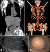
Tumoral calcinosis represents ectopic calcification in the skin and/or subcutaneous tissue in periarticular locations consisting primarily of hydroxyapatite [Boskey et al 1983, Slavin et al 1993] and – in some reports – calcium carbonate [Annamunthodo 1960]. The lesions often occur at sites of repeat trauma and/or pressure, with the lateral hips being the most frequently reported site. However, the clinical spectrum of tumoral calcinosis is widely variable, ranging from no lesions [Ramnitz et al 2016], to isolated eyelid calcifications [Ichikawa et al 2006], to periarticular calcifications of significant size (Figure 2A&B).

These lesions can be extremely painful and debilitating; some may progress in size, perforate the skin, and drain liquid hydroxyapatite (also known as "milk of calcium"), which is often confused with purulent drainage. Such lesions often heal poorly.

Depending on the size and location, tumoral calcinosis can significantly impair range of motion or lead to frozen joints.

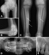
Hyperostosis typically presents as painful swelling in the areas overlying the diaphyses of the long bones (especially the tibiae), and can be the first observed manifestation of HFTC [Clarke et al 1984, Frishberg et al 2005, Ichikawa et al 2007a, Olauson et al 2008, Ichikawa et al 2010, Joseph et al 2010, Favia et al 2014, Jost et al 2016].

Some patients experience repeat episodes of diaphysitis (inflammation of the shaft of a long bone) involving a number of sites including the ulna, tibia, metacarpal bones, and radius [Clarke et al 1984, Campagnoli et al 2006b, Gok et al 2009].

Biopsies of hyperostosis lesions demonstrate reactive new bone surrounded by fibroblastic stroma infiltrated with plasma cells, lymphocytes, and polymorphonuclear cells; cultures are negative [Clarke et al 1984, Ichikawa et al 2010].

Vascular calcifications, identified in affected individuals ranging in age from three to 58 years, are rarely the presenting feature of HFTC. While the true frequency is unknown, the risk of vascular calcifications in HFTC is likely increased over that of the general population.

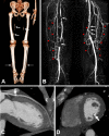
The calcifications are often incidental findings on radiologic studies, and patients are often asymptomatic. Some patients have significant symptoms of peripheral vascular insufficiency such as pain, cold extremities, and decreased peripheral pulses. Small to large vessel calcifications have been identified in variable anatomic locations including the cerebral vasculature, lower-extremity vasculature (Figure 3A&B), coronary arteries (Figure 3C), mesenteric arteries, renal arteries, carotid arteries, iliac vessels, and aorta [Martinez et al 1990, Li Voon Chong et al 1999, Ichikawa et al 2007c, Lammoglia & Mericq 2009, Rafaelsen et al 2014, Masi et al 2015, Ramnitz et al 2016]. The most severe reported consequences of vascular calcifications were below- and above-the-knee amputations in one man and digit amputations in the man's two affected sisters [Shah et al 2014].

Dental phenotype characteristic of HFTC includes enamel hypoplasia, short and bulbous roots, pulp chamber and canal obliterations, and pulp stones (Figure 2D&E) [Lyles et al 1985, Burkes et al 1991, Benet-Pagès et al 2005, Laleye et al 2008, Krstevska et al 2012, Favia et al 2014].

Dental findings, reported to be the presenting sign in some individuals [Dumitrescu et al 2009, Vieira et al 2015], may be the most common imaging findings of HFTC as they can occur in the absence of calcinosis.

Renal calcifications have been described in two individuals, one of whom had mild impairment in renal function [Chefetz et al 2005, Bergwitz et al 2009].

Inflammation. The cause and role of inflammation in the pathogenesis of the calcific tumors is unclear. Microscopy of the lesions reveals macrophage engulfment of hydroxyapatite crystals, which probably release inflammatory cytokines, the likely etiology of the observed systemic inflammation [Ramnitz et al 2016].

Five individuals have had elevated markers of inflammation; i.e., C-reactive protein (CRP) and/or erythrocyte sedimentation rate (ESR) [Garringer et al 2006, Masi et al 2009, Masi et al 2015, Ramnitz et al 2016]. In one individual clinical symptoms of inflammation included recurrent acute inflammatory polyarthritis of the shoulders, hands, and feet [Garringer et al 2006]. Two others experienced increased fatigue, inflammation-mediated anemia of chronic disease, and thrombocytosis; one also had intermittent fevers and a cutaneous inflammatory calcific reaction [Ramnitz et al 2016].

Testicular microlithiasis was reported in a boy age 14 years. The ultrasound examination showed increased echogenicity with scattered hyperechogenic lesions. A biopsy of one testicle revealed diffuse intratubular calcifications; oligo-azoospermia was noted on spermiogram [Campagnoli et al 2006b]. Similar findings were reported in a man age 45 years [Garringer et al 2007].

Angioid streaks of the retina were reported in four individuals with HFTC [McPhaul & Engel 1961, Yancovitch et al 2011]. One had symptomatic sudden onset of blurred vision attributed to choroidal neovascularization secondary to angioid streaks 14 years after the initial diagnosis of HFTC [McGrath et al 2010].

Phenotype Correlations by Gene

Vascular calcifications appear to be more frequent in persons with biallelic FGF23 loss-of-function variants (5/12) compared to persons with biallelic GALNT3 loss-of-function variants (5/37). However, this observation is limited because not all reported individuals were evaluated for the presence of vascular calcifications.

Pathophysiology

Deficiency of or resistance to FGF23 leads to hyperphosphatemia resulting in increased renal phosphate reabsorption and elevated or inappropriately normal 1,25D production, which promotes gastrointestinal absorption of phosphorus and calcium. Thereby increased blood calcium X phosphate product, calculated by multiplying the blood calcium (mg/dL) by the blood phosphate (mg/dL), predisposes to ectopic soft-tissue calcification in tissues exposed to trauma or inflammation. In persons with renal insufficiency (where this product is most commonly clinically utilized), the incidence of soft-tissue calcification is increased when the calcium X phosphate product exceeds 70 mg2/dL2 and decreased when the calcium X phosphate product is less than 50 mg2/dL2 [Langman & Salusky 2005].

Genotype-Phenotype Correlations

No genotype-phenotype correlations are known.

  • Sibs with the same GALNT3 pathogenic variants have been reported to have markedly different clinical courses [Ramnitz et al 2016].
  • Treatment response does not appear to vary by genotype.

Nomenclature

In the past, hyperphosphatemic familial tumoral calcinosis (HFTC) has been referred to as the following:

  • Lipocalcinogranulomatosis
  • Teutschlaender's lipocalcinogranulomatosis
  • Morbus Teutschländer

Prevalence

No formal study has estimated the prevalence of HFTC; the disorder is rare. To date, 75 individuals with a molecularly confirmed diagnosis of HFTC have been reported in the medical literature with an increased propensity in Africa and the Middle East and in populations originating from these regions [Sprecher 2010].

Differential Diagnosis

Table 2.

Genetic Disorders to Consider in the Differential Diagnosis of Hyperphosphatemic Familial Tumoral Calcinosis

DisorderGene(s)MOIFeatures of This Disorder
Overlapping w/HFTCDistinguishing from HFTC
BiochemicalClinical
Porphyria cutanea tarda UROD AD
  • Dystrophic calcification in areas of pseudoscleroderma
  • Ulcerated areas at pseudoscleroderma sites in some
Normal blood phosphorus
  • Blistering & skin fragility w/sun exposure
  • Hypertrichosis/ hyperpigmentation
  • Pseudoscleroderma
  • Increased urinary excretion of uroporphyrin
Normophosphatemic
familial tumoral
calcinosis
(OMIM 610455)
SAMD9 ARSkin/subcutaneous calcinosis
  • Normal blood phosphorus
  • Normal renal & intestinal reabsorption of phosphorus
  • Calcification often preceded by inflammatory rash
  • Lesions found in acral locations rather than periarticular
  • Severe conjunctivitis & gingivitis
Pseudohypopara-
thyroidism 1a, 1b, 1c
(see Disorders of GNAS Inactivation)
GNAS See footnote 1.
  • Elevated blood phosphorus
  • Subcutaneous ossifications
  • Elevated blood parathyroid hormone due to end-organ resistance
  • Low blood calcium, obesity
  • Resistance to other hormones incl TSH, LH/FSH, GHRH
Clinical features of Albright hereditary osteodystrophy incl short stature, round facies, brachydactyly, other skeletal anomalies
Pseudopseudohypo-
parathyroidism (see
Disorders of GNAS Inactivation)
GNAS See footnote 1.Subcutaneous ossificationsNormal blood phosphorus, parathyroid hormone, calciumClinical features of Albright hereditary osteodystrophy incl short stature, round facies, brachydactyly, other skeletal anomalies
Progressive osseous
heteroplasia (see
Disorders of GNAS Inactivation)
GNAS See footnote 1.Subcutaneous ossificationsNormal blood phosphorus, parathyroid hormone, calcium
  • Infantile onset of subcutaneous ossifications
  • Congenital papular rash
Fibrodysplasia ossificans progressiva ACVR1 ADSubcutaneous ossifications that can impair joint mobilityNormal blood phosphorus
  • Gradual replacement of muscle tissue & connective tissue by bone starting w/neck & shoulders & proceeding down the body
  • Typically, malformed big toes
Generalized arterial calcification of infancy ABCC6
ENPP1
AR
  • Extravascular periarticular calcifications
  • May have angioid streaks of the retina
Normal or low blood phosphorus (some develop hypophosphatemic rickets after infancy)
  • Extensive medium- & large-artery calcification in infancy resulting in cardiovascular findings
  • May have yellow papules in flexural body areas
  • Hearing loss
Cole disease
(OMIM 615522)
ENPP1 ADSkin/subcutaneous calcinosisNormal blood phosphorus
  • Congenital or early-onset punctate keratoderma of palms & soles
  • Diffuse, irregularly shaped hypopigmented skin macules typically over arms & legs
  • May have calcific tendinopathy
Arterial calcification due
to CD73 deficiency
(OMIM 211800)
NT5E AR
  • Periarticular calcifications of large & small joints of lower extremities
  • Symptomatic calcification of the large arteries of lower extremities
Normal blood phosphorusTypically present w/claudication & hemodynamically significant peripheral obstructive artery disease of lower extremities
Caffey disease COL1A1 ADCortical hyperostosis of long bonesNormal blood phosphorusOnset between birth & 5 mos; spontaneous resolution by age 2 yrs

Table 3.

Acquired Disorders to Consider in the Differential Diagnosis of Hyperphosphatemic Familial Tumoral Calcinosis

DisorderClinical Features of the Disorder
Overlapping w/HFTCDistinguishing from HFTC
BiochemicalClinical
Osteomyelitis 1
  • Erythema, warmth, & diaphyseal pain of the long bones
  • Fever
  • Ulceration & drainage of skin
  • Elevated inflammatory markers (ESR/CRP)
  • Normal blood phosphorus
  • Blood cultures may be positive
  • Positive bacterial culture from bone biopsy obtained via sterile technique
  • Distinct radiographic features of osteomyelitis
  • Distinct histopathologic features following biopsy
  • May present as septic arthritis
Autoimmune connective tissue disease
  • Skin/subcutaneous calcinosis
  • Calcinosis can occur over joints leading to functional impairment.
  • Ulcerated areas can occur at sites of calcinosis.
  • Elevated inflammatory markers (ESR/CRP)
Normal blood phosphorus
  • Other features of dermatomyositis (proximal muscle weakness, interstitial pulmonary disease, dysphagia, polyarthritis, Gottron's papules, heliotrope eruption)
  • Other features of systemic sclerosis (arthralgia, myalgia, skin thickening/hardening, sclerodactyly, Raynaud phenomenon, esophageal dysmotility, telangiectasia, & pulmonary, renal, or cardiac disease)
  • Other features of systemic lupus erythematosus (arthralgia, myalgia, fever, malar erythema, Raynaud phenomenon, vasculitis, & thromboembolic, pulmonary, renal, cardiac, or ophthalmologic disease)
Panniculitis
  • Skin/subcutaneous calcinosis
  • Ulcerated areas can occur at sites of calcinosis.
Normal blood phosphorus
  • Other features of pancreatic disease in pancreatic panniculitis (pancreatitis, elevated amylase/lipase)
  • Other features of subcutaneous fat necrosis of newborn (presents during 1st few weeks of life, typically history of trauma during delivery; may develop hypercalcemia)
TraumaSkin/subcutaneous calcinosisNormal blood phosphorusHistory of prior tissue injury (e.g., burns, trauma, surgery)
InfectionSkin/subcutaneous calcinosisNormal blood phosphorus
  • Other symptoms of onchocerciasis (river blindness) & travel to endemic areas (sub-Saharan Africa)
  • Other symptoms of cysticercosis & ingestion of undercooked meat
Chronic renal failure
  • Elevated blood phosphorus
  • Low blood 1,25D levels
May have decreased blood calcium & secondary hyperparathyroidismBiochemical evidence of renal insufficiency (elevated creatinine & BUN)
Iatrogenic calcinosis cutisSkin/subcutaneous calcinosisNormal blood phosphorusHistory of extravasation of intravenous solutions (e.g., calcium gluconate, calcium chloride, or phosphate-containing solutions at site of calcinosis)
Hypervitaminosis D
  • Skin/subcutaneous calcinosis
  • Low blood 1,25D levels
  • Normal or low blood phosphorus
  • Elevated blood calcium & renal calcium excretion
  • Elevated 25-OH vitamin D
Symptoms of hypercalcemia incl polyuria, polydipsia, confusion, anorexia, vomiting, muscle weakness
1.

Hyperostosis is often misdiagnosed as osteomyelitis resulting in unnecessary long-term treatment with intravenous antibiotics.

Management

Evaluations Following Initial Diagnosis

To establish the extent of disease and needs in an individual diagnosed with hyperphosphatemic familial tumoral calcinosis (HFTC), the following evaluations are recommended if not done at the time of diagnosis:

  • Physical examination with particular attention to:
    • Skin and joints to assess for tumoral calcinosis;
    • Lower extremities to assess for edema, erythema, and pain related to hyperostosis;
    • Peripheral pulses to assess for clinically significant vascular calcification.
    Use ankle-brachial systolic pressure index to confirm clinical suspicion of lower-extremity arterial occlusive disease.
  • Metabolic work up including blood phosphorus, blood creatinine, urine phosphorus, urine creatinine to evaluate renal phosphate handling by measuring the tubular reabsorption of phosphorus (TRP) and the ratio of the renal tubular maximum reabsorption rate of phosphate to the glomerular filtration rate (TmP/GFR)
  • Duplex ultrasound examination, CT angiography, and/or magnetic resonance angiography (Figure 3B) to evaluate for vascular calcifications if there is a history of claudication or atypical leg symptoms, or clinical signs of vascular disease on physical examination (e.g., diminished pulses or cold extremities)
  • Cardiac CT angiography (Figure 3C&D) to assess for coronary artery calcifications particularly in patients with documented vascular calcifications
  • Consultation with a vascular specialist and/or cardiologist for patients with symptomatic vascular calcifications
  • Complete blood count, erythrocyte sedimentation rate, and C-reactive protein to evaluate for evidence of systemic inflammation
  • Dental consultation including dental radiographs to assess for the HFTC dental phenotype (Figure 2D&E)
  • Testicular ultrasound examination to assess for microlithiasis (Figure 1C)
  • Renal ultrasound examination to evaluate for medullary nephrocalcinosis
  • Ophthalmologic consultation to evaluate for eyelid calcifications and angioid streaks of the retina
  • Consultation with a clinical geneticist and/or genetic counselor

Treatment of Manifestations

No standard treatment for HFTC exists. No randomized clinical trials have been performed, and studies of the treatment of HFTC consist of case reports or case series.

Most reported therapies attempt to lower blood phosphorus; most patients with HFTC are treated with a combination of medical therapies. Clinical response to these treatments varies. No treatment regimen appears to consistently or universally decrease lesion size or prevent progression or recurrence of lesions after surgery.

Tumoral calcinosis

  • Low-phosphate diet. Many patients are instructed to follow a low-phosphate diet in combination with other medical therapies. Limited benefit was reported in use of low-phosphate diet alone [Carmichael et al 2009].
  • Phosphate binders, such as sevelamer and aluminum hydroxide, are used to decrease absorption of dietary phosphorus. Response is mixed, including: (1) decrease or no change in blood phosphorus; (2) decrease, no change, or increase in the size of the tumoral calcinosis [Steinherz et al 1985, Yamaguchi et al 1995, Campagnoli et al 2006a, Garringer et al 2006, Ichikawa et al 2007b, Lammoglia and Mericq 2009, Ichikawa et al 2010, Finer et al 2014, Keskar et al 2015, Ramnitz et al 2016].
    Note: (1) Aluminum-containing phosphate binders can probably be used safely in patients with HFTC as opposed to patients with renal failure, in whom the risk of aluminum toxicity is increased due to impaired renal clearance. (2) Calcium salts, used in the past, have fallen out of favor because of their potential to increase the calcium X phosphate product, as defined in Pathophysiology.
  • Acetazolamide, a carbonic anhydrase inhibitor, increases urinary phosphate excretion. While some reports describe improvement or resolution of tumoral calcinosis following treatment with acetazolamide alone or – most often – in combination with phosphate binders, other reports describe no change in lesions. Similarly, blood phosphorus levels decreased or remained unchanged [Yamaguchi et al 1995, Garringer et al 2006, Dumitrescu et al 2009, Lammoglia & Mericq 2009, Ichikawa et al 2010, Finer et al 2014, Ramnitz et al 2016].
  • Probenecid (which promotes renal phosphate excretion) has been used along with other phosphate-lowering medications. Note: Because probenecid prolongs the half-life of a number of drugs, physicians should assess for drug interactions prior to starting this medication.
    • Treatment with probenecid did not decrease blood phosphorus or improve clinical status in two individuals [McPhaul & Engel 1961, Lammoglia & Mericq 2009].
    • Following surgical resection of tumoral calcinosis, one individual was recurrence free after six years of probenecid and phosphate binder therapy, despite no change in blood phosphate [Ichikawa et al 2010].
    • In one series, treatment that included probenecid was associated with complete resolution of tumoral calcinosis in one patient but no change in the size of lesions in others [Ramnitz et al 2016].
  • Niacinamide/nicotinamide, shown to decrease renal phosphate reabsorption, has been evaluated as a short-term treatment in two patients: one showed decreased blood phosphorus during a 48-hour treatment [Dumitrescu et al 2009]; one showed no change in blood phosphorus during an approximately three-week treatment [Ramnitz et al 2016]. Of note, there are no long-term treatment data.
  • Topical sodium thiosulfate applied to skin overlying the tumoral calcinosis lesions has been shown to decrease ectopic calcifications after at least five months of treatment in three patients [Jost et al 2016]. The mechanism by which topical or systemic sodium thiosulfate inhibits mineralization is unclear.
  • Risedronate, a bisphosphonate with anti-osteoclast activity, was used in one patient for one year, after which no change in blood phosphorus level or obvious improvement in tumoral calcinosis burden was observed [Ichikawa et al 2010]. Of note, there is no mechanistic support for the use of second-generation bisphosphonates in the treatment of HFTC.
  • Synthetic salmon calcitonin, a hormone that inhibits osteoclast activity and rental tubular reabsorption of calcium and phosphorus, was administered subcutaneously to one patient in combination with a low-phosphate diet and phosphate binder. The patient had a decrease in blood phosphorus on treatment; tumoral calcinosis lesions remained stable in size while no new ectopic calcifications developed [Candrina et al 1989].
  • Surgical resection of tumoral calcinosis lesions has variable outcomes. Some patients have complete resolution of the lesions while others have required multiple surgeries due to lesion recurrence [Carmichael et al 2009, Ichikawa et al 2010, Ramnitz et al 2016]. Given the risks of surgical morbidities and recurrence of lesions, surgical resection of tumoral calcinosis may be reserved for those with significant pain or functional impairment.

Hyperostosis

  • Nonsteroidal anti-inflammatory drugs (NSAIDs) have been used to treat the pain and edema associated with hyperostosis [Ichikawa et al 2007a].
  • Acetaminophen in combination with NSAIDs has been shown to improve the symptoms of hyperostosis [Ichikawa et al 2010].
  • Steroids – prescribed for a brief course in one patient with hyperostosis – reduced edema [Masi et al 2015].

Vascular calcification

  • No data are available on the management of vascular calcification in HFTC.
  • Patients with vascular calcification should be evaluated by a vascular specialist and/or cardiologist for further assessment and treatment including optimizing other modifiable cardiovascular risk factors.

Systemic inflammation. Interleukin-1 (IL-1) antagonists (canakinumab, a monoclonal IL-1β antibody; or anakinra, a recombinant IL-1 receptor antagonist) were used to treat two patients with overwhelming systemic inflammation after one patient failed treatment with NSAIDs and glucocorticoids. In both, inflammatory markers significantly decreased; in one, calcinosis cutis resolved and perilesional edema of hip tumoral calcinosis improved. Both subjectively reported improved energy, appetite, and overall sense of well-being [Ramnitz et al 2016].

Dental abnormalities. Patients should receive routine dental care.

Testicular microlithiasis

  • No data are available on the management of testicular microlithiasis.
  • Men with impaired fertility may be referred to a reproductive specialist for further evaluation.

Angioid streaks

  • Patients should be referred to an ophthalmologist for evaluation, treatment, and monitoring of angioid streaks of the retina.
  • Intravitrial ranibizumab injections improved visual acuity in the only individual reported to experience sudden worsening of vision due to choroidal neovascularization associated with an angioid streak [McGrath et al 2010].

Surveillance

No specific guidelines address the issue of surveillance in HFTC. The frequency of monitoring depends on clinical findings and medical treatment, as there are no data to support an appropriate monitoring interval.

Tumoral calcinosis. When initiating medical therapy the following should be monitored regularly:

  • Blood phosphorus to help manage medication doses
  • Blood calcium and intact parathyroid hormone because levels of 1,25D can be elevated
  • Complete blood count, ESR, and CRP to assess for evidence of inflammation
  • Renal function because hyperphosphatemia can contribute to development of kidney disease

In patients treated with acetazolamide, blood chemistries to assess for evidence of metabolic acidosis.

In patients treated with probenecid, blood uric acid levels to help manage medication doses. Probenecid should be increased until the blood uric acid level is suppressed or the maximum dose for age is reached.

Radiographs of affected areas can be performed intermittently to evaluate and assess the presence or change in size of lesions of tumoral calcinosis (with the goal of minimizing radiation exposure).

Hyperostosis. Routine surveillance of hyperostosis without clinical symptoms is unnecessary; however, if long-bone pain, erythema, and/or edema develop, obtain radiographs to assess for hyperostosis.

Vascular calcifications. No data are available regarding surveillance for vascular calcifications.

Dental abnormalities. Provide dental surveillance as part of routine dental care.

Testicular microlithiasis. No data are available regarding surveillance for testicular microlithiasis.

Angioid streaks of the retina. Patients with angioid streaks of the retina should be followed by an ophthalmologist.

Agents/Circumstances to Avoid

Because phosphorus-lowering medications are aimed at lowering the calcium X phosphate product, it is prudent to avoid the following:

  • Use of calcium salts as phosphate binders
  • Excessive dietary calcium intake
  • Excessive vitamin D intake and/or supplements
  • Foods high in phosphorus

Evaluation of Relatives at Risk

It is appropriate to evaluate apparently asymptomatic older and younger sibs of a proband in order to identify as early as possible those who would benefit from prompt initiation of treatment and preventive measures.

Evaluations can include:

  • Blood phosphorus, the most cost-effective approach
  • Molecular genetic testing if the pathogenic variants in the family are known
  • Evaluation of phosphorus and calcium metabolism including blood phosphorus, blood creatinine, blood calcium, parathyroid hormone, 1,25D, 25-OH vitamin D, C-terminal FGF23, urine phosphorus, and urine creatinine if initial blood phosphorus level is inconclusive or if the pathogenic variants in the family are not known

See Genetic Counseling for issues related to testing of at-risk relatives for genetic counseling purposes.

Pregnancy Management

While there is a lack of information regarding pregnancies in women with HFTC, calcifications of the placenta have been noted post delivery [Li Voon Chong et al 1999, McGrath et al 2010]. Therefore, pregnancy is best managed by a specialist in high-risk maternal-fetal obstetrics.

Therapies Under Investigation

Search ClinicalTrials.gov in the US and EU Clinical Trials Register in Europe for access to information on clinical studies for a wide range of diseases and conditions. Note: There may not be clinical trials for this disorder.

Genetic Counseling

Genetic counseling is the process of providing individuals and families with information on the nature, mode(s) of inheritance, and implications of genetic disorders to help them make informed medical and personal decisions. The following section deals with genetic risk assessment and the use of family history and genetic testing to clarify genetic status for family members; it is not meant to address all personal, cultural, or ethical issues that may arise or to substitute for consultation with a genetics professional. —ED.

Mode of Inheritance

Hyperphosphatemic familial tumoral calcinosis (HFTC) is inherited in an autosomal recessive manner.

Risk to Family Members

Parents of a proband

  • The normophosphatemic parents of an affected child are obligate heterozygotes (i.e., carriers of one FGF23, GALNT3, or KL pathogenic variant).
  • Heterozygotes (carriers) are asymptomatic and are not at risk of developing the disorder; however, one heterozygote from a large kindred with HFTC was reported to have hyperphosphatemia and elevated 1,25D without calcinosis lesions [Ichikawa et al 2005].
  • Rarely, when there is consanguinity this autosomal recessive condition can be reported in consecutive generations.

Siblings of a proband

  • At conception, each sib of an affected individual has a 25% chance of being affected, a 50% chance of being an asymptomatic carrier, and a 25% chance of being unaffected and not a carrier.
  • Because of intrafamilial clinical variability, it is not possible to predict the phenotype in sibs who have inherited biallelic pathogenic variants (see Clinical Description).
  • Although heterozygotes (carriers) are asymptomatic and are not at risk of developing the disorder, one heterozygote from a large kindred with HFTC was reported to have hyperphosphatemia and elevated 1,25D without calcinosis lesions [Ichikawa et al 2005].

Offspring of a proband

  • Unless an individual with HFTC has children with an affected individual or a carrier, his/her offspring will be obligate heterozygotes (carriers) for an FGF23, GALNT3, or KL pathogenic variant.
  • HFTC has been reported in many families with multigenerational consanguinity. Consanguinity increases the likelihood that an affected individual may have a reproductive partner who is heterozygous or homozygous for pathogenic variants in the same HFTC-related gene.
  • The offspring of a proband and an individual heterozygous for a pathogenic variant in the same HFTC-related gene have a 50% chance of being affected and 50% chance of being heterozygotes.

Other family members. Each sib of the proband's heterozygous parents is at a 50% risk of being a carrier of a FGF23, GALNT3, or KL pathogenic variant. If there is a history of consanguinity, the risk of being a carrier may be higher.

Carrier (Heterozygote) Detection

Carrier testing for at-risk relatives requires prior identification of the FGF23, GALNT3, or KL pathogenic variants in an affected family member.

Related Genetic Counseling Issues

See Management, Evaluation of Relatives at Risk for information on evaluating at-risk relatives for the purpose of early diagnosis and treatment.

Family planning

  • The optimal time for determination of genetic risk, clarification of carrier status, and discussion of the availability of prenatal/preimplantation genetic testing is before pregnancy.
  • It is appropriate to offer genetic counseling (including discussion of potential risks to offspring and reproductive options) to young adults who are affected, are carriers, or are at risk of being carriers.
  • Carrier testing for reproductive partners of known carriers is appropriate, particularly if consanguinity is likely.

DNA banking. Because it is likely that testing methodology and our understanding of genes, pathogenic mechanisms, and diseases will improve in the future, consideration should be given to banking DNA from probands in whom a molecular diagnosis has not been confirmed (i.e., the causative pathogenic mechanism is unknown).

Prenatal Testing and Preimplantation Genetic Testing

Once the FGF23, GALNT3, or KL pathogenic variants have been identified in an affected family member, prenatal testing for a pregnancy at increased risk and preimplantation genetic testing are possible.

Differences in perspective may exist among medical professionals and within families regarding the use of prenatal testing, particularly if the testing is being considered for the purpose of pregnancy termination rather than early diagnosis. While most centers would consider use of prenatal testing to be a personal decision, discussion of these issues may be helpful.

Resources

GeneReviews staff has selected the following disease-specific and/or umbrella support organizations and/or registries for the benefit of individuals with this disorder and their families. GeneReviews is not responsible for the information provided by other organizations. For information on selection criteria, click here.

Molecular Genetics

Information in the Molecular Genetics and OMIM tables may differ from that elsewhere in the GeneReview: tables may contain more recent information. —ED.

Table A.

Hyperphosphatemic Familial Tumoral Calcinosis: Genes and Databases

Data are compiled from the following standard references: gene from HGNC; chromosome locus from OMIM; protein from UniProt. For a description of databases (Locus Specific, HGMD, ClinVar) to which links are provided, click here.

Table B.

OMIM Entries for Hyperphosphatemic Familial Tumoral Calcinosis (View All in OMIM)

211900TUMORAL CALCINOSIS, HYPERPHOSPHATEMIC, FAMILIAL, 1; HFTC1
601756UDP-N-ACETYL-ALPHA-D-GALACTOSAMINE:POLYPEPTIDE N-ACETYLGALACTOSAMINYLTRANSFERASE 3; GALNT3
604824KLOTHO; KL
605380FIBROBLAST GROWTH FACTOR 23; FGF23

FGF23

Gene structure. FGF23 comprises three coding exons, spanning 10 kb of genomic sequence. See Table A, Gene for a detailed summary of gene and protein information.

Pathogenic variants. Nine FGF23 pathogenic variants have been identified: eight missense variants and one large intragenic deletion. The pathogenic variant c.211A>G has been identified most frequently (i.e., in 3/11 probands).

Table 4.

FGF23 Pathogenic Variants Discussed in This GeneReview

DNA Nucleotide ChangePredicted Protein ChangeReference Sequences
c.211A>Gp.Ser71Gly NM_020638​.2
NP_065689​.1
(5.25-Mb deletion1

Variants listed in the table have been provided by the authors. GeneReviews staff have not independently verified the classification of variants.

GeneReviews follows the standard naming conventions of the Human Genome Variation Society (varnomen​.hgvs.org). See Quick Reference for an explanation of nomenclature.

1.

Normal gene product. FGF23 has 251 amino acids and is secreted as an intact biologically active protein and also as processed inactive N-terminal and C-terminal fragments.

Abnormal gene product. The pathogenic variants in FGF23 causing HFTC result in reduced secretion of intact FGF23 and increased secretion of the inactive N-terminal and C-terminal fragments. Most (not all) reported missense FGF23 pathogenic variants affect putative glycosylation sites, either eliminating a serine or threonine residue or creating a serine or threonine residue.

GALNT3

Gene structure. GALNT3 transcript NM_004482.3 comprises 11 exons, one of which is noncoding. See Table A, Gene for a detailed summary of gene and protein information.

Pathogenic variants. Thirty-five GALNT3 pathogenic variants have been identified, including missense, nonsense, splice site, and frameshift variants as well as insertions and deletions. See Table A, Locus-Specific Databases and HGMD.

Normal gene product. GALNT3 encodes a 633-amino acid protein, which has a single membrane-spanning region. The protein performs O-glycosylation of serine and threonine residues on an array of glycoproteins including FGF23. When FGF23 is O-glycosylated by GALNT3, it is protected from cleavage by a pro-protein convertase and is secreted in the intact, biologically active form. When FGF23 is not O-glycosylated, it is readily cleaved by a pro-protein convertase into inactive N-terminal and C-terminal fragments.

Abnormal gene product. Pathogenic variants in GALNT3 result in a protein that is presumed to be unable to O-glycosylate FGF23. Therefore, the FGF23 protein is readily cleaved into biologically inactive N-terminal and C-terminal fragments, resulting in reduced circulating intact FGF23.

KL

Gene structure. KL comprises five exons and spans approximately 50 kb of genomic DNA. See Table A, Gene for a detailed summary of gene and protein information.

Pathogenic variants. One pathogenic variant in KL has been identified to date: a homozygous c.578A>G transition in exon 1 resulted in a p.His19Arg substitution in the first of two tandem putative glycosidase domains [Ichikawa et al 2007c].

Table 5.

KL Pathogenic Variants Discussed in This GeneReview

DNA Nucleotide ChangePredicted Protein ChangeReference Sequences
c.578A>Gp.His193Arg NM_004795​.3
NP_004786​.2

Variants listed in the table have been provided by the authors. GeneReviews staff have not independently verified the classification of variants.

GeneReviews follows the standard naming conventions of the Human Genome Variation Society (varnomen​.hgvs.org). See Quick Reference for an explanation of nomenclature.

Normal gene product. Klotho is a 1,012-amino-acid protein that binds to FGFR1(IIIc), and together they form the FGF23 receptor.

Abnormal gene product. The pathogenic variant in KL is predicted to destabilize the tertiary fold of the glycosidase domain, leading to decreased expression and secretion of the protein. Absence of Klotho results in FGF23 resistance, in which intact biologically active FGF23 is unable to bind to its receptor. This, in turn, results in increase in circulating intact FGF23 as well as its inactive fragments.

References

Literature Cited

  • Annamunthodo H. Calcinosis. Am J Surg. 1960;99:951–5. [PubMed: 13793741]
  • Benet-Pagès A, Orlik P, Strom TM, Lorenz-Depiereux B. An FGF23 missense mutation causes familial tumoral calcinosis with hyperphosphatemia. Hum Mol Genet. 2005;14:385–90. [PubMed: 15590700]
  • Bergwitz C, Banerjee S, Abu-Zahra H, Kaji H, Miyauchi A, Sugimoto T, Jüppner H. Defective O-glycosylation due to a novel homozygous S129P mutation is associated with lack of fibroblast growth factor 23 secretion and tumoral calcinosis. J Clin Endocrinol Metab. 2009;94:4267–74. [PMC free article: PMC2775647] [PubMed: 19837926]
  • Boskey AL, Vigorita VJ, Sencer O, Stuchin SA, Lane JM. Chemical, microscopic, and ultrastructural characterization of the mineral deposits in tumoral calcinosis. Clin Orthop Relat Res. 1983;(178):258–69. [PubMed: 6883859]
  • Burkes EJ Jr, Lyles KW, Dolan EA, Giammara B, Hanker J. Dental lesions in tumoral calcinosis. J Oral Pathol Med. 1991;20:222–7. [PubMed: 2066872]
  • Campagnoli MF, Pucci A, Garelli E, Carando A, Defilippi C, Lala R, Ingrosso G, Dianzani I, Forni M, Ramenghi U. Familial tumoral calcinosis and testicular microlithiasis associated with a new mutation of GALNT3 in a white family. J Clin Pathol. 2006b;59:440–2. [PMC free article: PMC1860360] [PubMed: 16567474]
  • Campagnoli M, Rosipal S, Debreová M, Rosipal R, Sala A, Romano A, Labò S, Galliano M, Minchiotti L. Analbuminemia in a Slovak Romany (gypsy) family: case report and mutational analysis. Clin Chim Acta. 2006a;365:188–93. [PubMed: 16183048]
  • Candrina R, Cerudelli B, Braga V, Salvi A. Effects of the acute subcutaneous administration of synthetic salmon calcitonin in tumoral calcinosis. J Endocrinol Invest. 1989;12:55–7. [PubMed: 2745932]
  • Carmichael KD, Bynum JA, Evans EB. Familial tumoral calcinosis: a forty-year follow-up on one family. J Bone Joint Surg Am. 2009;91:664–71. [PubMed: 19255228]
  • Chefetz I, Heller R, Galli-Tsinopoulou A, Richard G, Wollnik B, Indelman M, Koerber F, Topaz O, Bergman R, Sprecher E, Schoenau E. A novel homozygous missense mutation in FGF23 causes familial tumoral calcinosis associated with disseminated visceral calcification. Hum Genet. 2005;118:261–6. [PubMed: 16151858]
  • Clarke E, Swischuk LE, Hayden CK Jr. Tumoral calcinosis, diaphysitis, and hyperphosphatemia. Radiology. 1984;151:643–6. [PubMed: 6718723]
  • Demellawy DE, Chang N, de Nanassy J, Nasr A. GALNT3 gene mutation-associated chronic recurrent multifocal osteomyelitis and familial hyperphosphatemic familial tumoral calcinosis. Scand J Rheumatol. 2015;44:170–2. [PubMed: 25351881]
  • Dumitrescu CE, Kelly MH, Khosravi A, Hart TC, Brahim J, White KE, Farrow EG, Nathan MH, Murphey MD, Collins MT. A case of familial tumoral calcinosis/hyperostosis-hyperphosphatemia syndrome due to a compound heterozygous mutation in GALNT3 demonstrating new phenotypic features. Osteoporos Int. 2009;20:1273–8. [PMC free article: PMC2692468] [PubMed: 18982401]
  • Favia G, Lacaita MG, Limongelli L, Tempesta A, Laforgia N, Cazzolla AP, Maiorano E. Hyperphosphatemic familial tumoral calcinosis: odontostomatologic management and pathological features. Am J Case Rep. 2014;15:569–75. [PMC free article: PMC4278695] [PubMed: 25537063]
  • Finer G, Price HE, Shore RM, White KE, Langman CB. Hyperphosphatemic familial tumoral calcinosis: response to acetazolamide and postulated mechanisms. Am J Med Genet A. 2014;164A:1545–9. [PubMed: 24668887]
  • Foster BL, Ramnitz MS, Gafni RI, Burke AB, Boyce AM, Lee JS, Wright JT, Akintoye SO, Somerman MJ, Collins MT. Rare bone diseases and their dental, oral, and craniofacial manifestations. J Dent Res. 2014;93:7S–19S. [PMC free article: PMC4107543] [PubMed: 24700690]
  • Frishberg Y, Topaz O, Bergman R, Behar D, Fisher D, Gordon D, Richard G, Sprecher E. Identification of a recurrent mutation in GALNT3 demonstrates that hyperostosis-hyperphosphatemia syndrome and familial tumoral calcinosis are allelic disorders. J Mol Med (Berl). 2005;83:33–8. [PubMed: 15599692]
  • Garringer HJ, Fisher C, Larsson TE, Davis SI, Koller DL, Cullen MJ, Draman MS, Conlon N, Jain A, Fedarko NS, Dasgupta B, White KE. The role of mutant UDP-N-acetyl-alpha-D-galactosamine-polypeptide N-acetylgalactosaminyltransferase 3 in regulating serum intact fibroblast growth factor 23 and matrix extracellular phosphoglycoprotein in heritable tumoral calcinosis. J Clin Endocrinol Metab. 2006;91:4037–42. [PubMed: 16868048]
  • Garringer HJ, Mortazavi SM, Esteghamat F, Malekpour M, Boztepe H, Tanakol R, Davis SI, White KE. Two novel GALNT3 mutations in familial tumoral calcinosis. Am J Med Genet A. 2007;143A:2390–6. [PubMed: 17853462]
  • Gok F, Chefetz I, Indelman M, Kocaoglu M, Sprecher E. Newly discovered mutations in the GALNT3 gene causing autosomal recessive hyperostosis-hyperphosphatemia syndrome. Acta Orthop. 2009;80:131–4. [PMC free article: PMC2823226] [PubMed: 19297793]
  • Ichikawa S, Baujat G, Seyahi A, Garoufali AG, Imel EA, Padgett LR, Austin AM, Sorenson AH, Pejin Z, Topouchian V, Quartier P, Cormier-Daire V, Dechaux M, Malandrinou FCh, Singhellakis PN, Le Merrer M, Econs MJ. Clinical variability of familial tumoral calcinosis caused by novel GALNT3 mutations. Am J Med Genet A. 2010;152A:896–903. [PMC free article: PMC3392654] [PubMed: 20358599]
  • Ichikawa S, Guigonis V, Imel EA, Courouble M, Heissat S, Henley JD, Sorenson AH, Petit B, Lienhardt A, Econs MJ. Novel GALNT3 mutations causing hyperostosis-hyperphosphatemia syndrome result in low intact fibroblast growth factor 23 concentrations. J Clin Endocrinol Metab. 2007a;92:1943–7. [PubMed: 17311862]
  • Ichikawa S, Imel EA, Kreiter ML, Yu X, Mackenzie DS, Sorenson AH, Goetz R, Mohammadi M, White KE, Econs MJ. A homozygous missense mutation in human KLOTHO causes severe tumoral calcinosis. J Musculoskelet Neuronal Interact. 2007b;7:318–9. [PubMed: 18094491]
  • Ichikawa S, Imel EA, Kreiter ML, Yu X, Mackenzie DS, Sorenson AH, Goetz R, Mohammadi M, White KE, Econs MJ. A homozygous missense mutation in human KLOTHO causes severe tumoral calcinosis. J Clin Invest. 2007c;117:2684–91. [PMC free article: PMC1940239] [PubMed: 17710231]
  • Ichikawa S, Imel EA, Sorenson AH, Severe R, Knudson P, Harris GJ, Shaker JL, Econs MJ. Tumoral calcinosis presenting with eyelid calcifications due to novel missense mutations in the glycosyl transferase domain of the GALNT3 gene. J Clin Endocrinol Metab. 2006;91:4472–5. [PubMed: 16940445]
  • Ichikawa S, Lyles KW, Econs MJ. A novel GALNT3 mutation in a pseudoautosomal dominant form of tumoral calcinosis: evidence that the disorder is autosomal recessive. J Clin Endocrinol Metab. 2005;90:2420–3. [PubMed: 15687324]
  • Joseph L, Hing SN, Presneau N, O'Donnell P, Diss T, Idowu BD, Joseph S, Flanagan AM, Delaney D. Familial tumoral calcinosis and hyperostosis-hyperphosphataemia syndrome are different manifestations of the same disease: novel missense mutations in GALNT3. Skeletal Radiol. 2010;39:63–8. [PubMed: 19830424]
  • Jost J, Bahans C, Courbebaisse M, Tran TA, Linglart A, Benistan K, Lienhardt A, Mutar H, Pfender E, Ratsimbazafy V, Guigonis V. Topical sodium thiosulfate: a treatment for calcifications in hyperphosphatemic familial tumoral calcinosis? J Clin Endocrinol Metab. 2016;101:2810–5. [PubMed: 27163355]
  • Keskar VS, Imel EA, Kulkarni M, Mane S, Jamale TE, Econs MJ, Hase NK. The case: ectopic calcifications in a child. Kidney Int. 2015;87:1079–81. [PMC free article: PMC4431583] [PubMed: 25951075]
  • Krstevska A, Gale S, Blair F. Tumoral calcinosis: a dental literature review and case report. Dent Update. 2012;39:416–8, 421. [PubMed: 22928454]
  • Laleye A, Alao MJ, Gbessi G, Adjagba M, Marche M, Coupry I, Redonnet-Vernhet I, Lepreux S, Ayivi B, Darboux RB, Lacombe D, Arveiler B. Tumoral calcinosis due to GALNT3 C.516-2A >T mutation in a black African family. Genet Couns. 2008;19:183–92. [PubMed: 18618993]
  • Lammoglia JJ, Mericq V. Familial tumoral calcinosis caused by a novel FGF23 mutation: response to induction of tubular renal acidosis with acetazolamide and the non-calcium phosphate binder sevelamer. Horm Res. 2009;71:178–84. [PubMed: 19188744]
  • Langman CB, Salusky IB. K/DOQI Clinical practice guidelines for bone metabolism and disease in children with chronic kidney disease - foreword. Am J Kidney Dis. Available online. 2005. Accessed 4-6-22.
  • Li Voon Chong SW, Ah Kion S, Cullen MJ. A report of familial hyperphosphataemia in an Irish family. Ir J Med Sci. 1999;168:262–4. [PubMed: 10624367]
  • Lyles KW, Burkes EJ, Ellis GJ, Lucas KJ, Dolan EA, Drezner MK. Genetic transmission of tumoral calcinosis: autosomal dominant with variable clinical expressivity. J Clin Endocrinol Metab. 1985;60:1093–6. [PubMed: 3998061]
  • Martinez S, Vogler JB 3rd, Harrelson JM, Lyles KW. Imaging of tumoral calcinosis: new observations. Radiology. 1990;174:215–22. [PubMed: 2294551]
  • Masi L, Beltrami G, Ottanelli S, Franceschelli F, Gozzini A, Zonefrati R, Galli G, Ciuffi S, Mavilia C, Giusti F, Marcucci G, Cioppi F, Colli E, Fossi C, Franchi A, Casentini C, Capanna R, Brandi ML. Human preosteoblastic cell culture from a patient with severe tumoral calcinosis-hyperphosphatemia due to a new GALNT3 gene mutation: study of in vitro mineralization. Calcif Tissue Int. 2015;96:438–52. [PubMed: 25899975]
  • Masi L, Gozzini A, Franchi A, Campanacci D, Amedei A, Falchetti A, Franceschelli F, Marcucci G, Tanini A, Capanna R, Brandi ML. A novel recessive mutation of fibroblast growth factor-23 in tumoral calcinosis. J Bone Joint Surg Am. 2009;91:1190–8. [PubMed: 19411468]
  • McGrath E, Harney F, Kinsella F. An ocular presentation of familial tumoral calcinosis. BMJ Case Rep. 2010;2010:bcr0520103044. pii. [PMC free article: PMC3029978] [PubMed: 22778290]
  • McPhaul JJ Jr, Engel FL. Heterotopic calcification, hyperphosphatemia and angioid streaks of the retina. Am J Med. 1961;31:488–92. [PubMed: 13774168]
  • Olauson H, Krajisnik T, Larsson C, Lindberg B, Larsson TE. A novel missense mutation in GALNT3 causing hyperostosis-hyperphosphataemia syndrome. Eur J Endocrinol. 2008;158:929–34. [PubMed: 18322299]
  • Rafaelsen S, Johansson S, Ræder H, Bjerknes R. Long-term clinical outcome and phenotypic variability in hyperphosphatemic familial tumoral calcinosis and hyperphosphatemic hyperostosis syndrome caused by a novel GALNT3 mutation; case report and review of the literature. BMC Genet. 2014;15:98. [PMC free article: PMC4181413] [PubMed: 25249269]
  • Ramnitz MS, Gourh P, Goldbach-Mansky R, Wodajo F, Ichikawa S, Econs MJ, White KE, Molinolo A, Chen MY, Heller T, Del Rivero J, Seo-Mayer P, Arabshahi B, Jackson MB, Hatab S, McCarthy E, Guthrie LC, Brillante BA, Gafni RI, Collins MT. Phenotypic and genotypic characterization and treatment of a cohort with familial tumoral calcinosis/hyperostosis-hyperphosphatemia syndrome. J Bone Miner Res. 2016;31:1845–54. [PMC free article: PMC5071128] [PubMed: 27164190]
  • Shah A, Miller CJ, Nast CC, Adams MD, Truitt B, Tayek JA, Tong L, Mehtani P, Monteon F, Sedor JR, Clinkenbeard EL, White K, Mehrotra R, LaPage J, Dickson P, Adler SG, Iyengar SK. Severe vascular calcification and tumoral calcinosis in a family with hyperphosphatemia: a fibroblast growth factor 23 mutation identified by exome sequencing. Nephrol Dial Transplant. 2014;29:2235–43. [PMC free article: PMC4240183] [PubMed: 25378588]
  • Slavin RE, Wen J, Kumar D, Evans EB. Familial tumoral calcinosis. A clinical, histopathologic, and ultrastructural study with an analysis of its calcifying process and pathogenesis. Am J Surg Pathol. 1993;17:788–802. [PubMed: 8338191]
  • Sprecher E. Familial tumoral calcinosis: from characterization of a rare phenotype to the pathogenesis of ectopic calcification. J Invest Dermatol. 2010;130:652–60. [PMC free article: PMC3169303] [PubMed: 19865099]
  • Steinherz R, Chesney RW, Eisenstein B, Metzker A, DeLuca HF, Phelps M. Elevated serum calcitriol concentrations do not fall in response to hyperphosphatemia in familial tumoral calcinosis. Am J Dis Child. 1985;139:816–9. [PubMed: 3839626]
  • Vieira AR, Lee M, Vairo F, Loguercio Leite JC, Munerato MC, Visioli F, D'Ávila SR, Wang SK, Choi M, Simmer JP, Hu JC. Root anomalies and dentin dysplasia in autosomal recessive hyperphosphatemic familial tumoral calcinosis (HFTC). Oral Surg Oral Med Oral Pathol Oral Radiol. 2015;120:e235–9. [PMC free article: PMC4640955] [PubMed: 26337219]
  • Yamaguchi T, Sugimoto T, Imai Y, Fukase M, Fujita T, Chihara K. Successful treatment of hyperphosphatemic tumoral calcinosis with long-term acetazolamide. Bone. 1995;16:247S–250S. [PubMed: 7626311]
  • Yancovitch A, Hershkovitz D, Indelman M, Galloway P, Whiteford M, Sprecher E, Kılıç E. Novel mutations in GALNT3 causing hyperphosphatemic familial tumoral calcinosis. J Bone Miner Metab. 2011;29:621–5. [PubMed: 21347749]

Chapter Notes

Author Notes

Dr Collins' web page

Our research focuses on bone biology and mineral metabolism, which are studied through clinical and translational studies. Specific areas of interest include the role of PTH, G-proteins, and cAMP in bone cell biology, and FGF23 in mineral metabolism. The primary approach is the study and treatment of patients with rare disorders of bone and mineral metabolism as models through which to understand human bone and mineral biology and physiology. Current models of focus include fibrous dysplasia of bone, hypoparathyroidism, and disorders of FGF23 excess such as tumor-induced osteomalacia and X-linked hypophosphatemic rickets and deficiency such as hyperphosphatemic familial tumoral calcinosis.

Acknowledgments

This research was supported by the Intramural Research Program of the NIH, NIDCR.

Revision History

  • 1 February 2018 (bp) Review posted live
  • 14 November 2016 (msr) Original submission

Note: Pursuant to 17 USC Section 105 of the United States Copyright Act, the GeneReview "Hyperphosphatemic Familial Tumoral Calcinosis" is in the public domain in the United States of America.

Copyright © 1993-2024, University of Washington, Seattle. GeneReviews is a registered trademark of the University of Washington, Seattle. All rights reserved.

GeneReviews® chapters are owned by the University of Washington. Permission is hereby granted to reproduce, distribute, and translate copies of content materials for noncommercial research purposes only, provided that (i) credit for source (http://www.genereviews.org/) and copyright (© 1993-2024 University of Washington) are included with each copy; (ii) a link to the original material is provided whenever the material is published elsewhere on the Web; and (iii) reproducers, distributors, and/or translators comply with the GeneReviews® Copyright Notice and Usage Disclaimer. No further modifications are allowed. For clarity, excerpts of GeneReviews chapters for use in lab reports and clinic notes are a permitted use.

For more information, see the GeneReviews® Copyright Notice and Usage Disclaimer.

For questions regarding permissions or whether a specified use is allowed, contact: ude.wu@tssamda.

Bookshelf ID: NBK476672PMID: 29389098

Views

Tests in GTR by Gene

Related information

  • OMIM
    Related OMIM records
  • PMC
    PubMed Central citations
  • PubMed
    Links to PubMed
  • Gene
    Locus Links

Similar articles in PubMed

See reviews...See all...

Recent Activity

Your browsing activity is empty.

Activity recording is turned off.

Turn recording back on

See more...