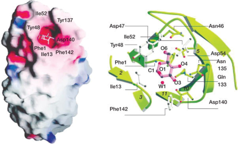
FIGURE 37.4.. The α anomer of mannose in the binding site of FimH.

FIGURE 37.4.

The α anomer of mannose in the binding site of FimH. The mannose residue is buried in a unique site at the tip of the carbohydrate-recognition domain (left) in a deep and negatively charged pocket (right). FimH prefers to bind D-mannose in the α-anomeric configuration. The hydroxyl groups of D-mannose interact with residues Phe1, Asn46, Asp47, Asp54, Gln133, Asn135, Asp140, and Phe142 by hydrogen-bonding and hydrophobic interactions. Residues in direct contact are shown as a ball-and-stick model. W1, Water. (Redrawn, with permission, from Hung CS, et al. 2002. Mol Microbiol 44: 903–915, © John Wiley and Sons.)

Download Teaching Slide (PPTX, 10M)

From: Chapter 37, Microbial Lectins: Hemagglutinins, Adhesins, and Toxins

Cover of Essentials of Glycobiology
Essentials of Glycobiology [Internet]. 4th edition.
Varki A, Cummings RD, Esko JD, et al., editors.
Cold Spring Harbor (NY): Cold Spring Harbor Laboratory Press; 2022.
Copyright © 2022 The Consortium of Glycobiology Editors, La Jolla, California; published by Cold Spring Harbor Laboratory Press; doi:10.1101/glycobiology.4e.37. All rights reserved.

The content of this book is licensed under a Creative Commons Attribution-NonCommercial-NoDerivs 4.0 Unported license. To view the terms and conditions of this license, visit https://creativecommons.org/licenses/by-nc-nd/4.0/

NCBI Bookshelf. A service of the National Library of Medicine, National Institutes of Health.