NCBI Bookshelf. A service of the National Library of Medicine, National Institutes of Health.
InformedHealth.org [Internet]. Cologne, Germany: Institute for Quality and Efficiency in Health Care (IQWiG); 2006-.
Whether during routine examinations or heart diagnostics, many people have already had an electrocardiogram (ECG or EKG). But what does it actually measure, and what does the ECG curve show us?
Our nerve and muscle cells communicate with each other using electrical and chemical signals. Regular electrical signals also control our heartbeat. These signals are sent by a group of cells in the right atrium of the heart known as the sinoatrial node (SA node), and they spread through the heart muscle tissue as tiny electrical impulses. This causes first the atria and then the ventricles of the heart to contract.
The way that these signals spread through the heart can also be measured on the surface of our skin. An ECG measures these changes in electrical signals (or, in fact, voltage) on different areas of skin and plots them as a graph. The resulting ECG graph is called an electrocardiogram.
When is an ECG done?
An ECG is used to check how the heart is functioning. It mainly records how often the heart beats (heart rate) and how regularly it beats (heart rhythm). It can give us important information, for instance about possible narrowing of the coronary arteries, a heart attack or an irregular heartbeat like atrial fibrillation.
What does an ECG show?
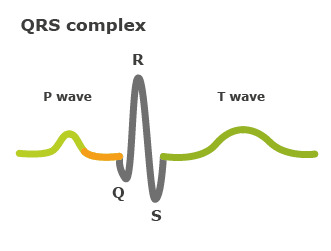
If the heart is beating steadily, it will produce the typical ECG pattern: The first peak (P wave) shows how the electrical impulse (excitation) spreads across the two atria of the heart. The atria contract (squeeze), pumping blood into the ventricles, and then immediately relax. The electrical impulse then reaches the ventricles. This can be seen in the Q, R and S waves of the ECG, which is called the QRS complex. The ventricles contract. Then the T wave shows that the electrical impulse has stopped spreading, and the ventricles relax once again.
Heart conditions such as heart attacks or irregular heartbeats can be detected in ECGs. They can help us find out what the underlying causes are.

The QRS complex between the P wave and the T wave in a normal heartbeat
What does the test involve?
The electrical activity of the heart can be measured on the surface of the skin – even on your arms or legs. The standard “12-lead ECG” uses a total of ten electrodes: six on your chest, and then one each on your forearms and calves. If there is too much body hair, these areas are shaved first; other than that, no preparation is needed. These electrodes are connected by cables to an ECG machine. The machine converts the signals it receives into an ECG graph and saves it. Some machines can also print the graphs out.
What types of ECG tests are done?
Resting ECG: This involves lying still on your back. It is important that you lie calmly and comfortably during the test because tensing your muscles, moving, coughing or shaking can affect the results. The actual measurement takes about one minute, or five minutes at most.
Exercise ECG: Here the electrical activity of your heart is measured while you are physically active. This usually involves riding an exercise bike. The amount of exertion is steadily increased by making it more and more difficult to turn the pedals. Your blood pressure is also checked regularly. The test is stopped if any irregularities in the ECG occur.
Holter monitor: The electrical activity of the heart is typically recorded over a period of 24 hours. Three or four electrodes are attached to your chest, and a small recording device is worn on a belt or hung around your neck. It is usually still possible to go about your day-to-day activities with the monitor. The ECG data are then transferred to a computer later on at the doctor's office for analysis. While you are wearing the Holter monitor, you also take notes about your daily schedule (such as unusual events, physical activity and sleep). The doctor needs this information to analyze the ECG. A Holter monitor may be used if, for instance, you only have an irregular heartbeat some of the time and it doesn't show up in a “normal” ECG. Special devices called event recorders can even document heartbeat irregularities over a period of several years. This way rare heartbeat irregularities can also be recorded. Event recorders are so small that they can be implanted under your skin.
What do the results of a 12-lead ECG show?
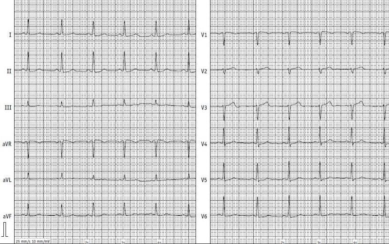
The 12-lead ECG compares the strength of the signals between two electrodes – doctors call these measurements “leads.” For example, one of the leads is measured based on the two electrodes on your arms. A 12-lead ECG, as its name implies, is used to measure twelve leads.
Depending on which lead shows deviations from a normal ECG, experts can find out things like in which part of the heart muscle an infarction has occurred, or whether a heart rhythm problem is coming from the atria or the ventricles.

Normal ECG – Left: the leads from the arms and legs, Right: the leads from the chest wall. Source: CCB Frankfurt a.M.
Sources
- Brandes R, Lang F, Schmidt R. Physiologie des Menschen: mit Pathophysiologie. Berlin: Springer; 2019.
- Pschyrembel Online. 2023.
- Trappe HJ, Schuster HP. EKG-Kurs für Isabel. Stuttgart: Thieme; 2017.
IQWiG health information is written with the aim of helping people understand the advantages and disadvantages of the main treatment options and health care services.
Because IQWiG is a German institute, some of the information provided here is specific to the German health care system. The suitability of any of the described options in an individual case can be determined by talking to a doctor. informedhealth.org can provide support for talks with doctors and other medical professionals, but cannot replace them. We do not offer individual consultations.
Our information is based on the results of good-quality studies. It is written by a team of health care professionals, scientists and editors, and reviewed by external experts. You can find a detailed description of how our health information is produced and updated in our methods.