From: Serotonin

NCBI Bookshelf. A service of the National Library of Medicine, National Institutes of Health.


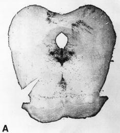
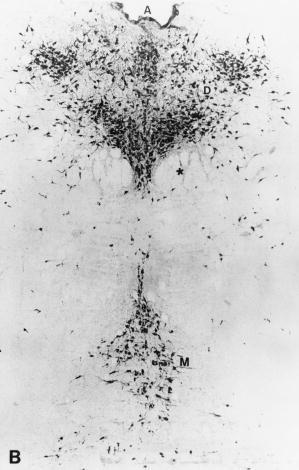
Serotonergic cell bodies in the midbrain raphe nuclei demonstrated by 5-hydroxytryptamine immunocytochemistry. A: Low magnification of transverse section through rat midbrain. The serotonergic cell body groups shown give rise to widespread serotonergic projections to cerebral cortex and forebrain structures. B: Higher magnification micrograph showing serotonergic cell bodies in dorsal and median raphe nuclei. The dorsal raphe nucleus lies in the central gray matter just beneath the cerebral aqueduct. In the transverse plane, the dorsal raphe can be subdivided further into a ventromedial cell cluster between and just above the medial longitudinal fasciculus (MLF)*, a smaller dorsomedial group just below the aqueduct and large bilateral cell groups. The median raphe nucleus lies in the central core of the midbrain, below the MLF. D, dorsal raphe; M, median raphe; A, aqueduct. (From [2], with permission.)
From: Serotonin

NCBI Bookshelf. A service of the National Library of Medicine, National Institutes of Health.