U.S. flag

An official website of the United States government

NCBI Bookshelf. A service of the National Library of Medicine, National Institutes of Health.

Adam MP, Feldman J, Mirzaa GM, et al., editors. GeneReviews® [Internet]. Seattle (WA): University of Washington, Seattle; 1993-2024.

Cover of GeneReviews®

GeneReviews® [Internet].

Show details

Mandibulofacial Dysostosis with Microcephaly

Synonym: Mandibulofacial Dysostosis, Guion-Almeida Type (MFDGA), EFTUD2-Related Mandibulofacial Dysostosis with Microcephaly (Guion-Almeida Type)

, MD, , MSc, , BSc, and , PhD, MD.

Author Information and Affiliations

Initial Posting: ; Last Revision: April 6, 2023.

Estimated reading time: 25 minutes

Summary

Clinical characteristics.

Mandibulofacial dysostosis with microcephaly (MFDM) is characterized by malar and mandibular hypoplasia, microcephaly (congenital or postnatal onset), intellectual disability (mild, moderate, or severe), malformations of the external ear, and hearing loss that is typically conductive. Associated craniofacial malformations may include cleft palate, choanal atresia, zygomatic arch cleft (identified on cranial CT scan), and facial asymmetry. Other relatively common findings (present in 25%-35% of individuals) can include cardiac anomalies, thumb anomalies, esophageal atresia/tracheoesophageal fistula, short stature, spine anomalies, and epilepsy.

Diagnosis/testing.

The diagnosis of MFDM is confirmed in a proband with typical clinical findings and a heterozygous pathogenic variant in EFTUD2 identified by genetic testing.

Management.

Treatment of manifestations: Individualized treatment of craniofacial manifestations is managed by a multidisciplinary team which may include: oromaxillofacial surgery, plastic surgery, otolaryngology, dentistry/orthodontics, and occupational and speech-language therapy. Newborn infants may have airway compromise at delivery due to choanal atresia and/or mandibular hypoplasia, requiring intubation and/or tracheostomy for initial stabilization. Esophageal atresia/tracheoesophageal fistula, cardiac defects, renal anomalies, and thumb anomalies are treated in a routine manner. Short stature is managed expectantly. Treatment of hearing loss is individualized, and may involve conventional hearing aid(s), bone-anchored hearing aid(s), and/or cochlear implant(s). Early individualized educational and therapy plans are devised as needed to optimize developmental outcome.

Surveillance: Annual growth assessment and periodic developmental assessment with evaluation for obstructive sleep apnea and epilepsy as needed.

Genetic counseling.

MFDM is an autosomal dominant disorder. Most individuals diagnosed with MFDM to date are presumed to have the disorder as the result of a de novo EFTUD2 pathogenic variant; in some individuals, the causative pathogenic variant was inherited from a parent with a milder phenotypic presentation. If a parent of the proband has the pathogenic variant identified in the proband, the risk to sibs of the proband (at conception) is 50%. Once the causative EFTUD2 pathogenic variant has been identified in an affected family member, prenatal testing and preimplantation genetic testing are possible.

Diagnosis

Suggestive Findings

Mandibulofacial dysostosis with microcephaly (MFDM) should be suspected in individuals with mandibulofacial dysostosis (a developmental disorder of the first and second branchial arches characterized by malar and maxillary hypoplasia) in the context of one or more additional features, including:

  • Microcephaly (defined here as occipitofrontal circumference ≥2 SD below mean), which may be either primary (i.e., congenital; present a birth) or secondary (i.e., postnatal onset)
  • Intellectual disability, which may be mild, moderate, or severe
  • Characteristic malformations of the external ear (see Figure 1), which include microtia (grades I-III), deficiency of the superior helix and antihelix, preauricular tags, and auditory canal atresia/stenosis. The posterior-inferior margin of the lobule may have a right-angle ("squared-off") configuration.
  • Hearing loss, typically conductive
Figure 1. . Range of external ear findings in MFDM.

Figure 1.

Range of external ear findings in MFDM. Microtia may be of any degree, and is frequently accompanied by preauricular tag(s) and/or auditory canal atresia/stenosis. The superior helix is relatively deficient. The posterior-inferior rim of the lobule may (more...)

Establishing the Diagnosis

The diagnosis of MFDM is established in a proband with typical clinical findings and a heterozygous pathogenic (or likely pathogenic) variant in EFTUD2 identified by genetic testing (see Table 1).

Note: (1) Per ACMG/AMP variant interpretation guidelines, the terms "pathogenic variants" and "likely pathogenic variants" are synonymous in a clinical setting, meaning that both are considered diagnostic and both can be used for clinical decision making [Richards et al 2015]. Reference to "pathogenic variants" in this section is understood to include any likely pathogenic variants. (2) Identification of a heterozygous EFTUD2 variant of uncertain significance does not establish or rule out the diagnosis.

Molecular genetic testing approaches can include a combination of gene-targeted testing (single-gene testing, multigene panel) and comprehensive genomic testing (exome sequencing, exome array, genome sequencing) depending on the phenotype.

Gene-targeted testing requires that the clinician determine which gene(s) are likely involved, whereas genomic testing does not. Because the clinical phenotype of MFDM varies, individuals with highly characteristic clinical findings are likely to be diagnosed using gene-targeted testing (see Option 1), whereas those with an atypical or nonspecific clinical phenotype overlapping other inherited syndromes are more likely to be diagnosed using comprehensive genomic testing (see Option 2).

Option 1

When the phenotypic and laboratory findings suggest the diagnosis of MFDM, molecular genetic testing approaches can include single-gene testing or use of a multigene panel:

  • Single-gene testing. Sequence analysis of EFTUD2 is performed first to detect small intragenic deletions/insertions and missense, nonsense, and splice site variants. Note: Depending on the sequencing method used, single-exon, multiexon, or whole-gene deletions/duplications may not be detected. If no variant is detected by the sequencing method used, the next step is to perform gene-targeted deletion/duplication analysis to detect exon and whole-gene deletions or duplications.
  • A multigene panel that includes EFTUD2 and other genes of interest (see Differential Diagnosis) is most likely to identify the genetic cause of the condition while limiting identification of variants of uncertain significance and pathogenic variants in genes that do not explain the underlying phenotype. Note: (1) The genes included in the panel and the diagnostic sensitivity of the testing used for each gene vary by laboratory and are likely to change over time. (2) Some multigene panels may include genes not associated with the condition discussed in this GeneReview. (3) In some laboratories, panel options may include a custom laboratory-designed panel and/or custom phenotype-focused exome analysis that includes genes specified by the clinician. (4) Methods used in a panel may include sequence analysis, deletion/duplication analysis, and/or other non-sequencing-based tests. For this disorder, a multigene panel that also includes deletion/duplication analysis is recommended (see Table 1).
    For an introduction to multigene panels click here. More detailed information for clinicians ordering genetic tests can be found here.

Option 2

When the phenotype is indistinguishable from many other inherited disorders with similar craniofacial features or is not considered because an individual has atypical phenotypic features, comprehensive genomic testing (which does not require the clinician to determine which gene[s] are likely involved) is the best option. Exome sequencing is most commonly used; genome sequencing is also possible.

If exome sequencing is not diagnostic, other techniques (when clinically available) may be considered to detect (multi)exon deletions or duplications that cannot be detected by sequence analysis (see Table 1).

For an introduction to comprehensive genomic testing click here. More detailed information for clinicians ordering genomic testing can be found here.

Table 1.

Molecular Genetic Testing Used in Mandibulofacial Dysostosis with Microcephaly

Gene 1MethodProportion of Probands with a Pathogenic Variant 2 Detectable by Method
EFTUD2 Sequence analysis 393% 4
Gene-targeted deletion/duplication analysis 57% 4
1.

See Table A. Genes and Databases for chromosome locus and protein.

2.

See Molecular Genetics for information on variants detected in this gene.

3.

Sequence analysis detects variants that are benign, likely benign, of uncertain significance, likely pathogenic, or pathogenic. Variants may include small intragenic deletions/insertions and missense, nonsense, and splice site variants; typically, exon or whole-gene deletions/duplications are not detected. For issues to consider in interpretation of sequence analysis results, click here.

4.
5.

Gene-targeted deletion/duplication analysis detects intragenic deletions or duplications. Methods used may include a range of techniques such as quantitative PCR, long-range PCR, multiplex ligation-dependent probe amplification (MLPA), and a gene-targeted microarray designed to detect single-exon deletions or duplications.

Clinical Characteristics

Clinical Description

Mandibulofacial dysostosis with microcephaly (MFDM) is a multiple malformation syndrome comprising craniofacial skeletal anomalies, microcephaly, developmental delay / intellectual disability, abnormalities of the ears and hearing, and, in some instances, extracranial malformations (esophageal atresia, congenital heart defects, thumb anomalies), and/or short stature.

To date, 126 individuals have been identified with a pathogenic variant in EFTUD2 [Gordon et al 2012, Lines et al 2012, Need et al 2012, Luquetti et al 2013, Voigt et al 2013, Lehalle et al 2014, Deml et al 2015, Gandomi et al 2015, Sarkar et al 2015, Smigiel et al 2015, Huang et al 2016, Vincent et al 2016, Bick et al 2017, Matsuo et al 2017, McDermott et al 2017, Rengasamy Venugopalan et al 2017, Williams et al 2017, Paderova et al 2018, Yu et al 2018, Lacour et al 2019, Silva et al 2019, Wu et al 2019]. The following description of the phenotypic features associated with this condition is based on these reports.

Table 2.

Mandibulofacial Dysostosis with Microcephaly: Frequency of Select Features

Feature% of Persons
w/Feature
Comment
Facial
structural
differences
Malar hypoplasia92%
Micrognathia / Mandibular
hypoplasia
93%
Cleft palate43%
Choanal atresia30%
Facial asymmetry58%
Microcephaly 87%Occipitofrontal circumference ≥2 SD below mean
Developmental delay / Intellectual disability 97%Severity varies (may be mild, moderate, or severe; critical sequelae (e.g. neonatal airway compromise, cardiac anomalies) may affect developmental outcome.
Ear
malformations
& hearing loss
Microtia / Dysplastic pinna(e)97%
Auditory canal atresia or stenosis68%
Preauricular tag50%
Hearing loss83%
Other findings Cardiac anomalies35%Typically atrial &/or ventricular septal defect
Thumb anomalies34%Typically proximally placed; uncommonly, preaxial polydactyly or hypoplasia
Esophageal atresia /
Tracheoesophageal fistula
33%
Short stature30%
Spine anomalies28%Incl scoliosis, kyphosis, hemivertebrae, & cervical segmentation anomalies
Epilepsy26%

Mandibulofacial dysostosis is characterized by malar and maxillary hypoplasia.

Accompanying findings in MFDM include micrognathia/mandibular hypoplasia, cleft palate, and/or choanal abnormality.

Cleft palate in MFDM occurs as a Pierre Robin sequence, characterized by a midline bony defect without accompanying cleft lip. Submucous cleft has also been described. Choanal atresia is generally osseous, being either unilateral or bilateral; choanal stenosis is also frequent.

Zygomatic arch cleft has been identified in ten of 19 individuals assessed (best done with cranial CT with 3-D reconstruction).

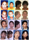
Characteristic dysmorphic features (Figure 2), distinct from those seen in the other mandibulofacial and acrofacial dysostoses (see Differential Diagnosis), are recognizable by early childhood. In addition to malar and maxillary hypoplasia, microcephaly, and the typical ear anomalies described in this section, features include metopic ridge, prominent glabella, broad nasal bridge with prominent ridge and bulbous tip, large oral aperture, everted lower lip, and/or (frequently) facial asymmetry.

Figure 2. . Typical craniofacial features of MFDM.

Figure 2.

Typical craniofacial features of MFDM. These include micrognathia, malar hypoplasia, a relatively high nasal root with prominent ridge, everted lower lip, and (frequently) facial asymmetry. Characteristic ear malformations, present in essentially all (more...)

Microcephaly is present in about 87% of reported individuals (n=33; median -3.5 SD; range -0.2 SD to -6.5 SD) [Huang et al 2016]. Cephalic growth curves for MFDM are published [Huang et al 2016]. In some instances, individuals have exhibited apparent cephalic "catch-up" growth, resulting in a normal adult occipitofrontal circumference despite microcephaly in childhood. Individuals whose head circumference falls within the normal range have also been reported to have intellectual disability [Luquetti et al 2013, Lehalle et al 2014].

Developmental delay and/or intellectual disability are present in almost all individuals. Among 30 persons on whom data are available, the degree of intellectual disability was reported as "mild" (~40%), "moderate" (~50%), or "severe" (~10%) [Gordon et al 2012, Lines et al 2012, Luquetti et al 2013, Voigt et al 2013].

Affected children are ambulatory but show delayed motor development, taking first steps at a median age of 26 months (n=38; range 13-60 months) [Huang et al 2016].

Among those who are verbal, the median reported age at first words is 27 months (n=32; range 12 months to 5.6 years); some affected persons remain nonverbal into adult life [Huang et al 2016]. Assessment of language skills may be confounded by the presence of hearing loss and/or cleft palate.

To date there have been no detailed or cross-sectional studies of long-term neuropsychological outcomes in MFDM. Developmental data in the few affected adults identified to date suggest a broad range of outcomes, with some affected persons achieving semi-independent living with paid employment [Huang et al 2016], whereas others are nonverbal and require extensive assistance with daily activities [Authors, unpublished data].

Ear malformations and hearing loss

  • External ear malformations. External ears are anomalous in virtually all affected individuals. Typical findings (see Figure 2) include microtia (grades I-III), deficiency of the superior helix and antihelix, preauricular tags, and auditory canal atresia/stenosis. The posterior-inferior margin of the lobule may have a right-angle ("squared-off") configuration.
  • Middle/inner ear malformations. Ear structures (ossicles, semicircular canals) are absent and/or malformed in some individuals; this is best assessed by temporal bone CT [Gordon et al 2012, Luquetti et al 2013, Voigt et al 2013].
  • Hearing loss. Hearing loss is typically conductive (~60%) as opposed to sensorineural or mixed, and is likely to result from malformation or absence of the middle ear ossicles, auditory canal atresia, or both.

Other relatively common findings

  • Cardiac anomalies are present in 35% of individuals. Hemodynamically insignificant atrial and ventricular septal defects are the most common; tetralogy of Fallot, patent ductus arteriosus, and aortic arch abnormalities (e.g., coarctation) have also been reported [Need et al 2012, Lehalle et al 2014].
  • Thumb anomalies (proximally placed, duplicated, or hypoplastic thumbs) are seen in about 35% of individuals.
  • Esophageal atresia / tracheoesophageal fistula (EA/TEF) is present in about 35% of affected individuals. EA/TEF is typically type C (the most common type), in which the upper esophageal pouch ends blindly and the lower esophageal pouch connects abnormally to the trachea (distal tracheoesophageal fistula). Laryngotracheal anomalies (tracheomalacia, posterior laryngotracheoesophageal clefts) may be seen in association with EA/TEF. It may be suspected antenatally because of polyhydramnios or absent stomach echolucency, or neonatally in the context of unexplained respiratory distress and/or failed nasogastric tube placement.
  • Short stature is present in 30% of individuals. Height growth curves for MFDM are published [Huang et al 2016].
  • Spine anomalies include scoliosis, kyphosis, hemivertebrae, and cervical segmentation anomalies.
  • Epilepsy is present in 26% of individuals. Detailed clinical data regarding the type of epilepsy have not been specifically reported. Matsuo et al [2017] report one individual with recurrent seizures for which EEG demonstrated occasional spike discharges originating from the right frontal area.

Additional malformations (low frequency)

Genotype-Phenotype Correlations

No genotype-phenotype correlations for EFTUD2 have been identified.

Individuals with microdeletions encompassing EFTUD2 and contiguous genes may have additional features or more severe intellectual disability [Lines et al 2012, Gandomi et al 2015].

Penetrance

MFDM is highly penetrant but variably expressive. Features may be subclinical in some affected individuals, as in the case of two non-mosaic, intellectually normal mothers – each with two affected children – in whom the only reported clinical findings were unilateral zygomatic cleft and facial asymmetry [Voigt et al 2013] and mild facial asymmetry and a preauricular tag [McDermott et al 2017].

Nomenclature

The descriptive term "mandibulofacial dysostosis with microcephaly" is synonymous with the eponym "mandibulofacial dysostosis, Guion-Almeida type" [Guion-Almeida et al 2009].

Some have suggested that MFDM be classified as an acrofacial dysostosis rather than a mandibulofacial dysostosis [Voigt et al 2013]. This is predominantly a clinical (rather than pathophysiologic) distinction based on the presence of limb anomalies in the former category, and their absence in the latter.

In the 2023 revision of the Nosology of Genetic Skeletal Disorders [Unger et al 2023], MFDM is referred to as EFTUD2-related mandibulofacial dysostosis with microcephaly (Guion-Almeida type) and included in the craniofacial dysostoses group.

Prevalence

The prevalence of MFDM has not been established. At least 126 cases caused by mutation of EFTUD2 have been reported to date (see Table 2 and references cited in Clinical Description).

Differential Diagnosis

Mandibulofacial Dysostosis

Table 3.

Genes of Interest in the Differential Diagnosis of Mandibulofacial Dysostosis with Microcephaly

Gene(s)DiffDx DisorderMOIClinical Characteristics of DiffDx Disorder Overlapping w/MFDMDistinguishing Features
CHD7 1CHARGE syndrome
(See CHD7 Disorder.)
ADMicrocephaly, ear anomalies, choanal atresia, TEF, CHDOcular coloboma & Mondini malformation are present in CHARGE but not in MFDM.
DHODH Miller acrofacial dysostosis
(OMIM 263750)
ARMFD w/postaxial limb defects ± other extracranial malformationsOFC & intelligence are typically normal in Miller acrofacial dysostosis.
POLR1C
POLR1D
TCOF1
Treacher Collins syndrome (TCS)AD
AR
  • MFD (may resemble MFD in MFDM)
  • Malformations occur in 1st & 2nd branchial arch-derived structures.
  • ID (w/o microcephaly) is not common but may be present in some persons. 2
  • Lower lid clefts, absent eyelashes, & lacrimal system anomalies may be present.
  • Cardiac & esophageal malformations are not assoc w/TCS.
  • Intellect & OFC are usually in average range in TCS.
  • Unlike TCS, palpebral fissures are not consistently downslanting in MFDM.
RPL5
RPL11
RPL35A
RPS10
RPS17
RPS19
RPS24
RPS26 3
Diamond-Blackfan anemia (DBA)AD
(XL 4)
~1/3 of persons exhibit MFD-like craniofacial anomalies ± cleft palate, anomalous thumbs, cardiac anomalies, &/or growth restriction.Moderate-to-severe anemia in DBA
SF3B4 Nager acrofacial dysostosis
(OMIM 154400)
ADMFD w/preaxial (typically upper) limb defects, ± other extracranial malformationsOFC & intelligence typically normal in Nager acrofacial dysostosis

AD = autosomal dominant; AR = autosomal recessive; CHD = congenital heart defect; DiffDx = differential diagnosis; ID = intellectual disability; MFD = mandibulofacial dysostosis; MFDM = mandibulofacial dysostosis with microcephaly; MOI = mode of inheritance; OFC = occipitofrontal circumference; TEF = tracheoesophageal fistula; XL = X-linked

1.

The finding of a CHD7 pathogenic variant is not equivalent to a diagnosis of CHARGE syndrome; the phenotypic spectrum of heterozygous CHD7 pathogenic variants encompasses CHARGE syndrome as well as subsets of features that comprise the CHARGE syndrome phenotype.

2.

Intellectual disability (without microcephaly) may be present in individuals with either (a) a history of neonatal airway compromise or (b) microdeletions encompassing TCOF1 and adjacent genes [Vincent et al 2016].

3.

Listed genes represent the most common genetic causes of Diamond-Blackfan anemia (DBA); more than 20 genes are known to be associated with DBA (see Diamond-Blackfan anemia).

4.

DBA is most often inherited in an autosomal dominant manner; GATA1- and TSR2-DBA are inherited in an X-linked manner (GATA1 and TSR2 are less common genetic causes of DBA and are not included in the table).

Craniofacial Microsomia

Craniofacial microsomia (CFM) is a first- and second-arch malformation spectrum encompassing several phenotypes, including oculo-auriculo-vertebral (OAV) syndrome and Goldenhar syndrome (OMIM 164210). CFM most frequently occurs as a simplex case (i.e., in a single individual in a family) with unknown etiology; recurrence risks are empiric.

CFM shares several major features with mandibulofacial dysostosis with microcephaly (MFDM), including preauricular tags, microtia, aural atresia, hearing loss, and – notably – facial asymmetry, present in approximately 65% of persons with CFM and also a frequent finding in MFDM (58%).

The spectrum of orofacial clefting differs between the two conditions: midline cleft palate is typical of MFDM, while CFM can be associated with any type of orofacial cleft, including lateral oral clefts. Although various extracranial anomalies may occur in either condition, vertebral anomalies in particular should suggest CFM.

At least two persons with an EFTUD2 pathogenic variant were diagnosed with "bilateral OAV syndrome" prior to the recognition of MFDM as a distinct syndrome [Authors, personal observation].

Tracheoesophageal Fistula

Tracheoesophageal fistula is a feature of several other recognized conditions, including Feingold syndrome and VACTERL association (OMIM 192350); clinical differentiation is generally straightforward.

Management

Evaluations Following Initial Diagnosis

To establish the extent of disease and needs in an individual diagnosed with mandibulofacial dysostosis with microcephaly (MFDM), the evaluations summarized in Table 4 (if not performed as part of the evaluation that led to the diagnosis) are recommended.

Table 4.

Recommended Evaluations Following Initial Diagnosis in Individuals with Mandibulofacial Dysostosis with Microcephaly

System/ConcernEvaluationComment
MFD
  • Airway assessment for evidence of upper-airway obstruction w/or w/o choanal atresia
  • Exam for midline cleft palate & referral to multidisciplinary cleft palate team as required
Important in newborns w/disorder
DD/ID Developmental assessmentIncl adaptive, cognitive, & speech-language evals
Hearing loss Audiologic evalAssess for hearing loss.
EA/TEF Urgent eval in newborns, esp in those w/history of polyhydramnios, unexplained respiratory distress, &/or failed nasogastric tube placement
Cardiac anomaly Echocardiogram & cardiologist eval
Renal anomaly Renal ultrasound
Skeletal
anomaly
Radiograph to assess for scoliosis, rib or thumb malformation as clinically indicated
Short stature Assess w/growth charts.Height growth curves for MFDM are published. 1
Genetic
counseling
By genetics professionals 2To inform affected persons & families re nature, MOI, & implications of MFDM to facilitate medical & personal decision making
Family support
& resources
Assess need for:

DD/ID = developmental delay / intellectual disability; EA/TF = esophageal atresia / tracheoesophageal fistula; MFD = mandibulofacial dysostosis; MFDM = mandibulofacial dysostosis with microcephaly; MOI = mode of inheritance

1.
2.

Medical geneticist, certified genetic counselor, or certified advanced genetic nurse

Treatment of Manifestations

There are no published management guidelines to date for MFDM.

Esophageal atresia / tracheoesophageal fistula, cardiac defects, renal anomalies, and thumb anomalies are treated in a routine manner. Short stature is managed expectantly. Of note, the response to human growth hormone has not been specifically reported.

Table 5.

Treatment of Manifestations in Individuals with MFDM

Manifestation/ConcernTreatmentConsiderations/Other
Mandibulofacial dysostosis
  • Neonates w/airway compromise at delivery may require intubation &/or tracheostomy for initial stabilization.
  • Treatment of craniofacial manifestations is individualized & managed by a multidisciplinary team incl oromaxillofacial surgery, plastic surgery, otolaryngology, dentistry/orthodontics, & occupational & speech-language therapy.
DD/ID See Developmental Delay / Intellectual Disability Management Issues.To optimize developmental outcome
Hearing loss Treatment is individualized & may involve conventional hearing aid(s), bone-anchored hearing aid(s), &/or cochlear implant(s).Community hearing services through early intervention or school district

DD/ID = developmental delay / intellectual disability

Developmental Delay / Intellectual Disability Management Issues

The following information represents typical management recommendations for individuals with developmental delay / intellectual disability in the United States; standard recommendations may vary from country to country.

Ages 0-3 years. Referral to an early intervention program is recommended for access to occupational, physical, speech, and feeding therapy as well as infant mental health services, special educators, and sensory impairment specialists. In the US, early intervention is a federally funded program available in all states that provides in-home services to target individual therapy needs.

Ages 3-5 years. In the US, developmental preschool through the local public school district is recommended. Before placement, an evaluation is made to determine needed services and therapies and an individualized education plan (IEP) is developed for those who qualify based on established motor, language, social, or cognitive delay. The early intervention program typically assists with this transition. Developmental preschool is center based; for children too medically unstable to attend, home-based services are provided.

All ages. Consultation with a developmental pediatrician is recommended to ensure the involvement of appropriate community, state, and educational agencies (US) and to support parents in maximizing quality of life. Some issues to consider:

  • IEP services:
    • An IEP provides specially designed instruction and related services to children who qualify.
    • IEP services will be reviewed annually to determine whether any changes are needed.
    • Special education law requires that children participating in an IEP be in the least restrictive environment feasible at school and included in general education as much as possible, when and where appropriate.
    • Hearing consultants should be a part of the child's IEP team to support access to academic material.
    • PT, OT, and speech services will be provided in the IEP to the extent that the need affects the child's access to academic material. Beyond that, private supportive therapies based on the affected individual's needs may be considered. Specific recommendations regarding type of therapy can be made by a developmental pediatrician.
    • As a child enters the teen years, a transition plan should be discussed and incorporated in the IEP. For those receiving IEP services, the public school district is required to provide services until age 21.
  • A 504 plan (Section 504: a US federal statute that prohibits discrimination based on disability) can be considered for those who require accommodations or modifications such as front-of-class seating, assistive technology devices, classroom scribes, extra time between classes, modified assignments, and enlarged text.
  • Developmental Disabilities Administration (DDA) enrollment is recommended. DDA is a US public agency that provides services and support to qualified individuals. Eligibility differs by state but is typically determined by diagnosis and/or associated cognitive/adaptive disabilities.
  • Families with limited income and resources may also qualify for supplemental security income (SSI) for their child with a disability.

Motor Dysfunction

Gross motor dysfunction

  • Physical therapy is recommended to maximize mobility and to reduce the risk for later-onset orthopedic complications (e.g., contractures, scoliosis, hip dislocation).
  • Consider use of durable medical equipment and positioning devices as needed (e.g., wheelchairs, walkers, bath chairs, orthotics, adaptive strollers).
  • For muscle tone abnormalities including hypertonia or dystonia, consider involving appropriate specialists to aid in management of baclofen, tizanidine, Botox®, antiparkinsonian medications, or orthopedic procedures.

Fine motor dysfunction. Occupational therapy is recommended for difficulty with fine motor skills that affect adaptive function such as feeding, grooming, dressing, and writing.

Oral motor dysfunction should be assessed at each visit and clinical feeding evaluations and/or radiographic swallowing studies should be obtained for choking/gagging during feeds, poor weight gain, frequent respiratory illnesses, or feeding refusal that is not otherwise explained. Assuming that the child is safe to eat by mouth, feeding therapy (typically from an occupational or speech therapist) is recommended to help improve coordination or sensory-related feeding issues. Feeds can be thickened or chilled for safety. When feeding dysfunction is severe, an NG-tube or G-tube may be necessary.

Communication issues. Consider evaluation for alternative means of communication (e.g., augmentative and alternative communication [AAC]) for individuals who have expressive language difficulties. An AAC evaluation can be completed by a speech-language pathologist who has expertise in the area. The evaluation will consider cognitive abilities and sensory impairments to determine the most appropriate form of communication. AAC devices can range from low-tech, such as picture exchange communication, to high-tech, such as voice-generating devices. Contrary to popular belief, AAC devices do not hinder verbal development of speech, but rather support optimal speech and language development.

Social/Behavioral Concerns

Children may qualify for and benefit from interventions used in treatment of autism spectrum disorder, including applied behavior analysis (ABA). ABA therapy is targeted to the individual child's behavioral, social, and adaptive strengths and weaknesses and typically performed one on one with a board-certified behavior analyst.

Consultation with a developmental pediatrician may be helpful in guiding parents through appropriate behavior management strategies or providing prescription medications, such as medication used to treat attention-deficit/hyperactivity disorder, when necessary.

Concerns about serious aggressive or destructive behavior can be addressed by a pediatric psychiatrist.

Surveillance

Table 6.

Recommended Surveillance for Individuals with MFDM

System/ConcernEvaluationFrequency
Mandibulofacial dysotosis Evaluate for obstructive sleep apnea.As needed
Developmental delay /
Intellectual disability
Developmental assessment & psychoeducational testing for childrenPeriodically throughout childhood
Short stature Growth parametersAnnually throughout childhood & adolescence
Epilepsy Neurologic eval w/EEG &/or brain imaging if appropriateAs needed

Evaluation of Relatives at Risk

See Genetic Counseling for issues related to testing of at-risk relatives for genetic counseling purposes.

Therapies Under Investigation

Search ClinicalTrials.gov in the US and EU Clinical Trials Register in Europe for access to information on clinical studies for a wide range of diseases and conditions. Note: There may not be clinical trials for this disorder.

Genetic Counseling

Genetic counseling is the process of providing individuals and families with information on the nature, mode(s) of inheritance, and implications of genetic disorders to help them make informed medical and personal decisions. The following section deals with genetic risk assessment and the use of family history and genetic testing to clarify genetic status for family members; it is not meant to address all personal, cultural, or ethical issues that may arise or to substitute for consultation with a genetics professional. —ED.

Mode of Inheritance

Mandibulofacial dysostosis with microcephaly (MFDM) is an autosomal dominant disorder often caused by a de novo pathogenic variant.

Risk to Family Members

Parents of a proband

  • More than 80% of individuals diagnosed with MFDM to date are presumed to have the disorder as the result of a de novo EFTUD2 pathogenic variant [Huang et al 2016].
  • In 12 of 64 individuals diagnosed with MFDM, the causative pathogenic variant was inherited from a parent with a milder phenotypic presentation [Gordon et al 2012, Voigt et al 2013, Lehalle et al 2014, Huang et al 2016].
  • Molecular genetic testing and clinical evaluation are recommended for the parents of a proband with an apparent de novo EFTUD2 pathogenic variant (i.e., the proband is the only family member known to have MFDM).
  • If the pathogenic variant identified in the proband is not identified in either parent, the following possibilities should be considered:
    • The proband has a de novo pathogenic variant. Note: A pathogenic variant is reported as "de novo" if: (1) the pathogenic variant found in the proband is not detected in parental DNA; and (2) parental identity testing has confirmed biological maternity and paternity. If parental identity testing is not performed, the variant is reported as "assumed de novo" [Richards et al 2015].
    • The proband inherited a pathogenic variant from a parent with germline (or somatic and germline) mosaicism. The incidence of parental germline mosaicism in MFDM is 6% [Huang et al 2016]. Note: Testing of parental leukocyte DNA may not detect all instances of somatic mosaicism.
  • The family history of some individuals diagnosed with MFDM may appear to be negative because of a failure to recognize the disorder in family members because of milder manifestations of the disorder in a heterozygous parent. Therefore, an apparently negative family history cannot be confirmed unless molecular genetic testing has demonstrated that neither parent is heterozygous for the pathogenic variant identified in the proband.

Sibs of a proband. The risk to the sibs of the proband depends on the clinical/genetic status of the proband's parents:

  • If a parent of the proband is affected and/or is known to have the pathogenic variant identified in the proband, the risk to sibs of the proband (at conception) is 50%. Although MFDM is thought to be highly penetrant, intrafamilial clinical variability is observed, and a sib who inherits an EFTUD2 pathogenic variant may be more or less severely affected than the proband [Huang et al 2016].
  • If the proband has a known MFDM-related pathogenic variant that cannot be detected in the leukocyte DNA of either parent, the recurrence risk to sibs is greater than that of the general population because of the possibility of parental germline mosaicism.
  • If the parents have not been tested for the EFTUD2 pathogenic variant but are clinically unaffected, sibs are still presumed to be at increased risk for MFDM because of the possibility of subclinical manifestations of the disorder in a heterozygous parent or the possibility of parental germline mosaicism.

Offspring of a proband. Each child of an individual with MFDM has a 50% chance of inheriting the EFTUD2 pathogenic variant.

Other family members. The risk to other family members depends on the genetic status of the proband's parents: if a parent has the EFTUD2 pathogenic variant, the parent's family members may be at risk.

Related Genetic Counseling Issues

Family planning

  • The optimal time for determination of genetic risk and discussion of the availability of prenatal/preimplantation genetic testing is before pregnancy.
  • It is appropriate to offer genetic counseling (including discussion of potential risks to offspring and reproductive options) to parents of affected individuals, as well as to young adults who are affected or at risk.

Prenatal Testing and Preimplantation Genetic Testing

Molecular genetic testing. Once the EFTUD2 pathogenic variant has been identified in an affected family member, prenatal testing and preimplantation genetic testing are possible. The phenotype of affected offspring cannot be accurately predicted based on the results of prenatal molecular genetic testing (see Penetrance).

Fetal ultrasound examination. Because the sensitivity of prenatal ultrasound for detection of MFDM has not been assessed, molecular genetic testing is the recommended mode of prenatal diagnosis. Many of the characteristic findings in MFDM have a low probability of detection by antenatal ultrasound.

Differences in perspective may exist among medical professionals and within families regarding the use of prenatal testing. While most centers would consider use of prenatal testing to be a personal decision, discussion of these issues may be helpful.

Resources

GeneReviews staff has selected the following disease-specific and/or umbrella support organizations and/or registries for the benefit of individuals with this disorder and their families. GeneReviews is not responsible for the information provided by other organizations. For information on selection criteria, click here.

  • American Cleft Palate-Craniofacial Association
    Phone: 919-933-9044
  • Children's Craniofacial Association
    Phone: 800-535-3643
    Email: contactCCA@ccakids.com
  • FACES: National Craniofacial Association
    Phone: 800-332-2373; 423-266-1632
    Email: info@faces-cranio.org
  • National Institute of Dental and Craniofacial Research (NIDCR)
    Bethesda MD 20892-2190
    Phone: 866-232-4528 (toll-free); 301-496-4261
    Fax: 301-480-4098
    Email: nidcrinfo@mail.nih.gov
  • World Craniofacial Foundation
    7777 Forest Lane
    Suite C-616
    Dallas TX 75230
    Phone: 800-533-3315
    Fax: 972-566-3850
    Email: info@worldcf.org

Molecular Genetics

Information in the Molecular Genetics and OMIM tables may differ from that elsewhere in the GeneReview: tables may contain more recent information. —ED.

Table A.

Mandibulofacial Dysostosis with Microcephaly: Genes and Databases

GeneChromosome LocusProteinLocus-Specific DatabasesHGMDClinVar
EFTUD2 17q21​.31 116 kDa U5 small nuclear ribonucleoprotein component EFTUD2 @ LOVD EFTUD2 EFTUD2

Data are compiled from the following standard references: gene from HGNC; chromosome locus from OMIM; protein from UniProt. For a description of databases (Locus Specific, HGMD, ClinVar) to which links are provided, click here.

Table B.

OMIM Entries for Mandibulofacial Dysostosis with Microcephaly (View All in OMIM)

603892ELONGATION FACTOR Tu GTP-BINDING DOMAIN-CONTAINING 2; EFTUD2
610536MANDIBULOFACIAL DYSOSTOSIS, GUION-ALMEIDA TYPE; MFDGA

Molecular Pathogenesis

EFTUD2 encodes a small GTPase that is one of several subunits belonging to the U5 small nuclear ribonucleoprotein particle (snRNP) [Fabrizio et al 1997]. The U5 snRNP is a component of the major and minor spliceosomes, two large macromolecular machines that mediate canonic (U2-dependent) and minor (U12-introns) intron splicing [Wahl et al 2009]. The Saccharomyces cerevisiae homolog of EFTUD2, Snu114p, is essential for (1) the dissociation of the U4 and U6 RNAs during pre-spliceosomal activation and (2) subunit disassembly and recycling after catalytic splicing is complete [Fabrizio et al 1997, Bartels et al 2002, Small et al 2006]. An EFTUD2 haploinsufficient zebrafish model displayed abnormal brain development with neuronal apoptosis. No other systems were affected. RNA sequencing and functional analyses demonstrated RNA splicing deficiency which led to inadequate nonsense-mediated RNA decay and activation of the p53 pathway [Lei et al 2017].

Mechanism of disease causation. Reported EFTUD2 pathogenic variants include missense, nonsense, frameshift, and splice site variants, as well as whole- or partial-gene deletions, consistent with haploinsufficiency as the underlying mechanism [Lines et al 2012].

Chapter Notes

Author Notes

Matthew Lines, MD; matthew.lines@albertahealthservices.ca

Taila Hartley, MSc; thartley@cheo.on.ca

Stella K MacDonald, BSc; smacdonald@cheo.on.ca

Kym M Boycott, PhD, MD; kboycott@cheo.on.ca

Acknowledgments

The authors wish to gratefully acknowledge the patients and their families from around the world who have contributed to the understanding of this condition. Our knowledge of MFDM has been facilitated by research support from the Care4Rare Canada Consortium.

Revision History

  • 6 April 2023 (sw) Revision: "EFTUD2-Related Mandibulofacial Dysostosis with Microcephaly (Guion-Almeida Type)" added as a synonym; Nosology of Genetic Skeletal Disorders: 2023 Revision [Unger et al 2023] added to Nomenclature
  • 12 November 2020 (ha) Comprehensive update posted live
  • 3 July 2014 (me) Review posted live
  • 21 January 2014 (ml) Original submission

References

Literature Cited

  • Bartels C, Klatt C, Lührmann R, Fabrizio P. The ribosomal translocase homologue Snu114p is involved in unwinding U4/U6 RNA during activation of the spliceosome. EMBO Rep. 2002;3:875–80. [PMC free article: PMC1084225] [PubMed: 12189173]
  • Bick D, Fraser P, Gutzeit M, Harris J, Hambuch T, Helbling D, Jacob H, Kersten JN, Leuthner SR, May T, North PE, Prisco SZ, Schuler BA, Shimoyama M, Strong KA, Van Why SK, Veith R, Verbsky J, Weborg AM Jr, Wilk BM, Willoughby RE Jr, Worthey EA, Dimmock DP. Successful application of whole genome sequencing in a medical genetics clinic. J Pediatr Genet. 2017;6:61–76. [PMC free article: PMC5423809] [PubMed: 28496993]
  • Deml B, Reis LM, Muheisen S, Bick D, Semina EV. EFTUD2 deficiency in vertebrates: identification of a novel human mutation and generation of a zebrafish model. Birth Defects Res A Clin Mol Teratol. 2015;103:630–40. [PMC free article: PMC4487781] [PubMed: 26118977]
  • Fabrizio P, Laggerbauer B, Lauber J, Lane WS, Lührmann R. An evolutionarily conserved U5 snRNP-specific protein is a GTP-binding factor closely related to the ribosomal translocase EF-2. EMBO J. 1997;16:4092–106. [PMC free article: PMC1170032] [PubMed: 9233818]
  • Gandomi SK, Parra M, Reeves D, Yap V, Gau CL. Array-CGH is an effective first-tier diagnostic test for EFTUD2-associated congenital mandibulofacial dysostosis with microcephaly. Clin Genet. 2015;87:80–4. [PubMed: 24266672]
  • Gordon CT, Petit F, Oufadem M, Decaestecker C, Jourdain AS, Andrieux J, Malan V, Alessandri JL, Baujat G, Baumann C, Boute-Benejean O, Caumes R, Delobel B, Dieterich K, Gaillard D, Gonzales M, Lacombe D, Escande F, Manouvrier-Hanu S, Marlin S, Mathieu-Dramard M, Mehta SG, Simonic I, Munnich A, Vekemans M, Porchet N, de Pontual L, Sarnacki S, Attie-Bitach T, Lyonnet S, Holder-Espinasse M, Amiel J. EFTUD2 haploinsufficiency leads to syndromic oesophageal atresia. J Med Genet. 2012;49:737–46. [PubMed: 23188108]
  • Guion-Almeida ML, Vendramini-Pittoli S, Passos-Bueno MR, Zechi-Ceide RM. Mandibulofacial syndrome with growth and mental retardation, microcephaly, ear anomalies with skin tags, and cleft palate in a mother and her son: autosomal dominant or X-linked syndrome? Am J Med Genet A. 2009;149A:2762–4. [PubMed: 19921636]
  • Huang L, Vanstone MR, Hartley T, Osmond M, Barrowman N, Allanson J, Baker L, Dabir TA, Dipple KM, Dobyns WB, Estrella J, Faghfoury H, Favaro FP, Goel H, Gregersen PA, Gripp KW, Grix A, Guion-Almeida ML, Harr MH, Hudson C, Hunter AG, Johnson J, Joss SK, Kimball A, Kini U, Kline AD, Lauzon J, Lildballe DL, López-González V, Martinezmoles J, Meldrum C, Mirzaa GM, Morel CF, Morton JE, Pyle LC, Quintero-Rivera F, Richer J, Scheuerle AE, Schönewolf-Greulich B, Shears DJ, Silver J, Smith AC, Temple IK, van de Kamp JM, van Dijk FS, Vandersteen AM, White SM, Zackai EH, Zou R, Bulman DE, Boycott KM, Lines MA, et al. Mandibulofacial dysostosis with microcephaly: mutation and database update. Hum Mutat. 2016;37:148–54. [PMC free article: PMC5512564] [PubMed: 26507355]
  • Lacour JC, McBride L, St Hilaire H, Mundinger GS, Moses M, Koon J, Torres JI, Lacassie Y. Novel de novo EFTUD2 mutations in 2 cases with MFDM, initially suspected to have alternative craniofacial diagnoses. Cleft Palate Craniofac J. 2019;56:674–8. [PubMed: 30343593]
  • Lehalle D, Gordon CT, Oufadem M, Goudefroye G, Boutaud L, Alessandri JL, Baena N, Baujat G, Baumann C, Boute-Benejean O, Caumes R, Decaestecker C, Gaillard D, Goldenberg A, Gonzales M, Holder-Espinasse M, Jacquemont ML, Lacombe D, Manouvrier-Hanu S, Marlin S, Mathieu-Dramard M, Morin G, Pasquier L, Petit F, Rio M, Smigiel R, Thauvin-Robinet C, Vasiljevic A, Verloes A, Malan V, Munnich A, de Pontual L, Vekemans M, Lyonnet S, Attié-Bitach T, Amiel J. Delineation of EFTUD2 haploinsufficiency-related phenotypes through a series of 36 patients. Hum Mutat. 2014;35:478–85. [PubMed: 24470203]
  • Lei L, Yan SY, Yang R, Chen JY, Li Y, Bu Y, Chang N, Zhou Q, Zhu X, Li CY, Xiong JW. Spliceosomal protein EFTUD2 mutation leads to p53-dependent apoptosis in zebrafish neural progenitors. Nucleic Acids Res. 2017;45:3422–36. [PMC free article: PMC5389467] [PubMed: 27899647]
  • Lines MA, Huang L, Schwartzentruber J, Douglas SL, Lynch DC, Beaulieu C, Guion-Almeida ML, Zechi-Ceide RM, Gener B, Gillessen-Kaesbach G, Nava C, Baujat G, Horn D, Kini U, Caliebe A, Alanay Y, Utine GE, Lev D, Kohlhase J, Grix AW, Lohmann DR, Hehr U, Böhm D, Majewski J, Bulman DE, Wieczorek D, Boycott KM, et al. Haploinsufficiency of a spliceosomal GTPase encoded by EFTUD2 causes mandibulofacial dysostosis with microcephaly. Am J Hum Genet. 2012;90:369–77. [PMC free article: PMC3276671] [PubMed: 22305528]
  • Luquetti DV, Hing AV, Rieder MJ, Nickerson DA, Turner EH, Smith J, Park S, Cunningham ML. Mandibulofacial dysostosis with microcephaly" caused by EFTUD2 mutations: expanding the phenotype. Am J Med Genet A. 2013;161A:108–13. [PMC free article: PMC3535578] [PubMed: 23239648]
  • Matsuo M, Yamauchi A, Ito Y, Sakauchi M, Yamamoto T, Okamoto N, Tsurusaki Y, Miyake N, Matsumoto N, Saito K. Mandibulofacial dysostosis with microcephaly: a case presenting with seizures. Brain Dev. 2017;39:177–81. [PubMed: 27670155]
  • McDermott JH, Study DDD, Clayton-Smith J. Sibling recurrence of total anomalous pulmonary venous drainage. Eur J Med Genet. 2017;60:265–67. [PubMed: 28286254]
  • Need AC, Shashi V, Hitomi Y, Schoch K, Shianna KV, McDonald MT, Meisler MH, Goldstein DB. Clinical application of exome sequencing in undiagnosed genetic conditions. J Med Genet. 2012;49:353–61. [PMC free article: PMC3375064] [PubMed: 22581936]
  • Paderova J, Drabova J, Holubova A, Vlckova M, Havlovicova M, Gregorova A, Pourova R, Romankovaa V, Moslerovaa V, Geryka J, Norambuenaa P, Krulisovaa V, Krepelovaa A, Macek M Sr, Macek M. Jr. Under the mask of kabuki syndrome: elucidation of genetic-and phenotypic heterogeneity in patients with kabuki-like phenotype. Eur J Med Genet. 2018;61:315–21. [PubMed: 29307790]
  • Rengasamy Venugopalan S, Farrow SEG, Lypka M. Whole-exome sequencing identified a variant in EFTUD2 gene in establishing a genetic diagnosis. Orthod Craniofac Res. 2017;20:50–6. [PubMed: 28643921]
  • Richards S, Aziz N, Bale S, Bick D, Das S, Gastier-Foster J, Grody WW, Hegde M, Lyon E, Spector E, Voelkerding K, Rehm HL., ACMG Laboratory Quality Assurance Committee. Standards and guidelines for the interpretation of sequence variants: a joint consensus recommendation of the American College of Medical Genetics and Genomics and the Association for Molecular Pathology. Genet Med. 2015;17:405–24. [PMC free article: PMC4544753] [PubMed: 25741868]
  • Sarkar A, Emrick LT, Smith EM, Austin EG, Yang Y, Hunter JV, Scaglia F, Lalani SR. Novel de novo mutations in EFTUD2 detected by exome sequencing in mandibulofacial dysostosis with microcephaly syndrome. Am J Med Genet A. 2015;167A:914–8. [PubMed: 25735261]
  • Silva JB, Soares D, Leão M, Santos H. Mandibulofacial dysostosis with microcephaly: a syndrome to remember. BMJ Case Rep. 2019;12:e229831.
  • Small EC, Leggett SR, Winans AA, Staley JP. The EF-G-like GTPase Snu114p regulates spliceosome dynamics mediated by Brr2p, a DExD/H box ATPase. Mol Cell. 2006;23:389–99. [PMC free article: PMC3777414] [PubMed: 16885028]
  • Smigiel R, Bezniakow N, Jakubiak A, Błoch M, Patkowski D, Obersztyn E, Sasiadek MM. Phenotype analysis of polish patients with mandibulofacial dysostosis type guion-almeida associated with esophageal atresia and choanal atresia caused by EFTUD2 gene mutations. J Appl Genet. 2015;56:199–204. [PubMed: 25387991]
  • Unger S, Ferreira CR, Mortier GR, Ali H, Bertola DR, Calder A, Cohn DH, Cormier-Daire V, Girisha KM, Hall C, Krakow D, Makitie O, Mundlos S, Nishimura G, Robertson SP, Savarirayan R, Sillence D, Simon M, Sutton VR, Warman ML, Superti-Furga A. Nosology of genetic skeletal disorders: 2023 revision. Am J Med Genet A. 2023. Epub ahead of print.
  • Vincent M, Geneviève D, Ostertag A, Marlin S, Lacombe D, Martin-Coignard D, Coubes C, David A, Lyonnet S, Vilain C, Dieux-Coeslier A, Manouvrier S, Isidor B, Jacquemont ML, Julia S, Layet V, Naudion S, Odent S, Pasquier L, Pelras S, Philip N, Pierquin G, Prieur F, Aboussair N, Attie-Bitach T, Baujat G, Blanchet P, Blanchet C, Dollfus H, Doray B, Schaefer E, Edery P, Giuliano F, Goldenberg A, Goizet C, Guichet A, Herlin C, Lambert L, Leheup B, Martinovic J, Mercier S, Mignot C, Moutard ML, Perez MJ, Pinson L, Puechberty J, Willems M, Randrianaivo H, Szakszon K, Toutain A, Verloes A, Vigneron J, Sanchez E, Sarda P, Laplanche JL, Collet C. Treacher collins syndrome: a clinical and molecular study based on a large series of patients. Genet Med. 2016;18:49–56. [PubMed: 25790162]
  • Voigt C, Mégarbané A, Neveling K, Czeschik JC, Albrecht B, Callewaert B, von Deimling F, Hehr A, Falkenberg Smeland M, König R, Kuechler A, Marcelis C, Puiu M, Reardon W, Riise Stensland HM, Schweiger B, Steehouwer M, Teller C, Martin M, Rahmann S, Hehr U, Brunner HG, Lüdecke HJ, Wieczorek D. Oto-facial syndrome and esophageal atresia, intellectual disability and zygomatic anomalies - expanding the phenotypes associated with EFTUD2 mutations. Orphanet J Rare Dis. 2013;8:110. [PMC free article: PMC3727992] [PubMed: 23879989]
  • Wahl MC, Will CL, Lührmann R. The spliceosome: design principles of a dynamic RNP machine. Cell. 2009;136:701–18. [PubMed: 19239890]
  • Williams LA, Quinonez SC, Uhlmann WR. The genetics journey: a case report of a genetic diagnosis made 30 years later. J Genet Couns. 2017;26:894–901. [PubMed: 28612151]
  • Wu J, Yang Y, He Y, Li Q, Wang X, Sun C, Wang L, An Y, Luo F. EFTUD2 gene deficiency disrupts osteoblast maturation and inhibits chondrocyte differentiation via activation of the p53 signaling pathway. Hum Genomics. 2019;13:63. [PMC free article: PMC6894506] [PubMed: 31806011]
  • Yu KPT, Luk HM, Gordon CT, Fung G, Oufadem M, Garcia-Barcelo MM, Amiel J, Chung BHY, Lo IFM, Tiong YT. Mandibulofacial dysostosis guion-almeida type caused by novel EFTUD2 splice site variants in two Asian children. Clin Dysmorphol. 2018;27:31–5. [PubMed: 29381487]
Copyright © 1993-2024, University of Washington, Seattle. GeneReviews is a registered trademark of the University of Washington, Seattle. All rights reserved.

GeneReviews® chapters are owned by the University of Washington. Permission is hereby granted to reproduce, distribute, and translate copies of content materials for noncommercial research purposes only, provided that (i) credit for source (http://www.genereviews.org/) and copyright (© 1993-2024 University of Washington) are included with each copy; (ii) a link to the original material is provided whenever the material is published elsewhere on the Web; and (iii) reproducers, distributors, and/or translators comply with the GeneReviews® Copyright Notice and Usage Disclaimer. No further modifications are allowed. For clarity, excerpts of GeneReviews chapters for use in lab reports and clinic notes are a permitted use.

For more information, see the GeneReviews® Copyright Notice and Usage Disclaimer.

For questions regarding permissions or whether a specified use is allowed, contact: ude.wu@tssamda.

Bookshelf ID: NBK214367PMID: 24999515

Views

  • PubReader
  • Print View
  • Cite this Page
  • PDF version of this page (1.5M)

Tests in GTR by Gene

Related information

  • OMIM
    Related OMIM records
  • PMC
    PubMed Central citations
  • PubMed
    Links to PubMed
  • Gene
    Locus Links

Similar articles in PubMed

See reviews...See all...

Recent Activity

Your browsing activity is empty.

Activity recording is turned off.

Turn recording back on

See more...