U.S. flag

An official website of the United States government

NCBI Bookshelf. A service of the National Library of Medicine, National Institutes of Health.

Adam MP, Feldman J, Mirzaa GM, et al., editors. GeneReviews® [Internet]. Seattle (WA): University of Washington, Seattle; 1993-2024.

Cover of GeneReviews®

GeneReviews® [Internet].

Show details

Treacher Collins Syndrome

Synonyms: Mandibulofacial Dysostosis, Treacher Collins-Franceschetti Syndrome

, MS and , MD.

Author Information and Affiliations

Initial Posting: ; Last Revision: August 20, 2020.

Estimated reading time: 31 minutes

Summary

Clinical characteristics.

Treacher Collins syndrome (TCS) is characterized by bilateral and symmetric downslanting palpebral fissures, malar hypoplasia, micrognathia, and external ear abnormalities. Hypoplasia of the zygomatic bones and mandible can cause significant feeding and respiratory difficulties. About 40%-50% of individuals have conductive hearing loss attributed most commonly to malformation of the ossicles and hypoplasia of the middle ear cavities. Inner ear structures tend to be normal. Other, less common abnormalities include cleft palate and unilateral or bilateral choanal stenosis or atresia. Typically intellect is normal.

Diagnosis/testing.

The diagnosis of TCS is established in about 97% of probands by detection of a heterozygous (autosomal dominant) pathogenic variant in TCOF1, POLR1D, or POLR1B or biallelic (autosomal recessive) pathogenic variants in POLR1C or POLR1D using molecular genetic testing and in about 3% of probands by clinical findings when molecular genetic testing has not been performed or does not identify pathogenic variants in one of the known genes.

Management.

Treatment of manifestations: Treatment should be tailored to the specific needs of each individual, preferably by a multidisciplinary craniofacial management team. Neonates with airway issues may require special positioning or tracheostomy to facilitate ventilation. Hearing loss is treated with bone conduction amplification, speech therapy, and educational intervention. Craniofacial reconstruction is often necessary. Cleft palate repair (if needed) occurs at about age one year; zygomatic and orbital reconstruction at about age five to seven years; and bilateral microtia and/or narrow ear canal reconstruction after age six years. The age of maxillomandibular reconstruction varies by severity; orthognathic therapies are typically before age 16 years.

Genetic counseling.

Treacher Collins syndrome (TCS) can be inherited in an autosomal dominant or autosomal recessive manner.

  • Autosomal dominant TCS. About 55%-61% of probands have the disorder as the result of a de novo TCOF1, POLR1D, or POLR1B pathogenic variant. Each child of an individual with TCS has a 50% chance of inheriting the pathogenic variant.
  • Autosomal recessive TCS. The parents of a child with autosomal recessive TCS are obligate heterozygotes (i.e., carriers of one POLR1C or POLR1D pathogenic variant). At conception, each sib of an affected individual has a 25% chance of being affected, a 50% chance of being an asymptomatic carrier, and a 25% chance of being unaffected and not a carrier.

Once the TCS-related pathogenic variant(s) have been identified in an affected family member, prenatal and preimplantation genetic testing are possible.

Diagnosis

Suggestive Findings

Treacher Collins syndrome (TCS) should be suspected in individuals with the following craniofacial features (see Teber et al [2004], Figure 2; Trainor et al [2009], Figure 2; Dauwerse et al [2011], Figure 1; and Vincent et al [2016], Figure 1), hearing loss, and radiographic findings.

Craniofacial features

  • Midface hypoplasia with a bilaterally symmetric convex facial profile, prominent nose, and characteristic downward slant of the eyes secondary to hypoplasia of the zygomatic arch and lateral aspects of the orbits
  • Micrognathia and retrognathia with variable effects on the temporomandibular joints and jaw muscles
  • External ear abnormalities including absent, small, malformed, and/or rotated ears, and atresia or stenosis of the external auditory canals
  • Lower eyelid abnormalities including the following:
    • Coloboma (notching)
    • Sparse, partially absent, or totally absent lashes and tear ducts

Preauricular hair displacement, in which hair growth extends in front of the ear to the lateral cheekbones

Conductive hearing loss is attributed most commonly to ankylosis, hypoplasia, or absence of the ossicles and hypoplasia of the middle ear cavities. Inner ear structures are typically normal.

Radiographic features

  • Hypoplasia or aplasia (discontinuity) of the zygomatic arch, detected by occipitomental radiographs (Waters' view) [Posnick & Ruiz 2000]
  • Mandibular retrognathia, detected by orthopantogram [Posnick & Ruiz 2000]

Establishing the Diagnosis

The diagnosis of TCS is established in about 97% of probands by detection of a heterozygous (autosomal dominant) pathogenic (or likely pathogenic) variant in TCOF1, POLR1D, or POLR1B or biallelic (autosomal recessive) pathogenic (or likely pathogenic) variants in POLR1C or POLR1D using molecular genetic testing (Table 1) and in about 3% of probands by clinical findings when molecular genetic testing has not been performed or does not identify pathogenic variants in one of the known genes.

Note: (1) Per ACMG/AMP variant interpretation guidelines, the terms "pathogenic variants" and "likely pathogenic variants" are synonymous in a clinical setting, meaning that both are considered diagnostic and both can be used for clinical decision making [Richards et al 2015]. Reference to "pathogenic variants" in this section is understood to include any likely pathogenic variants. (2) The identification of variant(s) of uncertain significance cannot be used to confirm or rule out the diagnosis.

Molecular Diagnosis of TCS

Molecular genetic testing approaches can include a combination of gene-targeted testing (concurrent or serial single-gene testing, multigene panel) and comprehensive genomic testing (chromosomal microarray analysis, exome sequencing, exome array, genome sequencing).

  • Serial single-gene testing. Sequence analysis detects small intragenic deletions/insertions and missense, nonsense, and splice site variants; typically, whole-exon or whole-gene deletions/duplications are not detected. Perform sequence analysis of TCOF1 first, since the majority of individuals with TCS will have a pathogenic variant in this gene (Table 1). If no pathogenic variant is found in TCOF1, perform sequence analysis of POLR1B, POLR1C, and POLR1D, and gene-targeted deletion/duplication analysis to detect intragenic deletions or duplications for TCOF1 and POLR1D.
  • Chromosomal microarray analysis (CMA), which uses oligonucleotide or SNP arrays to detect genome-wide large deletions/duplications (including TCOF1 as well as surrounding genes) that cannot be detected by sequence analysis, could be considered when an individual with clinical features of TCS also has intellectual disability.
  • A multigene panel that includes TCOF1, POLR1B, POLR1C, and POLR1D, and other genes of interest (see Differential Diagnosis) is most likely to identify the genetic cause of the condition while limiting identification of variants of uncertain significance and pathogenic variants in genes that do not explain the underlying phenotype. Note: (1) The genes included in the panel and the diagnostic sensitivity of the testing used for each gene vary by laboratory and are likely to change over time. (2) Some multigene panels may include genes not associated with the condition discussed in this GeneReview. (3) In some laboratories, panel options may include a custom laboratory-designed panel and/or custom phenotype-focused exome analysis that includes genes specified by the clinician. (4) Methods used in a panel may include sequence analysis or deletion/duplication analysis. For this disorder a multigene panel that also includes deletion/duplication analysis is recommended (see Table 1).
    For an introduction to multigene panels click here. More detailed information for clinicians ordering genetic tests can be found here.
  • Comprehensive genomic testing (which does not require the clinician to determine which gene[s] are likely involved) is an option when TCOF1 sequence analysis does not reveal a pathogenic variant. Exome sequencing could be considered to assess variants in POLR1B, POLR1C, POLR1D, and the rest of the genome. If exome sequencing is not diagnostic, exome array (when clinically available) may be considered to detect (multi)exon deletions or duplications that cannot be detected by sequence analysis. Genome sequencing is also possible.
    For an introduction to comprehensive genomic testing click here. More detailed information for clinicians ordering genomic testing can be found here.

Table 1.

Molecular Genetic Testing Used in Treacher Collins Syndrome

Gene 1, 2Proportion of TCS Attributed to Pathogenic Variants in GeneMOIProportion of Pathogenic Variants 3 Detectable by Method
Sequence analysis 4Gene-targeted deletion/duplication analysis 5CMA 6
POLR1B 1.3% 7AD (3/3) 75/5 7No multiexon or whole-gene deletions / duplications reported
POLR1C 1.2% 8AR (3/3) 83/3 8No multiexon or whole-gene deletions / duplications reportedNo whole-gene deletions / duplications reported
POLR1D 6% 9AD (28/31) 1028/31 10, 113/31 10
AR (3/31) 11
TCOF1 63%-93% 12
86% w/typical features 13
AD>97% 1214 deletions reported 142 large deletions 14
Unknown3% 7, 9NA
1.

Genes are listed in alphabetic order.

2.
3.

See Molecular Genetics for information on allelic variants detected in this gene.

4.

Sequence analysis detects variants that are benign, likely benign, of uncertain significance, likely pathogenic, or pathogenic. Variants may include small intragenic deletions/insertions and missense, nonsense, and splice site variants; typically, exon or whole-gene deletions/duplications are not detected. For issues to consider in interpretation of sequence analysis results, click here.

5.

Gene-targeted deletion/duplication analysis detects intragenic deletions or duplications. Methods used may include a range of techniques such as quantitative PCR, long-range PCR, multiplex ligation-dependent probe amplification (MLPA), and a gene-targeted microarray designed to detect single-exon deletions or duplications. Gene-targeted deletion/duplication testing will detect deletions ranging from a single exon to the whole gene; however, breakpoints of large deletions and/or deletion of adjacent genes (e.g., those described by Vincent et al [2014]) may not be detected by these methods.

6.

Chromosomal microarray analysis (CMA) uses oligonucleotide or SNP arrays to detect genome-wide large deletions/duplications (including TCOF1 or POLR1D) that cannot be detected by sequence analysis. The ability to determine the size of the deletion/duplication depends on the type of microarray used and the density of probes in the 5q32-q33.1 or 13q12.2 regions.

7.

Sanchez et al [2020] reported three new missense POLR1B variants in five families with classic TCS clinical signs and lacking genetic variants in TCOF1, POLR1C, and POLR1D. Three of the individuals had de novo variants; one family showed autosomal dominant inheritance in a mother and daughter; one family showed mosaicism in the father of the affected individual. The mosaic father presented with mild features characteristic of TCS.

8.
9.

Individuals with typical clinical signs of TCS who do not have pathogenic variants in TCOF1, POLR1B, POLR1C, or POLR1D [Vincent et al 2016, Sanchez et al 2020]

10.
11.

The same homozygous variant, c.163C>G (p.Leu55Val), was reported in three families [Schaefer et al 2014, Vincent et al 2016].

12.

The majority of individuals with TCS are heterozygous for a pathogenic variant in TCOF1. Differences in reported percentages could reflect variations in populations or clinical criteria for typical TCS. Splendore et al [2000] reported 93% sensitivity (26/28 individuals with clinical diagnosis of TCS); Teber et al [2004] reported 78% sensitivity (8/36 individuals who had unequivocal features of TCS had no pathogenic variant in TCOF1); Bowman et al [2012] identified a TCOF1 pathogenic variant in 70.6% (84/119) of unrelated individuals with a strong suspicion of TCS; Vincent et al [2016] reported 63% sensitivity among individuals (92/146) with typical and atypical clinical features of TCS.

13.

Vincent et al [2016] reported 86% (72/84) of those with typical TCS features had a TCOF1 variant.

14.

Reported deletions range from single exon to whole gene [Beygo et al 2012, Bowman et al 2012, Vincent et al 2014, Vincent et al 2016]. Although >97% of reported cases had a pathogenic variant detectable by sequencing, Bowman et al [2012] reported 5% of cases (5/92) with a large deletion; therefore, the rate of large deletions may be higher than current data suggest.

Clinical Diagnosis of TCS

TCS is generally characterized by bilaterally symmetric abnormalities of the facial and mandibular structures; downward-slanting palpebral fissures, and hypoplasia of the zygomatic complex and mandible are almost always evident (see Suggestive Findings and Clinical Description).

Clinical Characteristics

Clinical Description

Treacher Collins syndrome (TCS) is characterized by facial features of bilateral and symmetric downslanting palpebral fissures, malar hypoplasia, and micrognathia. The hypoplasia of the zygomatic bones and mandible can cause significant feeding and respiratory difficulties. Ear abnormalities are associated with conductive hearing loss. Other, less common abnormalities include cleft palate and unilateral or bilateral choanal stenosis or atresia.

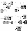
Significant inter- and intrafamilial clinical variability is common in TCS [Posnick & Ruiz 2000, Teber et al 2004]. While some individuals may be so mildly affected as to go undiagnosed (Figure 1), others can have severe facial involvement and life-threatening airway compromise [Trainor & Andrews 2013].

Figure 1. . Intrafamilial variation in families with a confirmed TCOF1 pathogenic variant.

Figure 1.

Intrafamilial variation in families with a confirmed TCOF1 pathogenic variant. a. The proband (arrow) has the characteristic facial phenotype with downward-slanting palpebral fissures, hypoplastic zygomatic complex, hypoplasia of mandible, slightly dysplastic (more...)

Classic features of TCS are bilaterally symmetric and evident at birth (Table 2). Table 3 presents a scoring system to rate the severity of clinical findings.

Table 2.

Classic Features of Treacher Collins Syndrome

Classic Feature% (n) of Affected Individuals w/Feature
Vincent et al [2016] Teber et al [2004] Splendore et al [2000]
Very
frequent
Downward-slanting palpebral fissures99% (76/77)100% (35/35)89%
Malar hypoplasia / hypoplasia of zygomatic complex97% (76/78)97% (34/35)81%
Conductive hearing loss92% (69/75)83% (25/30)
Mandibular hypoplasia / micrognathia88% (69/78)91% (32/35)78%
Frequent Atresia of external ear canal71% (46/65)68% (23/34)
Microtia70% (55/79)71% (25/35)77%
Coloboma (notching) of the lower lid63% (46/73)54% (19/35)69%
Delayed speech development57% (16/28)
Asymmetry52% (34/65)
Preauricular hair displacement49% (25/51)24% (8/33)
Rare Nasogastric tube or gastrostomy in neonates28% (17/60)
Cleft palate21% (15/70)33% (11/33)28%
Intubation or tracheostomy in neonates18% (12/65)12% (4/34)
Choanal stenosis/atresia13% (8/64)25% (8/32)
Cardiac malformation11% (7/65)
Very rare Rachis malformation7% (3/42)
Renal malformation4% (2/50)
Microcephaly3% (2/66)
Intellectual disability / delayed motor development1.7% (1/58)10% (3/30)
Limb anomaly1.5% (1/67)

Table 3.

Scoring Systems to Rate Severity of Clinical Findings

FeaturePoints Assigned 1
Teber et al [2004] Vincent et al [2016]
Downward-slanting palpebral fissures21
Malar hypoplasia / hypoplasia of zygomatic complex21
Conductive hearing loss12
Mandibular hypoplasia / micrognathia21
Atresia of external ear canal11
Microtia21
Coloboma (notching) of the lower lid21
Delayed speech developmentNot assignedNot assigned
AsymmetryNot assigned1
Preauricular hair displacement11
Nasogastric tube or gastrostomy in neonatesNot assigned2
Cleft palate11
Intubation or tracheostomy in neonates12
Choanal stenosis/atresia12
Cardiac malformationNot assignedNot assigned
Rachis malformationNot assignedNot assigned
Renal malformationNot assignedAtypical
MicrocephalyNot assignedAtypical
Intellectual disability / delayed motor development1Atypical
Limb anomalyNot assignedAtypical
Total Points
1.

Maximum score is 17 for both systems. Scores ≤8 = mildly affected; scores ≥9 = severely affected.

In newborns with TCS, airway management may be required to address narrowing of the airway or extreme shortening of the mandible with severe micrognathia. Choanal atresia or stenosis, or severe micrognathia with glossoptosis can also obstruct the airway in an infant. Neonatal death is usually associated with obstructive sleep apnea as a result of these malformations.

External ear anomalies including absent, small, or rotated ears are typical. Conductive hearing loss is usually attributed to middle ear anomalies including hypoplasia or absence of the ossicles or middle ear cavities.

In those with atresia or stenosis of the external auditory canals, the presence and severity of external auditory canal defects correlate highly with the presence and severity of middle ear defects [Posnick 1997]. The inner ear structures are typically normal.

Ophthalmologic defects, including coloboma of the lower eyelid, can result in corneal exposure. Other possible causes of vision impairment include refractive errors, anisometropia, and strabismus [Hertle et al 1993].

Dental anomalies, found in 60% of individuals with TCS reported, include tooth agenesis (33.3%), enamel opacities (20%), and ectopic eruption of the maxillary first molars (13.3%) [da Silva Dalben et al 2006]. One to eight anomalies per individual have been identified.

Although craniosynostosis is not a feature of TCS, the cranium may have an abnormal shape (brachycephaly with bitemporal narrowing) [Posnick 1997].

Cardiac malformations are reported in some individuals with typical TCS caused by pathogenic variants in either TCOF1 or POLR1D.

Altered function of the upper digestive tract has been reported [Giabicani et al 2017].

Although mild developmental delay has been reported [Teber et al 2004, Vincent et al 2014], intelligence is usually normal.

Fertility is normal.

Less frequently observed features in individuals with TCS:

  • Nasal deformity
  • High-arched palate
  • Angle class II anterior open-bite malocclusion
  • Macrostomia

Abnormalities occasionally observed in individuals with TCS:

Genotype-Phenotype Correlations

In most cases, the phenotype cannot be predicted by the genotype [Splendore et al 2000, Teber et al 2004, Vincent et al 2016, Sanchez et al 2020].

Penetrance

While the penetrance of pathogenic variants associated with TCS is high, reduced penetrance in TCOF1 has also been reported [Dixon et al 2004, Vincent et al 2016] and POLR1D [Dauwerse et al 2011, Vincent et al 2016].

Nomenclature

Autosomal dominant TCS has variably been termed Fransceschetti-Zwahlen-Klein syndrome and zygoauromandibular dysplasia.

Prevalence

The prevalence of TCS is estimated at 1:50,000 [Fazen et al 1967, Trainor et al 2009].

Differential Diagnosis

Table 4.

Disorders with Mandibulofacial Dysostosis to Consider in the Differential Diagnosis of Treacher Collins Syndrome

DisorderGeneMOIDistinguishing Clinical Features
Mandibulofacial dysostosis with microcephaly EFTUD2 AD
  • Microcephaly
  • ID
  • Asymmetry of facial features
  • Esophageal atresia
  • Thumb abnormalities
Nager syndrome
(OMIM 154400)
SF3B4 ADLimb deformities, preaxial abnormalities (e.g., small or absent thumbs, triphalangeal thumbs, radial hypoplasia or aplasia, radioulnar synostosis)
Miller syndrome
(OMIM 263750)
DHODH ARLimb deformities, postaxial abnormalities (e.g., small or absent 5th digit incl 5th metacarpal, ulnar hypoplasia, absent 5th toe)
Toriello-Carey syndrome
(OMIM 217980)
AR
  • Failure to grow
  • Microcephaly
  • Agenesis of corpus callosum
  • ID
  • Urogenital anomalies in affected males
  • Facial dysmorphisms (hypertelorism, flattened nasal bridge, anteverted nares)
  • Short neck
Branchial arch syndrome
(OMIM 301950)
XL
  • Microcephaly
  • ID
  • High-arched palate
  • Webbed neck
Bauru syndrome
(OMIM 604830)
AD
  • Upslanting palpebral fissures
  • Hypoplastic tragus & ear lobes
Hedera-Toriello-Petty syndrome
(OMIM 608257)
ADPtosis
Hemifacial macrosomia, Goldenhar syndrome, Oculoauriculovertebral spectrum
(OMIM 164210)
UnknownAD
  • Asymmetric
  • Preauricular tags & pits
  • Ocular epibulbar dermoid cyst
  • Cleft lip & palate
  • Vertebral anomalies, Kippel-Feil syndrome
Pierre Robin sequence
(OMIM 261800)
Cases of disruption of long-range cis-regulatory element → misregulation of SOX9ARMicrognathia, glossoptosis, & airway obstruction w/cleft palate deformity may self-correct w/growth & w/o intervention. 1
Nonsyndromic mandibular hypoplasia
  • Severe mandibular deficiencies (e.g., temporomandibular joint, ankylosis, aglossia/microglossia, rare craniofacial cleft)
  • Progressive micrognathia or retrognathia 1, 2

AD = autosomal dominant; AR = autosomal recessive; ID = intellectual disability; MOI = mode of inheritance; XL = X-linked

1.
2.

In one study, 52 of 266 individuals with congenital mandibular hypoplasia had TCS [Singh & Bartlett 2005]. Molecular diagnosis was not confirmed on these individuals.

Management

Evaluations Following Initial Diagnosis

To establish the extent of disease in an infant diagnosed with Treacher Collins syndrome, the evaluations summarized in this section (if not performed as part of the evaluation that led to the diagnosis) are recommended:

  • The airway for evidence of choanal atresia/stenosis and/or micrognathia and glossoptosis predisposing to obstruction of the oropharynx
  • The palate for clefts
  • Swallowing function
  • Hearing through formal audiologic examination (see Hereditary Hearing Loss and Deafness Overview). If conductive hearing loss is documented during the first six months of life, a craniofacial CT scan (axial and coronal slices) can be performed to document the anatomy of the head and neck and the external auditory canal, middle ear, and inner ear.
  • Ophthalmologic evaluation with attention to extraocular movement, corneal exposure, and visual acuity
  • Assessment for dental anomalies when teeth have erupted
  • Cardiology and echocardiogram evaluation for structural heart defects
  • Consultation with a clinical geneticist and/or genetic counselor

Treatment of Manifestations

Treatment should be tailored to the specific needs of each individual, preferably by a multidisciplinary craniofacial management team that typically comprises a clinical geneticist, plastic surgeon, head and neck surgeon, otolaryngologist, oral surgeon, orthodontist, audiologist, speech pathologist, and psychologist.

Major management issues can be stratified by three age groups and graded for severity [Thompson et al 2009, Trainor & Andrews 2013]:

Birth to age two years. Major management issues are airway and feeding difficulties.

  • If a diagnosis of TCS is suspected prenatally, a detailed ("level II") fetal ultrasound examination and consultation(s) with a high-risk obstetrician and/or neonatologist should be considered. The delivering team should be aware of the potential for life-threatening neonatal airway compromise [Trainor & Andrews 2013]. Management of the airway in neonates typically includes special positioning of the infant called an EXIT (ex-utero intrapartum treatment), in which the neonate's head and neck are partially delivered by cesarean section in order to perform an oral intubation or tracheostomy during birth. With proper management, life expectancy approximates that of the general population.
  • Procedures for surgical intervention for the airway, if needed, are standard, primarily for improving respiratory function or restoring patency of the nostrils and distraction of the mandible. Intubation techniques other than direct laryngoscopy may be required during surgeries [Hosking et al 2012].
  • Repair of cleft palate (if present) at age one to two years is recommended [Kobus & Wojcicki 2006].
  • Assess nutrition and feeding to determine if gastrostomy or tracheostomy is needed to assure adequate caloric intake while protecting the airway [Trainor & Andrews 2013].
  • Bone conduction amplification, speech therapy, and educational intervention are indicated for treatment of hearing loss. The bone-anchored hearing aid (BAHA) is an alternative for individuals with ear anomalies [Trainor & Andrews 2013].
  • Ophthalmologic evaluation is indicated to assess eye movement, vision, lower eyelid coloboma, and diminished tearing. When present, corneal exposure keratitis should be medically treated [Hertle et al 1993].
  • Structural heart defects (e.g., patent ductus arteriosus, atrial septal defects, hypertrophic myopathy) are managed as per standard practice.

Age three to 12 years. Major management issues are speech therapy and integration into education system.

Craniofacial reconstruction is often necessary [Posnick 1997, Trainor & Andrews 2013]. Generally, bone reconstruction precedes soft tissue corrections. Reconstruction can prevent the progression of facial asymmetry:

  • Zygomatic and orbital reconstruction can be undertaken once cranio-orbitozygomatic bony development is complete (age ~5-7 years).
  • External ear reconstruction should be performed after age six years and should precede reconstruction of the external auditory canal or middle ear.
  • External auditory canal and middle ear reconstruction should be performed for individuals with bilateral microtia and/or narrow ear canals.
  • Coloboma of the lower eyelid can be treated with botulinum toxin and subsequent surgery [Warner et al 2008].
  • Eyelid reconstruction to correct downward-slanting fissure can use redundant upper eyelid skin [Trainor & Andrews 2013].
  • Misaligned teeth often require orthodonture.

Age 13 to 18 years. Major management issues:

  • Orthognathic therapies are typically indicated before age 16 years.
  • Nasal reconstruction, if needed, should follow orthognathic surgeries.
  • Maxillomandibular reconstruction is recommended as follows:
    • Type I (mild) and type IIA (moderate) malformation at age 13 to 16 years
    • Type IIB (moderate to severe) malformation at skeletal maturity (~16 years)
    • Type III (severe) malformation at age six to ten years

Surveillance

The following are appropriate:

  • Periodic assessment of growth and development (preferably by a pediatrician) with inquiry into symptoms of obstructive sleep apnea.
  • Routine follow up by an audiologist, ophthalmologist, and dentist

Evaluation of Relatives at Risk

See Genetic Counseling for issues related to testing of at-risk relatives for genetic counseling purposes.

Therapies Under Investigation

Search ClinicalTrials.gov in the US and EU Clinical Trials Register in Europe for information on clinical studies for a wide range of diseases and conditions.

Genetic Counseling

Genetic counseling is the process of providing individuals and families with information on the nature, mode(s) of inheritance, and implications of genetic disorders to help them make informed medical and personal decisions. The following section deals with genetic risk assessment and the use of family history and genetic testing to clarify genetic status for family members; it is not meant to address all personal, cultural, or ethical issues that may arise or to substitute for consultation with a genetics professional. —ED.

Mode of Inheritance

Mode of inheritance of Treacher Collins syndrome (TCS) can be autosomal dominant or autosomal recessive (Table 1).

Autosomal Dominant Inheritance – Risk to Family Members

Parents of a proband

Sibs of a proband. The risk to the sibs of the proband depends on the clinical/genetic status of the proband's parents:

  • If a parent of the proband is affected and/or is known to have the pathogenic variant identified in the proband, the risk to the sibs is 50%. The specific malformations or their severity cannot be predicted in sibs who inherit a pathogenic variant because significant intrafamilial clinical variability and reduced penetrance have been reported.
  • If the proband has a known TCOF1, POLR1B, or POLR1D pathogenic variant that cannot be detected in the leukocyte DNA of either parent, the recurrence risk to sibs is slightly greater than that of the general population because of the possibility of parental germline mosaicism [Shoo et al 2004, Vincent et al 2016, Sanchez et al 2020].
  • If the parents have not been tested for the pathogenic variant identified in the proband but are clinically unaffected, the risk to the sibs of a proband appears to be low. However, sibs of a proband with clinically unaffected parents are still presumed to be at increased risk for TSC because of the possibility of reduced penetrance in a parent or parental germline mosaicism.

Offspring of a proband

Other family members. The risk to other family members depends on the clinical/genetic status of the proband's parents: if a parent is affected or has a TCS-related pathogenic variant, the parent's family members may at risk.

Autosomal Recessive Inheritance – Risk to Family Members

Parents of a proband

Sibs of a proband

  • If both parents are known to be heterozygous for a TCS-causing pathogenic variant, each sib of an affected individual has at conception a 25% chance of being affected, a 50% chance of being an asymptomatic carrier, and a 25% chance of being unaffected and not a carrier.
  • Heterozygotes (carriers) are asymptomatic.

Offspring of a proband. The offspring of an individual with autosomal recessive TCS are obligate heterozygotes (carriers) for a POLR1C or POLR1D pathogenic variant.

Other family members. Each sib of the proband's parents is at a 50% risk of being a carrier of a POLR1C or POLR1D pathogenic variant.

Carrier detection. Carrier testing for at-risk relatives requires prior identification of the POLR1C or POLR1D pathogenic variants in the family.

Related Genetic Counseling Issues

Considerations in families with an apparent de novo pathogenic variant. When neither parent of a proband with an autosomal dominant condition has the pathogenic variant identified in the proband or clinical evidence of the disorder, the pathogenic variant is likely de novo. However, non-medical explanations including alternate paternity or maternity (e.g., with assisted reproduction) and undisclosed adoption could also be explored.

Family planning

  • The optimal time for determination of genetic risk and discussion of the availability of prenatal/preimplantation genetic testing is before pregnancy.
  • It is appropriate to offer genetic counseling (including discussion of potential risks to offspring and reproductive options) to young adults who are affected or at risk.

DNA banking. Because it is likely that testing methodology and our understanding of genes, pathogenic mechanisms, and diseases will improve in the future, consideration should be given to banking DNA from probands in whom a molecular diagnosis has not been confirmed (i.e., the causative pathogenic mechanism is unknown). For more information, see Huang et al [2022].

Prenatal Testing and Preimplantation Genetic Testing

Molecular genetic testing. Once the TCS-related pathogenic variant(s) have been identified in an affected family member, prenatal and preimplantation genetic testing are possible.

Note: (1) The presence of a TCS-causing pathogenic variant detected by prenatal testing does not predict the specific malformation(s) or their severity. (2) The possibility of reduced penetrance (particularly of the common TCOF1 c.4369_4373delAAGAA pathogenic variant) in the fetus needs to be considered (see Penetrance).

Ultrasound examination. In pregnancies known to be at risk for TCS, prenatal diagnosis using ultrasound examination to detect anomalies such as polyhydramnios, microcephaly, abnormal fetal facial features (micrognathia), and abnormal fetal swallowing is possible [Rotten et al 2002, Konstantinidou et al 2013]. Diagnostic features in a mildly affected fetus are likely to be missed. Three-dimensional imaging can assist with differential diagnosis prior to birth [Pereira et al 2013].

Differences in perspective may exist among medical professionals and within families regarding the use of prenatal testing. While most centers would consider use of prenatal testing to be a personal decision, discussion of these issues may be helpful.

Resources

GeneReviews staff has selected the following disease-specific and/or umbrella support organizations and/or registries for the benefit of individuals with this disorder and their families. GeneReviews is not responsible for the information provided by other organizations. For information on selection criteria, click here.

Molecular Genetics

Information in the Molecular Genetics and OMIM tables may differ from that elsewhere in the GeneReview: tables may contain more recent information. —ED.

Table A.

Treacher Collins Syndrome: Genes and Databases

Data are compiled from the following standard references: gene from HGNC; chromosome locus from OMIM; protein from UniProt. For a description of databases (Locus Specific, HGMD, ClinVar) to which links are provided, click here.

Table B.

OMIM Entries for Treacher Collins Syndrome (View All in OMIM)

154500TREACHER COLLINS SYNDROME 1; TCS1
248390TREACHER COLLINS SYNDROME 3; TCS3
602000POLYMERASE I, RNA, SUBUNIT B; POLR1B
606847TREACLE RIBOSOME BIOGENESIS FACTOR 1; TCOF1
610060POLYMERASE I, RNA, SUBUNIT C; POLR1C
613715POLYMERASE I, RNA, SUBUNIT D; POLR1D
613717TREACHER COLLINS SYNDROME 2; TCS2
618939TREACHER COLLINS SYNDROME 4; TCS4

Molecular Pathogenesis

Cartilage and bone making up the craniofacial complex is primarily derived from neural crest cells [Trainor & Andrews 2013]. Thus, TCS features can be explained by disturbances in neural crest cell development during embryogenesis. These disturbances can be attributed to pathogenic variants in the genetic pathway activating cell development.

TCOF1, POLR1B, POLR1C, and POLR1D are all expressed in neural crest cells, and their gene products (treacle; subunits C and D for RNA polymerase I and RNA polymerase III) colocalize to the nucleolus and are involved in ribogenesis. It is hypothesized that the variants in the three key proteins are disrupting cell division by triggering p53-directed apoptosis of neuroepithelial cells [Gonzales et al 2005]. Variants affecting RNA polymerase I and/or III result in a deficiency of ribosomal RNA and/or transfer RNA [Dauwerse et al 2011], potentially leading to an insufficient number of mature ribosomes in the neuroepithelium and neural crest cells during embryogenesis [Dixon et al 2000, Dauwerse et al 2011].

POLR1B

Gene structure. POLR1B comprises 15 coding exons, with 17 splice variants. The most common transcript contains an open reading frame of 7,527 nucleotides. For a detailed summary of gene and protein information, see Table A, Gene.

Pathogenic variants. To date, three heterozygous POLR1B pathogenic variants have been identified in five affected individuals lacking pathogenic variants in TCOF1, POLR1C, and POLR1D [Sanchez et al 2020]. These variants were all missense, two of them at the same nucleotide (c.3007C>T and c.3007C>A) in the last exon (exon 15), and one variant in exon 12 (c.2046T>A). See Table 5.

Table 5.

POLR1B Pathogenic Variants Discussed in This GeneReview

DNA Nucleotide ChangePredicted Protein ChangeReference Sequences
c.2046T>Ap.Ser682Arg NM_019014​.6
NP_061887​.2
c.3007C>Tp.Arg1003Cys
c.3007C>Ap.Arg1003Ser

Variants listed in the table have been provided by the authors. GeneReviews staff have not independently verified the classification of variants.

GeneReviews follows the standard naming conventions of the Human Genome Variation Society (varnomen​.hgvs.org). See Quick Reference for an explanation of nomenclature.

Normal gene product. POLR1B encodes the 128-kd RNA polymerase I subunit B, one of the two largest subunits (with POLR1A) that comprises the polymerase I complex. The most common isoform of the 17 transcripts codes 1,135 amino-acids.

Abnormal gene product. Pathogenic variants in POLR1B are in highly conserved amino acids in the catalytic domain of the POLR1B protein. POLR1B is associated with the POLR1A subunit of RNA polymerase. Variants may affect the hydrogen bonding that restricts interaction of the two subunits, which is suspected to cause p53-dependent apoptosis in the neuroepithelium, altering neural crest cell migration and affecting cranioskeletal formations [Sanchez et al 2020].

POLR1C

Gene structure. POLR1C comprises nine coding exons, with two isoforms. The longest transcript contains an open reading frame of 1,355 nucleotides with an 88-bp 5' UTR and a 226-bp 3' UTR. For a detailed summary of gene and protein information, see Table A, Gene.

Pathogenic variants. To date, six compound heterozygous POLR1C pathogenic variants have been identified in three affected individuals without a TCOF1 pathogenic variant [Dauwerse et al 2011]. These variants included three missense, one splice site, one nonsense, and one frameshift.

Normal gene product. POLR1C encodes the 16-kd (133-amino acid) subunit that comprises both RNA polymerase I and RNA polymerase III complexes. RNA polymerase I and RNA polymerase III are involved in ribosomal RNA transcription [Dauwerse et al 2011].

Abnormal gene product. Compound heterozygous pathogenic variants in POLR1C lead to functional depletion of POLR1C at a critical time during embryogenesis [Dauwerse et al 2011, Noack Watt et al 2016].

POLR1D

Gene structure. POLR1D comprises three exons, with two isoforms. The longest transcript contains an open reading frame of 1,945 nucleotides with a 118-bp 5' UTR and a 1,458-bp 3' UTR. For a detailed summary of gene and protein information, see Table A, Gene.

Pathogenic variants. More than 30 heterozygous POLR1D pathogenic variants have been identified in individuals with TCS without a TCOF1 pathogenic variant [Dauwerse et al 2011, Vincent et al 2016]. These include loss-of-function (e.g., nonsense) variants, splice site and missense variants, and small deletions and duplications. Whole-gene deletions have also been described [Dauwerse et al 2011, Vincent et al 2016].

Table 6.

POLR1D Pathogenic Variants Discussed in This GeneReview

DNA Nucleotide ChangePredicted Protein ChangeReference Sequences
c.163C>Gp.Leu55Val NM_015972​.3
NP_057056​.1

Variants listed in the table have been provided by the authors. GeneReviews staff have not independently verified the classification of variants.

GeneReviews follows the standard naming conventions of the Human Genome Variation Society (varnomen​.hgvs.org). See Quick Reference for an explanation of nomenclature.

Normal gene product. POLR1D encodes the 39-kd (346-amino acid) subunit that comprises both RNA polymerase I and RNA polymerase III complexes. RNA polymerase I and RNA polymerase III are involved in ribosomal RNA transcription [Dauwerse et al 2011].

Abnormal gene product. Pathogenic variants in POLR1D lead to haploinsufficiency of POLR1D [Dauwerse et al 2011, Noack Watt et al 2016].

TCOF1

Gene structure. TCOF1 comprises 27 coding exons, three of which are alternatively spliced in-frame (6A, 16A, and 19) [Splendore et al 2005], and an additional exon containing the 3' UTR [So et al 2004]. The longest transcript (NM_001135243.1) contains an open reading frame of 4,467 nucleotides starting in the first exon. The open reading frame is preceded by a 93-bp 5' untranslated region (UTR) and followed by a 507-bp 3' UTR [Dixon et al 1997a]. For a detailed summary of gene and protein information, see Table A, Gene.

Pathogenic variants. Hundreds of pathogenic variants in TCOF1 have been reported in individuals with TCS, with novel variants being identified in a significant proportion of families [Gladwin et al 1996, Treacher Collins Syndrome Collaborative Group 1996, Edwards et al 1997, Wise et al 1997, Splendore et al 2000, Ellis et al 2002, Splendore et al 2002, Dixon et al 2004, Horiuchi et al 2005, Trainor et al 2009, Bowman et al 2012, Vincent et al 2016]. The majority of pathogenic variants found to date are frameshift variants leading to a premature termination of the transcript. Pathogenic variants have been found throughout the gene.

Of TCOF1 sequencing variants, 57%-60% are small deletions or insertions, 9%-16% are splice site variants, 19%-23% nonsense variants, and 3%-4% missense variants [Bowman et al 2012, Vincent et al 2016]. Large deletions of one or more exons have also been identified in up to 5% of individuals with TCS [Beygo et al 2012, Bowman et al 2012, Vincent et al 2016]. In one case, a synonymous pathogenic TCOF1 variant led to missplicing of a constitutive exon [Macaya et al 2009].

While several pathogenic variants have occurred more than once, only one variant in TCOF1, c.4369_4373delAAGAA, has been identified as commonly recurrent. This variant is present in 16% of individuals with an identifiable pathogenic variant.

Table 7.

TCOF1 Pathogenic Variants Discussed in This GeneReview

DNA Nucleotide Change
(Alias 1)
Predicted Protein Change
(Alias 1)
Reference Sequences
c.1021_1022delAG
(790_791delAG)
p.Ser341GlnfsTer7
(Ser264GlnfsTer7)
NM_001135243​.1
NP_001128715​.1
c.2490delAp.Val831Ter
c.2853dupT
(2853_2854insT)
p.Ala952CysfsTer5
c.4369_4373delAAGAAp.Lys1457GlufsTer12

Variants listed in the table have been provided by the authors. GeneReviews staff have not independently verified the classification of variants.

GeneReviews follows the standard naming conventions of the Human Genome Variation Society (varnomen​.hgvs.org). See Quick Reference for an explanation of nomenclature.

1.

Variant designation that does not conform to current naming conventions

Normal gene product. The 144-kd treacle protein comprises 1,488 amino acids. Treacle is a low-complexity, three-domain nucleolar protein having unique N and C termini that is structurally related to the nucleolar phosphoprotein Nopp140 [Isaac et al 2000]. A central ten-repeat motif contains protein kinase C and casein kinase 2 phosphorylation sites [Dixon et al 1997b, Winokur & Shiang 1998]. The protein has at least two functional nuclear localization signals and a nucleolar localization signal in the C terminus. Both Nopp140 and treacle contain LIS1 motifs, leading to speculation of involvement in microtubule dynamics [Emes & Ponting 2001].

Treacle interacts with the small nucleolar ribonucleoprotein hNop56p, suggesting that it is involved in ribosomal biogenesis [Hayano et al 2003]. Treacle is involved in rDNA transcription, nucleologenesis, or trafficking of proteins or ribosomal subunits between the nucleolus and cytoplasm [Winokur & Shiang 1998, Dauwerse et al 2011]; and perhaps neural crest cell migration [Sakai & Trainor 2009, Trainor & Andrews 2013, Calo et al 2018].

Abnormal gene product. Pathogenic variants in TCOF1 lead to haploinsufficiency of the treacle protein [Isaac et al 2000]. Missense variants that allow production of an abnormal protein can disrupt either the N- or C-terminus nuclear localization signals and affect the protein's ability to transport into the nucleus during first and second branchial arch development, causing cephalic neural crest cells to undergo apoptosis during embryogenesis [Dixon et al 2000, Isaac et al 2000, Trainor & Andrews 2013]. Downregulation of TCOF1 and POLR1D leads to relocalization of DDX21, a nucleolar protein involved in ribosome biogenesis [Calo et al 2018].

Chapter Notes

Acknowledgments

We acknowledge Mac Hays for his background research.

Author History

Garry R Cutting, MD; Johns Hopkins University (2004-2011)
Sara Huston Katsanis, MS (2004-present)
Ethylin Wang Jabs, MD (2011-present)

Revision History

  • 20 August 2020 (aa) Revision: added POLR1B and related citation (Sanchez et al [2020])
  • 27 September 2018 (bp) Comprehensive update posted live
  • 30 August 2012 (cd) Revision: TCOF1 and POLR1D deletions reported in individuals with Treacher Collins syndrome; deletion/duplication analysis available clinically for POLR1C and POLR1D
  • 27 October 2011 (me) Comprehensive update posted live
  • 27 October 2006 (me) Comprehensive update posted live
  • 20 July 2004 (me) Review posted live
  • 1 March 2004 (shk,gc) Original submission

References

Literature Cited

  • Beygo J, Buiting K, Seland S, Lüdecke HJ, Hehr U, Lich C, Prager B, Lohmann DR, Wieczorek D. First report of a single exon deletion in TCOF1 causing Treacher Collins syndrome. Mol Syndromol. 2012;2:53–9. [PMC free article: PMC3326662] [PubMed: 22712005]
  • Bowman M, Oldridge M, Archer C, O'Rourke A, McParland J, Brekelmans R, Seller A, Lester T. Gross deletions in TCOF1 are a cause of Treacher-Collins-Franceschetti syndrome. Eur J Hum Genet. 2012;20:769–77. [PMC free article: PMC3376267] [PubMed: 22317976]
  • Calo E, Gu B, Bowen ME, Aryan F, Zalc A, Liang J, Flynn RA, Swigut T, Chang HY, Attardi LD, Wysocka J. Tissue-selective effects of nucleolar stress and rDNA damage in developmental disorders. Nature. 2018;554:112–7. [PMC free article: PMC5927778] [PubMed: 29364875]
  • da Silva Dalben G, Costa B, Gomide MR. Prevalence of dental anomalies, ectopic eruption and associated oral malformations in subjects with Treacher Collins syndrome. Oral Surg Oral Med Oral Pathol Oral Radiol Endod. 2006;101:588–92. [PubMed: 16632269]
  • Dauwerse JG, Dixon J, Seland S, Ruivenkamp CA, van Haeringen A, Hoefsloot LH, Peters DJ, Boers AC, Daumer-Haas C, Maiwald R, Zweier C, Kerr B, Cobo AM, Toral JF, Hoogeboom AJ, Lohmann DR, Hehr U, Dixon MJ, Breuning MH, Wieczorek D. Mutations in genes encoding subunits of RNA polymerases I and III cause Treacher Collins syndrome. Nat Genet. 2011;43:20–2. [PubMed: 21131976]
  • Dixon J, Brakebusch C, Fassler R, Dixon MJ. Increased levels of apoptosis in the prefusion neural folds underlie the craniofacial disorder, Treacher Collins syndrome. Hum Mol Genet. 2000;9:1473–80. [PubMed: 10888597]
  • Dixon J, Edwards SJ, Anderson I, Brass A, Scambler PJ, Dixon MJ. Identification of the complete coding sequence and genomic organization of the Treacher Collins syndrome gene. Genome Res. 1997a;7:223–34. [PubMed: 9074926]
  • Dixon J, Ellis I, Bottani A, Temple K, Dixon MJ. Identification of mutations in TCOF1: use of molecular analysis in the pre- and postnatal diagnosis of Treacher Collins syndrome. Am J Med Genet A. 2004;127A:244–8. [PubMed: 15150774]
  • Dixon J, Hovanes K, Shiang R, Dixon MJ. Sequence analysis, identification of evolutionary conserved motifs and expression analysis of murine tcof1 provide further evidence for a potential function for the gene and its human homologue, TCOF1. Hum Mol Genet. 1997b;6:727–37. [PubMed: 9158147]
  • Edwards SJ, Gladwin AJ, Dixon MJ. The mutational spectrum in Treacher Collins syndrome reveals a predominance of mutations that create a premature-termination codon. Am J Hum Genet. 1997;60:515–24. [PMC free article: PMC1712503] [PubMed: 9042910]
  • Ellis PE, Dawson M, Dixon MJ. Mutation testing in Treacher Collins Syndrome. J Orthod. 2002;29:293–7. [PubMed: 12444270]
  • Emes RD, Ponting CP. A new sequence motif linking lissencephaly, Treacher Collins and oral-facial-digital type 1 syndromes, microtubule dynamics and cell migration. Hum Mol Genet. 2001;10:2813–20. [PubMed: 11734546]
  • Fazen LE, Elmore J, Nadler HL. Mandibulo-facial dysostosis. (Treacher-Collins syndrome). Am J Dis Child. 1967;113:405–10. [PubMed: 6024864]
  • Giabicani E, Lemale J, Dainese L, Boudjemaa S, Coulomb A, Tounian P, Dubern B. Chronic intestinal pseudo-obstruction in a child with Treacher Collins syndrome. Archives de Pédiatrie. 2017;24:1000–4. [PubMed: 28927774]
  • Gladwin AJ, Dixon J, Loftus SK, Edwards S, Wasmuth JJ, Hennekam RC, Dixon MJ. Treacher Collins syndrome may result from insertions, deletions or splicing mutations, which introduce a termination codon into the gene. Hum Mol Genet. 1996;5:1533–8. [PubMed: 8894686]
  • Gonzales B, Henning D, So RB, Dixon J, Dixon MJ, Valdez BC. The Treacher Collins syndrome (TCOF1) gene product is involved in pre-rRNA methylation. Hum Mol Genet. 2005;14:2035–43. [PubMed: 15930015]
  • Hayano T, Yanagida M, Yamauchi Y, Shinkawa T, Isobe T, Takahashi N. Proteomic analysis of human Nop56p-associated pre-ribosomal ribonucleoprotein complexes. Possible link between Nop56p and the nucleolar protein treacle responsible for Treacher Collins syndrome. J Biol Chem. 2003;278:34309–19. [PubMed: 12777385]
  • Hertle RW, Ziylan S, Katowitz JA. Ophthalmic features and visual prognosis in the Treacher-Collins syndrome. Br J Ophthalmol. 1993;77:642–5. [PMC free article: PMC504607] [PubMed: 8218033]
  • Horiuchi K, Ariga T, Fujioka H, Kawashima K, Yamamoto Y, Igawa H, Sugihara T, Sakiyama Y. Mutational analysis of the TCOF1 gene in 11 Japanese patients with Treacher Collins Syndrome and mechanism of mutagenesis. Am J Med Genet A. 2005;134:363–7. [PubMed: 15759264]
  • Hosking J,Zoanetti D, Carlyle A, Anderson P, Costi D (2012) Anesthesia for Treacher Collins syndrome: a review of airway management in 240 pediatric cases. [PubMed: 22394325]
  • Huang SJ, Amendola LM, Sternen DL. Variation among DNA banking consent forms: points for clinicians to bank on. J Community Genet. 2022;13:389–97. [PMC free article: PMC9314484] [PubMed: 35834113]
  • Isaac C, Marsh KL, Paznekas WA, Dixon J, Dixon MJ, Jabs EW, Meier UT. Characterization of the nucleolar gene product, treacle, in Treacher Collins syndrome. Mol Biol Cell. 2000;11:3061–71. [PMC free article: PMC14975] [PubMed: 10982400]
  • Jónsson H, Sulem P, Kehr B, Kristmundsdottir S, Zink F, Hjartarson E, Hardarson MT, Hjorleifsson KE, Eggertsson HP, Gudjonsson SA, Ward LD, Arnadottir GA, Helgason EA, Helgason H, Gylfason A, Jonasdottir A, Jonasdottir A, Rafnar T, Frigge M, Stacey SN, Th Magnusson O, Thorsteinsdottir U, Masson G, Kong A, Halldorsson BV, Helgason A, Gudbjartsson DF, Stefansson K. Parental influence on human germline de novo mutations in 1,548 trios from Iceland. Nature. 2017;549:519–22. [PubMed: 28959963]
  • Kobus K, Wojcicki P. Surgical treatment of Treacher Collins syndrome. Ann Plast Surg. 2006;56:549–54. [PubMed: 16641634]
  • Konstantinidou AE, Tasoulas J, Kallipolitis G, Gasparatos S, Velissariou V, Paraskevakou H. Mandibulofacial dysostosis (Treacher-Collins syndrome) in the fetus: novel association with Pectus carinatum in a molecularly confirmed case and review of the fetal phenotype. Birth Defects Res A Clin Mol Teratol. 2013;97:774–80. [PubMed: 24288143]
  • Macaya D, Katsanis SH, Hefferon TW, Audlin S, Mendelsohn NJ, Roggenbuck J, Cutting GR. A synonymous mutation in TCOF1 causes Treacher Collins syndrome due to mis-splicing of a constitutive exon. Am J Med Genet. 2009;149A:1624–7. [PubMed: 19572402]
  • Marres HA. Hearing loss in the Treacher-Collins syndrome. Adv Otorhinolaryngol. 2002;61:209–15. [PubMed: 12408086]
  • Marszałek B, Wojcicki P, Kobus K, Trzeciak WH. Clinical features, treatment and genetic background of Treacher Collins syndrome. J Appl Genet. 2002;43:223–33. [PubMed: 12080178]
  • Noack Watt KE, Achilleos A, Neben CL, Merrill AE, Trainor PA. The roles of RNA polymerase I and III subunits Polr1c and Polr1d in craniofacial development and in zebrafish models of Treacher Collins syndrome. PLoS Genet. 2016;12:e1006187. [PMC free article: PMC4957770] [PubMed: 27448281]
  • Pereira DC, Bussamra LC, Araujo Júnior E, Drummond CL, Nardozza LM, Moron AF, Aldrighi JM. Prenatal diagnosis of Treacher-Collins syndrome using three-dimensional ultrasonography and differential diagnosis with other acrofacial dysostosis syndromes. Case Rep Obstet Gynecol. 2013;2013;2013:203976. [PMC free article: PMC3638565] [PubMed: 23653874]
  • Posnick JC. Treacher Collins syndrome: perspectives in evaluation and treatment. J Oral Maxillofac Surg. 1997;55:1120–33. [PubMed: 9331237]
  • Posnick JC, Ruiz RL. Treacher Collins syndrome: current evaluation, treatment, and future directions. Cleft Palate Craniofac J. 2000;37:434. [PubMed: 11034023]
  • Richards S, Aziz N, Bale S, Bick D, Das S, Gastier-Foster J, Grody WW, Hegde M, Lyon E, Spector E, Voelkerding K, Rehm HL, et al. Standards and guidelines for the interpretation of sequence variants: a joint consensus recommendation of the American College of Medical Genetics and Genomics and the Association for Molecular Pathology. Genet Med. 2015;17:405–24. [PMC free article: PMC4544753] [PubMed: 25741868]
  • Rotten D, Levaillant JM, Martinez H, Ducou le Pointe H, Vicaut E. The fetal mandible: a 2D and 3D sonographic approach to the diagnosis of retrognathia and micrognathia. Ultrasound Obstet Gynecol. 2002;19:122–30. [PubMed: 11876802]
  • Sakai D, Trainor PA. Treacher Collins syndrome: unmasking the role of Tcof1/treacle. Int J Biochem Cell Biol. 2009;41:1229–32. [PMC free article: PMC3093759] [PubMed: 19027870]
  • Sanchez E, Laplace-Builhé B, Mau-Them FT, Richard E, Goldenberg A, Toler TL, Guignard T, Gatinois V, Vincent M, Blanchet C, Boland A, Bihoreau MT, Deleuze JF, Olaso R, Nephi W, Lüdecke HJ, Verheij JBGM, Moreau-Lenoir F, Denoyelle F, Rivière JB, Laplanche JL, Willing M, Captier G, Apparailly F, Wieczorek D, Collet C, Djouad F, Geneviève D. POLR1B and neural crest cell anomalies in Treacher Collins syndrome type 4. Genet Med. 2020;22:547–56. [PMC free article: PMC7056642] [PubMed: 31649276]
  • Schaefer E, Collet C, Genevieve D, Vincent M, Lohmann DR, Sanchez E, Bolender C, Eliot MM, Nürnberg G, Passos-Bueno MR, Wieczorek D, van Maldergem L, Doray B. Autosomal recessive POLR1D mutation with decrease of TCOF1 mRNA is responsible for Treacher Collins syndrome. Genet Med. 2014;16:720–4. [PubMed: 24603435]
  • Shoo BA, McPherson E, Jabs EW. Mosaicism of a TCOF1 mutation in an individual clinically unaffected with Treacher Collins syndrome. Am J Med Genet. 2004;126A:84–8. [PubMed: 15039977]
  • Singh DJ, Bartlett SP. Congenital mandibular hypoplasia: analysis and classification. J Craniofac Surg. 2005;16:291–300. [PubMed: 15750428]
  • So RB, Gonzales B, Henning D, Dixon J, Dixon MJ, Valdez BC. Another face of the Treacher Collins syndrome (TCOF1) gene: identification of additional exons. Gene. 2004;328:49–57. [PubMed: 15019983]
  • Splendore A, Fanganiello RD, Masotti C, Morganti LS, Passos-Bueno MR. TCOF1 mutation database: novel mutation in the alternatively spliced exon 6A and update in mutation nomenclature. Hum Mutat. 2005;25:429–34. [PubMed: 15832313]
  • Splendore A, Jabs EW, Passos-Bueno MR. Screening of TCOF1 in patients from different populations: confirmation of mutational hot spots and identification of a novel missense mutation that suggests an important functional domain in the protein treacle. J Med Genet. 2002;39:493–5. [PMC free article: PMC1735178] [PubMed: 12114482]
  • Splendore A, Silva EO, Alonso LG, Richieri-Costa A, Alonso N, Rosa A, Carakushanky G, Cavalcanti DP, Brunoni D, Passos-Bueno MR. High mutation detection rate in TCOF1 among Treacher Collins syndrome patients reveals clustering of mutations and 16 novel pathogenic changes. Hum Mutat. 2000;16:315–22. [PubMed: 11013442]
  • Teber OA, Gillessen-Kaesbach G, Fischer S, Bohringer S, Albrecht B, Albert A, Arslan-Kirchner M, Haan E, Hagedorn-Greiwe M, Hammans C, Henn W, Hinkel GK, Konig R, Kunstmann E, Kunze J, Neumann LM, Prott EC, Rauch A, Rott HD, Seidel H, Spranger S, Sprengel M, Zoll B, Lohmann DR, Wieczorek D. Genotyping in 46 patients with tentative diagnosis of Treacher Collins syndrome revealed unexpected phenotypic variation. Eur J Hum Genet. 2004;12:879–90. [PubMed: 15340364]
  • Thompson JT, Anderson PJ, David DJ. Treacher Collins syndrome: protocol management from birth to maturity. J Craniofac Surg. 2009;20:2028–35. [PubMed: 19881372]
  • Trainor PA, Andrews BT. Facial dysostoses: etiology, pathogenesis and management. Am J Med Genet C Semin Med Genet. 2013;163C:283–94. [PMC free article: PMC3870197] [PubMed: 24123981]
  • Trainor PA, Dixon J, Dixon MJ. Treacher Collins syndrome: etiology, pathogenesis and prevention. Eur J Hum Genet. 2009;17:275–83. [PMC free article: PMC2986179] [PubMed: 19107148]
  • Treacher Collins Syndrome Collaborative Group. Positional cloning of a gene involved in the pathogenesis of Treacher Collins syndrome. Nat Genet. 1996;12:130–6. [PubMed: 8563749]
  • Vincent M, Collet C, Verloes A, Lambert L, Herlin C, Blanchet C, Sanchez E, Drunat S, Vigneron J, Laplanche JL, Puechberty J, Sarda P, Geneviève D. Large deletions encompassing the TCOF1 and CAMK2A genes are responsible for Treacher Collins syndrome with intellectual disability. Eur J Hum Genet. 2014;22:52–6. [PMC free article: PMC3865420] [PubMed: 23695276]
  • Vincent M, Geneviève D, Ostertag A, Marlin S, Lacombe D, Martin-Coignard D, Coubes C, David A, Lyonnet S, Vilain C, Dieux-Coeslier A, Manouvrier S, Isidor B, Jacquemont ML, Julia S, Layet V, Naudion S, Odent S, Pasquier L, Pelras S, Philip N, Pierquin G, Prieur F, Aboussair N, Attie-Bitach T, Baujat G, Blanchet P, Blanchet C, Dollfus H, Doray B, Schaefer E, Edery P, Giuliano F, Goldenberg A, Goizet C, Guichet A, Herlin C, Lambert L, Leheup B, Martinovic J, Mercier S, Mignot C, Moutard ML, Perez MJ, Pinson L, Puechberty J, Willems M, Randrianaivo H, Szakszon K, Toutain A, Verloes A, Vigneron J, Sanchez E, Sarda P, Laplanche JL, Collet C. Treacher Collins syndrome: a clinical and molecular study based on a large series of patients. Genet Med. 2016;18:49–56. [PubMed: 25790162]
  • Warner RM, Fagan JM, Das-Gupta R. Treacher Collins syndrome: an undescribed characteristic of the condition and its management with botulinum toxin and surgery. J Craniofac Surg. 2008;19:1600–2. [PubMed: 19098560]
  • Winokur ST, Shiang R. The Treacher Collins syndrome (TCOF1) gene product, treacle, is targeted to the nucleolus by signals in its C-terminus. Hum Mol Genet. 1998;7:1947–52. [PubMed: 9811939]
  • Wise CA, Chiang LC, Paznekas WA, Sharma M, Musy MM, Ashley JA, Lovett M, Jabs EW. TCOF1 gene encodes a putative nucleolar phosphoprotein that exhibits mutations in Treacher Collins syndrome throughout its coding region. Proc Natl Acad Sci U S A. 1997;94:3110–5. [PMC free article: PMC20330] [PubMed: 9096354]
Copyright © 1993-2024, University of Washington, Seattle. GeneReviews is a registered trademark of the University of Washington, Seattle. All rights reserved.

GeneReviews® chapters are owned by the University of Washington. Permission is hereby granted to reproduce, distribute, and translate copies of content materials for noncommercial research purposes only, provided that (i) credit for source (http://www.genereviews.org/) and copyright (© 1993-2024 University of Washington) are included with each copy; (ii) a link to the original material is provided whenever the material is published elsewhere on the Web; and (iii) reproducers, distributors, and/or translators comply with the GeneReviews® Copyright Notice and Usage Disclaimer. No further modifications are allowed. For clarity, excerpts of GeneReviews chapters for use in lab reports and clinic notes are a permitted use.

For more information, see the GeneReviews® Copyright Notice and Usage Disclaimer.

For questions regarding permissions or whether a specified use is allowed, contact: ude.wu@tssamda.

Bookshelf ID: NBK1532PMID: 20301704

Views

Tests in GTR by Gene

Related information

  • MedGen
    Related information in MedGen
  • OMIM
    Related OMIM records
  • PMC
    PubMed Central citations
  • PubMed
    Links to PubMed
  • Gene
    Locus Links

Similar articles in PubMed

See reviews...See all...

Recent Activity

Your browsing activity is empty.

Activity recording is turned off.

Turn recording back on

See more...