Learning Outcome
- List the risk factors for aspiration pneumonia.
- Describe the presentation of aspiration pneumonia.
- Recall the nursing care in patients with aspiration pneumonia.
- Describe the complications of aspiration pneumonia.
Introduction
The infectious pulmonary process that occurs after abnormal entry of fluids into the lower respiratory tract is termed aspiration pneumonia. The aspirated fluid can be formed from oropharyngeal secretions or particulate matter or can also be gastric content. The term aspiration pneumonitis refers to inhalational acute lung injury that occurs after aspiration of sterile gastric contents. In an observational study, it is found that the risk of patients hospitalized for community-acquired pneumonia in developing aspiration pneumonia is found to be about 13.8%. The mortality rate from aspiration pneumonia is largely dependent on the volume and content of aspirate and can range up to 70%.[1][2][3][4]
Nursing Diagnosis
- Dyspnea
- Chest discomfort
- Cough
- Decreased oxygen saturation
- Tachycardia
- Tachypnea
- Fever
- Foul sputum
Causes
Failure of the natural defense mechanisms like the closure of the glottis and cough reflex increases the risk of aspiration. The common risk factors for aspiration include altered mental status, neurologic disorders, esophageal motility disorders, protracted vomiting, and gastric outlet obstruction. Although the common organisms involved in the etiology of community-acquired pneumonia are Streptococci, Haemophilus, and gram-negative bacilli, the etiology of aspiration pneumonia depends on the content of aspirate. A prospective study in 95 patients showed that gram-negative bacilli contributed to 49%, followed by anaerobes (16%). The major anaerobes isolated were Fusobacterium, Bacteroides, and Peptostreptococcus. In hospital-acquired aspiration pneumonia, common etiology includes gram-negative organisms, specifically Pseudomonas aeruginosa, which has to be considered.[5][6][7]
Risk Factors
Due to the lack of biomarkers, epidemiological studies to find the incidence of aspiration pneumonia have been difficult. Several studies showed that aspiration pneumonia contributes to 5 to 15% of all community-acquired pneumonia. A retrospective study done on 628 patients with aspiration pneumonia by Lanspa et al. showed 30-day mortality of 21%. The study also showed that CURB 65, a predictor of mortality in Community-acquired pneumonia, is not a reliable tool for aspiration pneumonia. Aspiration pneumonia remains one of the common complications following general anesthesia and occurs in one in every 2000 to 30000 cases. In a study conducted on the elderly population aged older than 65 who underwent cardiovascular surgery, the incidence of aspiration pneumonia was found to be 9.8%, with 12 out of 123 patients having aspiration pneumonia post-extubation. A case-control study conducted by Marrie T.J. et al. showed that the incidence of aspiration pneumonia was 18% in nursing home patients and 15% in community-acquired aspiration pneumonia. Since most cases of aspiration pneumonia are silent or unwitnessed, the true incidence rate is difficult to calculate.
Assessment
The common clinical features that should raise suspicion for aspiration include sudden onset dyspnea, fever, hypoxemia, radiological findings of bilateral infiltrates, and crackles on lung auscultation in a hospitalized patient. The common site involved depends on the position at the time of aspiration, commonly the lower lobes are involved in an upright position, and superior lobes can be involved in the recumbent position. The radiological findings will develop within 2 hours after aspiration, and bronchoscopy can reveal erythematous bronchi.
Evaluation
A high level of suspicion should be made in diagnosing aspiration pneumonia, especially in critically ill hospitalized patients and antibiotic treatment should be initiated immediately, and imaging studies should not delay the treatment. The commonly utilized imaging studies are chest x-ray, computed tomography of the chest, which will help in localizing the site of aspiration. However, most cases are unwitnessed aspiration, making it very hard to distinguish between aspiration pneumonitis and aspiration pneumonia. Clinical features may help to distinguish. In aspiration pneumonitis, a large volume of gastric content has to be aspirated to produce chemical pneumonitis and can quickly progress to acute lung injury with subsequent development of acute respiratory distress syndrome (ARDS). Whereas, in aspiration pneumonia, aspiration can be of smaller volumes and can be unwitnessed, which with inoculation of bacteria progress to features of pneumonia and subsequent development of acute respiratory distress syndrome (ARDS).
Medical Management
The treatment varies between aspiration pneumonia and aspiration pneumonitis. To prevent further aspiration, the patient’s position should be adjusted, followed by suction of oropharyngeal contents with the placement of the nasogastric tube. In patients who are not intubated, humidified oxygen is administered, and the head end of the bed should be raised by 45 degrees. Close monitoring of the patient's oxygen saturation is important and immediate intubation with mechanical ventilation should be provided if hypoxia is noted. Flexible bronchoscopy is usually indicated for large volume aspiration to clear the secretion and also for obtaining the sample of bronchoalveolar lavage for quantitative bacteriological studies. In general practice, antibiotics are initiated immediately even though they are not required in aspiration pneumonitis to prevent the progression of the disease. The choice of antibiotics for community-acquired aspiration pneumonia are ampicillin-sulbactam, or a combination of metronidazole and amoxicillin can be used. In patients with penicillin allergy, clindamycin is preferred. However, in hospital-acquired aspiration pneumonia, antibiotics that cover resistant Gram-negative bacteria and S. aureus, so the use of a combination of vancomycin and piperacillin-tazobactam is most widely used. Once the culture results are obtained, the antibiotic regimen should be narrowed to organism-specific.[8][9][10][11]
Nursing Management
Protect airway
Suction if thick secretions
Provide oxygen
Position patient upright
Monitor vitals
Listen to lung sounds
Ensure swallow screen done before feeding
If NG tube placed for feeding, make sure chest x-ray is done to confirm the position
Check arterial blood gas to ensure oxygenation is adequate
Administer antibiotics as ordered
When To Seek Help
- If oxygen desaturation
- Hypotension
- Respiratory distress
- Coughing
- Fever
Outcome Identification
Able to eat food without aspiration
No coughing spells
No respiratory distress
Normal vitals
Chest x-ray is clear
Monitoring
- Vitals
- Respiration, oxygenation
- Labs
- Urine output
- Dietary intake
- Lung sounds
Coordination of Care
The management of aspiration pneumonia is with an interprofessional team that consists of a nurse practitioner, primary care provider, internist, infectious disease specialist, radiologist, and pulmonologist. Besides treating pneumonia, it is important to educate the staff looking after the patient on further prevention of aspiration. This means having the head of the bed elevated, close monitoring of oxygen status, and regularly suctioning the oral cavity in patients with swallowing difficulties. The choice of antibiotics for community-acquired aspiration pneumonia are ampicillin-sulbactam, or a combination of metronidazole and amoxicillin can be used. In patients with penicillin allergy, clindamycin is preferred. However, in hospital-acquired aspiration pneumonia, antibiotics that cover resistant gram-negative bacteria and S. aureus; so use of a combination of vancomycin and piperacillin-tazobactam is most widely used. Once the culture results are obtained the antibiotic regimen should be narrowed to organism-specific. The outlook for patients with aspiration pneumonia depends on the severity of the infection, comorbidity, age, presence of ARDS, and response to antibiotics. If there is treatment delay, mortality rates are high.[5] [Level 5]
Health Teaching and Health Promotion
Educate caregiver on feeding
Educate caregiver on patient positioning
Risk Management
- Not recognizing that aspiration is occurring
- Feeding patients at high risk for aspiration
- Starting feeding without assessing the risk of aspiration
- Feeding via an NG tube prior to getting a chest x-ray
Discharge Planning
Eat and sleep with the head of the bed elevated
Take antibiotics as prescribed
Pearls and Other issues
The disease syndrome that occurs after macro aspiration of gastric and oropharyngeal contents leading to acute pulmonary infection is aspiration pneumonia. It is important to understand and distinguish between aspiration pneumonitis which is an inflammatory process that occurs post-aspiration and aspiration pneumonia for guiding treatment. Since the major complication of this disease syndrome results in ARDS, prompt treatment with supplemental oxygen and antibiotics and measures to prevent further aspiration have to be undertaken. Close monitoring of the patient for hypoxia is mandated and if the present patient should be intubated and mechanically ventilated. In a meta-analysis conducted by Kosaku K et al. it is found that aspiration pneumonia is associated with high-30-day and in-hospital, but outside the intensive care setting, mortality rates in patients with community-acquired pneumonia.
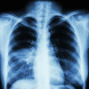
Figure
Ventilator-Associated Aspiration Pneumonia. Chest radiograph showing ventilator-associated aspiration pneumonia. Melvil, Public Domain, via Wikimedia Commons.
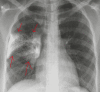
Figure
Aspiration Pneumonia. Chest radiograph demonstrating alveolar infiltrates in the superior segments of the right lower lobe in aspiration pneumonia. Contributed by O Chaigasame
References
- 1.
- Mandell LA, Niederman MS. Aspiration Pneumonia. N Engl J Med. 2019 Feb 14;380(7):651-663. [PubMed: 30763196]
- 2.
- Kudo H, Ide H, Nakabayashi M, Goto T, Wakakuri A, Iwata N, Kuroki Y. [The effectiveness of the complete lateral position method in elderly patients with severe dysphagia]. Nihon Ronen Igakkai Zasshi. 2019;56(1):59-66. [PubMed: 30760684]
- 3.
- Cicala G, Barbieri MA, Spina E, de Leon J. A comprehensive review of swallowing difficulties and dysphagia associated with antipsychotics in adults. Expert Rev Clin Pharmacol. 2019 Mar;12(3):219-234. [PubMed: 30700161]
- 4.
- Neill S, Dean N. Aspiration pneumonia and pneumonitis: a spectrum of infectious/noninfectious diseases affecting the lung. Curr Opin Infect Dis. 2019 Apr;32(2):152-157. [PubMed: 30676341]
- 5.
- Bowerman TJ, Zhang J, Waite LM. Antibacterial treatment of aspiration pneumonia in older people: a systematic review. Clin Interv Aging. 2018;13:2201-2213. [PMC free article: PMC6214417] [PubMed: 30464429]
- 6.
- Marin S, Serra-Prat M, Ortega O, Clavé P. Cost of oropharyngeal dysphagia after stroke: protocol for a systematic review. BMJ Open. 2018 Dec 14;8(12):e022775. [PMC free article: PMC6303570] [PubMed: 30552255]
- 7.
- Liu C, Cao Y, Lin J, Ng L, Needleman I, Walsh T, Li C. Oral care measures for preventing nursing home-acquired pneumonia. Cochrane Database Syst Rev. 2018 Sep 27;9(9):CD012416. [PMC free article: PMC6513285] [PubMed: 30264525]
- 8.
- Barrett Mørk FC, Gade C, Thielsen M, Frederiksen MS, Arpi M, Johannesen J, Jimenez-Solem E, Holst H. Poor compliance with antimicrobial guidelines for childhood pneumonia. Dan Med J. 2018 Nov;65(11) [PubMed: 30382022]
- 9.
- Jam R, Mesquida J, Hernández Ó, Sandalinas I, Turégano C, Carrillo E, Pedragosa R, Valls J, Parera A, Ateca B, Salamero M, Jane R, Oliva JC, Delgado-Hito P. Nursing workload and compliance with non-pharmacological measures to prevent ventilator-associated pneumonia: a multicentre study. Nurs Crit Care. 2018 Nov;23(6):291-298. [PubMed: 30182383]
- 10.
- Kazachkov M, Palma JA, Norcliffe-Kaufmann L, Bar-Aluma BE, Spalink CL, Barnes EP, Amoroso NE, Balou SM, Bess S, Chopra A, Condos R, Efrati O, Fitzgerald K, Fridman D, Goldenberg RM, Goldhaber A, Kaufman DA, Kothare SV, Levine J, Levy J, Lubinsky AS, Maayan C, Moy LC, Rivera PJ, Rodriguez AJ, Sokol G, Sloane MF, Tan T, Kaufmann H. Respiratory care in familial dysautonomia: Systematic review and expert consensus recommendations. Respir Med. 2018 Aug;141:37-46. [PMC free article: PMC6084453] [PubMed: 30053970]
- 11.
- Son YG, Shin J, Ryu HG. Pneumonitis and pneumonia after aspiration. J Dent Anesth Pain Med. 2017 Mar;17(1):1-12. [PMC free article: PMC5564131] [PubMed: 28879323]
Disclosure: Raghavendra Sanivarapu declares no relevant financial relationships with ineligible companies.
Disclosure: Joshua Gibson declares no relevant financial relationships with ineligible companies.
Disclosure: Karen Overmeyer declares no relevant financial relationships with ineligible companies.
Publication Details
Author Information and Affiliations
Authors
Raghavendra R. Sanivarapu1; Joshua Gibson2; Karen A. Overmeyer.Affiliations
Publication History
Last Update: May 8, 2023.
Copyright
This book is distributed under the terms of the Creative Commons Attribution-NonCommercial-NoDerivatives 4.0 International (CC BY-NC-ND 4.0) ( http://creativecommons.org/licenses/by-nc-nd/4.0/ ), which permits others to distribute the work, provided that the article is not altered or used commercially. You are not required to obtain permission to distribute this article, provided that you credit the author and journal.
Publisher
StatPearls Publishing, Treasure Island (FL)
NLM Citation
Sanivarapu RR, Gibson J, Overmeyer KA. Aspiration Pneumonia (Nursing) [Updated 2023 May 8]. In: StatPearls [Internet]. Treasure Island (FL): StatPearls Publishing; 2024 Jan-.

