Continuing Education Activity
Glomangioma or glomuvenous malformation is a rare cutaneous venous malformation that shows glomus cells (undifferentiated smooth muscle cells which are thermoregulatory units) along with the venous system in histology. Glomus cells are specialized smooth muscle cells who regulates the temperature in the body. Masson described glomangioma at first and Papoff studied it extensively. This activity describes the evaluation, and treatment of glomangioma, and highlights the role of the interprofessional team in improving care for patients with this condition.
Objectives:
Identify the etiology of glomangioma medical conditions and emergencies.
Review the appropriate history, physical, and evaluation of glomangioma.
Summarize the management options available for glomangioma.
Outline some interprofessional team strategies for improving care coordination and communication to advance glomangioma and improve outcomes.
Access free multiple choice questions on this topic.
Introduction
Glomangiomas, or glomuvenous malformations (GVM), are rare cutaneous venous malformations that show glomus cells (undifferentiated smooth muscle cells, which are thermoregulatory units), along with the venous system in histology.[1] Glomus cells are specialized smooth muscle cells that regulate the temperature in the body.[2] Masson first described glomangiomas and Papoff further extensively studied.[3] There are three types of glomus tumors, classified based on their dominant component:
Solid: mainly glomus cells.
Glomangioma: mainly blood vessels.
Glomangiomyoma: mainly smooth muscle cells.
[4] Glomangimyomas are further divided into (a) regional, (b) disseminated, and (c) congenital plaque-like.
[5]
Glomangiomas usually present in multiples, often at birth or during childhood, and they do not involve the subungual region. A majority of glomangiomas are benign, although malignant cases have also been reported.[6][7] Rarely seen, the disseminated type distributes throughout the body.
Etiology
1. Inherited or familial (38% to 68% of glomangiomas):
Generally, it is autosome dominant with incomplete penetrance and variable expression, located in a 4--6--cm region of chromosome 1p21-22. Glomulin is the mutated gene that is located on the YAC and PAC maps. This gene includes 14 mutations in patients with this medical condition, which results in a loss of function. This loss of function increases cyclin E and c-Myc levels. In 60% of cases, one other family member is also affected. It can present at birth or later during adolescence.[8][7][9][10] 157_161del mutation is another documented mutation that may have a role in GVM malformations.[11]
Segmental type 2 is a variant of inherited glomangiomas. Initially they present with one primary lesion, followed by multiple distal lesions.
2. Sporadic or de novo mutation[12]:
It presents during birth.
Epidemiology
Glomangiomas are responsible for 1.6% to 2.0% of soft skin tumors and 20% of all glomus tumors. Plaque-like glomangiomas are very rare, with only 4 cases reported so far. It is more predominant in the male gender. About one-third of the patients present before the age of 20.[8][13][14] 10% of cases are of the disseminated type.[10]
The most common reason for referral among vascular anomalies is venous malformations.[15]
Histopathology
These lesions are distinguishable by glomus cells surrounded by enlarged, dilated vein-like tubes. Glomus cells are poorly differentiated smooth muscle cells. They stain positive for vimentin, calponin, and SMC alfa-actin. They are negative for S-100, von Willebrand factor, and desmin.[1][7]
History and Physical
It typically presents as purple skin lesions with a cobblestone pattern at birth. Lesions are usually bluish-purple, papular or nodular, hyperkeratotic, and 2 to 10 mm in size. The size and number of them are variable. These lesions are tender on palpation.[1][12] Pressure and cold trigger the pain. Areas rich with glomus bodies include the involved sites such as distal extremities, especially palms, wrists, forearms, feet, and subungual regions. 75% of cases present in hands.[8] Visceral organ involvement is very rare, although it has been reported in the nasal cavity, mediastinum, gastrointestinal tract, respiratory tract, urogenital tract, and hepatobiliary system. Ventricular septal defects and transposition of the great vessels were reported in patients with GVM.[6][16][17] There is a case report of the involvement of nerves with glomangioma, although normal human nerve is without glomus bodies.[18] Tracheal involvement is associated with dyspnea, hemoptysis, and retrosternal chest pain.[19]
Glomangiomas are larger and less well-circumscribed than solitary tumors. They have slow blood flow and usually grow over time.
The classic triad consists of the following : (a) hypersensitivity, (b) Intermittent pain, and (c) pinpoint pain.[20] Most of the time, glomangiomas do not present the classic triad.
The plaque-like type presents as indurated, nodular, or discolored lesions, which are non-tender and bleed with minor trauma. They are larger than the other types of glomangiomas.[14] This is the rarest type of glomangiomas.
Evaluation
The confirmation of the diagnosis is through histopathology.[8] If a tumor has atypical histology, immunohistochemistry assists in diagnosis. The role of smoothelin should be considered as it is an indicator of the smooth muscle cell.[21]
Electron microscopy shows glomus cells with dense bodies and smooth muscle myofibrils [1].
An X-ray may show osseous defects.
MRI and color Doppler ultrasonography help define shape, size, and accurate location.[20][13] Dynamic time-resolved contrast-enhanced MR angiography can define vascularity.[22]
Treatment / Management
The goal of treatment is to decrease the symptoms. For asymptomatic lesions, monitoring and observation are recommended. Surgery, electron-beam radiation, sclerotherapy with hypertonic saline or sodium tetradecyl sulfate, argon, flash lamp tunable dye laser (for multiple lesions), and CO2 lasers are different treatment modalities.[8][7] Excision therapy is the preferred treatment for painful lesions. Sclerotherapy was shown to be more effective in venous malformation than glomuvenous malformations. In cases of nasal involvement, endoscopic excision or surgery is recommended.[23][9][24] In cases of large glomangiomas that are difficult to excise, 1064-nm Nd: YAG laser is effective.[25] Also, positive results with Nd: YAG laser has been reported in symptomatic familial cases.[26][27]
External compression by elastic compressive garments worsens the pain.[12]
Differential Diagnosis
Venous malformations: Glomangiomas are limited to the skin and mucosa. In contrast, other types of venous malformations can extend to deeper layers like muscles.
[12] Blue rubber bleb nevus syndrome (BRBNS)
[7]: multiple visceral and cutaneous venous malformations, compressible lesions, sporadic, gastrointestinal bleeding is the reason for death
Neuroma
Hemangiopericytoma
Angioleiomyoma
Hamartoma
Subdermal mass
Carcinoid tumors
Paraganglioma
Maffucci syndrome: multiple subcutaneous vascular nodules on the toes and fingers
Glomus tumor: (seen in the adult population), painful, more commonly involve nail beds, and genetic/histology is cellular dominant with glomus cell infiltration
Spiradenoma
Leiomyoma
Venous malformation: compressible, painful
Prognosis
If glomangioma fully excised, the prognosis is favorable.[3] Metastasis is a poor prognostic marker.[10]
Complications
Recurrence after surgical excision is seen in 10% to 33% of cases.[9][13]
The chance of malignancy is very low. Risk factors for malignancy are the following: size greater than 2 cm, deep lesions, muscle, and bone invasion, and high mitotic activity.[14] Cases of metastasis were reported but are exceedingly rare.[10]
Nerve compression[29]
These lesions can be life-threatening due to the risk of growth, bleeding, or vital organ obstruction.[12]
One case reported Spitz nevi growing upon a congenital glomuvenous malformation. There is a theory that hyperemia in glomangioma can nourish the hair follicles. Mutated glomulin may also have a role in this case.[30]
Deterrence and Patient Education
It is recommended that patients return to their physician in case of further growth, bleeding, or recurrence.
Pearls and Other Issues
Sometimes it is challenging to diagnose GVM as there are different similar medical conditions, as discussed above.
Enhancing Healthcare Team Outcomes
Glomangiomas may exhibit signs and symptoms similar to that of papule or nodule.
It is important to consult with specialists, including dermatologists or surgery for the management of this medical condition. Specialty care nurses coordinate care and provide patient education. Radiologists may be involved during the assessment. Care can be enhanced by the interprofessional team. [Level 5]
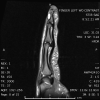
STIR sagittal image of the left index finger demonstrates a markedly hyperintense subungual mass. Contributed by Hassana Barazi, MD.
References
- 1.
Brouillard P, Boon LM, Mulliken JB, Enjolras O, Ghassibé M, Warman ML, Tan OT, Olsen BR, Vikkula M. Mutations in a novel factor, glomulin, are responsible for glomuvenous malformations ("glomangiomas").
Am J Hum Genet. 2002 Apr;70(4):866-74. [
PMC free article: PMC379115] [
PubMed: 11845407]
- 2.
Abbas A, Braswell M, Bernieh A, Brodell RT. Glomuvenous malformations in a young man.
Dermatol Online J. 2018 Oct 15;24(10) [
PubMed: 30677819]
- 3.
Arens C, Dreyer T, Eistert B, Glanz H. Glomangioma of the nasal cavity. Case report and literature review.
ORL J Otorhinolaryngol Relat Spec. 1997 May-Jun;59(3):179-81. [
PubMed: 9186975]
- 4.
Chatterjee JS, Youssef AH, Brown RM, Nishikawa H. Congenital nodular multiple glomangioma: a case report.
J Clin Pathol. 2005 Jan;58(1):102-3. [
PMC free article: PMC1770555] [
PubMed: 15623496]
- 5.
Munoz C, Bobadilla F, Fuenzalida H, Goldner R, Sina B. Congenital glomangioma of the breast: type 2 segmental manifestation.
Int J Dermatol. 2011 Mar;50(3):346-9. [
PubMed: 21342169]
- 6.
Tewattanarat N, Srinakarin J, Wongwiwatchai J, Areemit S, Komvilaisak P, Ungarreevittaya P, Intarawichian P. Imaging of a glomus tumor of the liver in a child.
Radiol Case Rep. 2020 Apr;15(4):311-315. [
PMC free article: PMC6971342] [
PubMed: 31988680]
- 7.
Leger M, Patel U, Mandal R, Walters R, Cook K, Haimovic A, Franks AG. Glomangioma.
Dermatol Online J. 2010 Nov 15;16(11):11. [
PubMed: 21163162]
- 8.
Souza NGA, Nai GA, Wedy GF, Abreu MAMM. Congenital plaque-like glomangioma: report of two cases.
An Bras Dermatol. 2017;92(5 Suppl 1):43-46. [
PMC free article: PMC5726674] [
PubMed: 29267443]
- 9.
Cabral CR, Oliveira Filho Jd, Matsumoto JL, Cignachi S, Tebet AC, Nasser Kda R. Type 2 segmental glomangioma--Case report.
An Bras Dermatol. 2015 May-Jun;90(3 Suppl 1):97-100. [
PMC free article: PMC4540520] [
PubMed: 26312686]
- 10.
Jha A, Khunger N, Malarvizhi K, Ramesh V, Singh A. Familial Disseminated Cutaneous Glomuvenous Malformation: Treatment with Polidocanol Sclerotherapy.
J Cutan Aesthet Surg. 2016 Oct-Dec;9(4):266-269. [
PMC free article: PMC5227083] [
PubMed: 28163461]
- 11.
Suárez-Magdalena O, Monteagudo B, Figueroa O, Gómez-Pérez MI. Glomulin gene c.157_161del mutation in a family with multiple glomuvenous malformations.
Int J Dermatol. 2019 Feb;58(2):e43-e45. [
PubMed: 30460983]
- 12.
Boon LM, Mulliken JB, Enjolras O, Vikkula M. Glomuvenous malformation (glomangioma) and venous malformation: distinct clinicopathologic and genetic entities.
Arch Dermatol. 2004 Aug;140(8):971-6. [
PubMed: 15313813]
- 13.
Gonçalves R, Lopes A, Júlio C, Durão C, de Mello RA. Knee glomangioma: a rare location for a glomus tumor.
Rare Tumors. 2014 Oct 27;6(4):5588. [
PMC free article: PMC4274446] [
PubMed: 25568752]
- 14.
Tony G, Hauxwell S, Nair N, Harrison DA, Richards PJ. Large plaque-like glomangioma in a patient with multiple glomus tumours: review of imaging and histology.
Clin Exp Dermatol. 2013 Oct;38(7):693-700. [
PubMed: 24073652]
- 15.
Boon LM, Brouillard P, Irrthum A, Karttunen L, Warman ML, Rudolph R, Mulliken JB, Olsen BR, Vikkula M. A gene for inherited cutaneous venous anomalies ("glomangiomas") localizes to chromosome 1p21-22.
Am J Hum Genet. 1999 Jul;65(1):125-33. [
PMC free article: PMC1378082] [
PubMed: 10364524]
- 16.
Cullen RD, Hanna EY. Intranasal glomangioma.
Am J Otolaryngol. 2000 Nov-Dec;21(6):402-4. [
PubMed: 11115526]
- 17.
Goujon E, Cordoro KM, Barat M, Rousseau T, Brouillard P, Vikkula M, Frieden IJ, Vabres P. Congenital plaque-type glomuvenous malformations associated with fetal pleural effusion and ascites.
Pediatr Dermatol. 2011 Sep-Oct;28(5):528-31. [
PubMed: 21133993]
- 18.
Scheithauer BW, Rodriguez FJ, Spinner RJ, Dyck PJ, Salem A, Edelman FL, Amrami KK, Fu YS. Glomus tumor and glomangioma of the nerve. Report of two cases.
J Neurosurg. 2008 Feb;108(2):348-56. [
PubMed: 18240933]
- 19.
Parker KL, Zervos MD, Donington JS, Shukla PS, Bizekis CS. Tracheal glomangioma in a patient with asthma and chest pain.
J Clin Oncol. 2010 Jan 10;28(2):e9-e10. [
PubMed: 19858390]
- 20.
Larsen DK, Madsen PV. [Glomus tumour of the distal phalanx].
Ugeskr Laeger. 2018 Jul 23;180(30) [
PubMed: 30037386]
- 21.
Aneiros-Fernandez J, Retamero JA, Husein-ElAhmed H, Carriel V, Ruiz Villaverde R, O'Valle F, Aneiros-Cachaza J. Smoothelin and WT-1 expression in glomus tumors and glomuvenous malformations.
Histol Histopathol. 2017 Feb;32(2):153-160. [
PubMed: 27184662]
- 22.
Flors L, Norton PT, Hagspiel KD. Glomuvenous malformation: magnetic resonance imaging findings.
Pediatr Radiol. 2015 Feb;45(2):286-90. [
PubMed: 24996811]
- 23.
Chirila M, Rogojan L. Glomangioma of the nasal septum: a case report and review.
Ear Nose Throat J. 2013 Apr-May;92(4-5):E7-9. [
PubMed: 23599117]
- 24.
Sharma JK, Miller R. Treatment of multiple glomangioma with tuneable dye laser.
J Cutan Med Surg. 1999 Jan;3(3):167-8. [
PubMed: 10082598]
- 25.
Rivers JK, Rivers CA, Li MK, Martinka M. Laser Therapy for an Acquired Glomuvenous Malformation (Glomus Tumour): A Nonsurgical Approach.
J Cutan Med Surg. 2016 Jan;20(1):80-3. [
PubMed: 26177926]
- 26.
Phillips CB, Guerrero C, Theos A. Nd:YAG laser offers promising treatment option for familial glomuvenous malformation.
Dermatol Online J. 2015 Apr 16;21(4) [
PubMed: 25933083]
- 27.
Jha A, Ramesh V, Singh A. Disseminated cutaneous glomuvenous malformation.
Indian J Dermatol Venereol Leprol. 2014 Nov-Dec;80(6):556-8. [
PubMed: 25382523]
- 28.
Babeau F, Knafo S, Rigau V, Lonjon N. Paravertebral glomangioma mimicking a schwannoma.
Neurochirurgie. 2013 Aug-Oct;59(4-5):187-90. [
PubMed: 24367799]
- 29.
Jiga LP, Rata A, Ignatiadis I, Geishauser M, Ionac M. Atypical venous glomangioma causing chronic compression of the radial sensory nerve in the forearm. A case report and review of the literature.
Microsurgery. 2012 Mar;32(3):231-4. [
PubMed: 22407591]
- 30.
Arica DA, Arica IE, Yayli S, Cobanoglu U, Akay BN, Anadolu R, Bahadir S. Spitz nevus arising upon a congenital glomuvenous malformation.
Pediatr Dermatol. 2013 May-Jun;30(3):e25-6. [
PubMed: 22304367]
Disclosure: Oranus Mohammadi declares no relevant financial relationships with ineligible companies.
Disclosure: Manuel Suarez declares no relevant financial relationships with ineligible companies.