Introduction
The dexamethasone suppression test (DST) is used in the evaluation of endogenous Cushing syndrome (CS) by assessing for the lack of suppression of the hypothalamic-pituitary-adrenal (HPA) axis by exogenous corticosteroids.[1] The first use of dexamethasone for assessing CS was in 1960 by Liddle. He developed a test based on the lack of suppression of endogenous cortisol production in CS versus the physiologic suppression in nonaffected individuals achieved by dexamethasone.[2]
Dexamethasone is a potent synthetic corticosteroid (dexamethasone 0.75 mg = prednisone 5 mg = methylprednisolone 4 mg = hydrocortisone 20 mg) with high affinity for the glucocorticoid receptors and long duration of action (biological half-life 36 to 54 hours; plasma half-life 4 to 5 hours).[3] It possesses minimal mineralocorticoid activity, and unlike other glucocorticoids, it does not interfere with cortisol measurement in the plasma, urine, or saliva. These qualities make dexamethasone the preferred steroid for assessing the HPA axis.[4]
The dexamethasone suppression test is also used to investigate mild autonomous cortisol excess (MACE) in patients with incidentalomas. For the diagnosis of CS and also MACE, a serum cortisol concentration of 1.8 µg/dL (50 nmol/L) is the widely recommended cut-off that increases the diagnostic sensitivity of the test to approximately 95%.[5] Serum cortisol concentrations <1.8 µg/dL suggest adequate HPA axis suppression post dexamethasone and exclude CS.[6]
Pathophysiology
The HPA axis, a primary neuroendocrine system, helps maintain homeostatic function and the stress response.[7] The neurons in the paraventricular nucleus of the hypothalamus synthesize corticotropin-releasing hormone (CRH), which is transported via the hypophyseal portal blood to the anterior pituitary, wherein it stimulates the production of adrenocorticotropic hormone (ACTH).[8] ACTH is transported via the systemic vasculature to the adrenal glands and stimulates the synthesis and secretion of cortisol by zona fasciculata of the adrenal cortex.[9]
Under basal conditions, only about 5% to 10% of circulating cortisol is free.[10] The rest is bound to proteins: 85% to transcortin (cortisol-binding globulin) and 5% to 10% to albumin. The free fraction is physiologically active, although the albumin-bound fraction is also available because the binding affinity of albumin for cortisol is low.[11]
Unbound cortisol is filtered at the glomerulus and excreted. This fraction constitutes less than 1% of the total cortisol synthesized daily; the rest is excreted as soluble metabolites and glucuronide conjugates.[12] The excretory products of cortisol that contain the dihydroxyacetone group are known as 17-hydroxycorticosteroids (17-OH-CS).[13] Urinary 17-OH-CS was previously used as an indirect measure of cortisol secretion rate. Urinary cortisol reflects free, circulating cortisol in the blood.[14]
Serum cortisol, also called the stress hormone, exerts negative feedback on both the hypothalamus and the anterior pituitary, inhibiting the secretion of CRH and ACTH, respectively. This positive/negative feedback mechanism helps regulate serum cortisol levels and the stress response.[15]When the HPA axis is intact, exogenously administered corticosteroids exert feedback inhibition on serum CRH and ACTH production by binding to the hypothalamic and pituitary glucocorticoid receptors, respectively, which subsequently causes suppression of the synthesis and secretion of serum cortisol.[7] However, in pathological hypercortisolism, the HPA axis becomes partially or entirely resistant to feedback inhibition by exogenous steroids.[16]
Specimen Requirements and Procedure
Both serum and heparinized plasma are suitable specimens for measuring cortisol in the blood.[17] Specimens can be stored at 2^oC to 8^oC for two days. For more extended storage, specimens must be frozen. Freeze/thaw cycles have not been found to alter cortisol concentrations significantly.[18]
Urinary free cortisol can be measured by immunoassay or high-performance liquid chromatography (HPLC) methods. These procedures require an extraction step using methylene chloride, and it is customary to monitor the efficiency of the extraction technique by extraction of a urine control with known cortisol concentration.[19] A 24-hour urine sample is preferred. Urine collected for a 24-hour sample should be refrigerated during the collection period.[20] Some laboratories use boric acid (approximately 10 g of boric acid per liter) or acetic acid (approximately 7 mL of glacial acetic acid per liter), placed in the collection container at the start of the collection, to preserve the specimen.[21] If the sample cannot be analyzed within a day of collection, it should be frozen at −20^oC.[22]
Immunoassays are the main methods for the estimation of cortisol in clinical laboratories. These immunoassays are competitive immunoassays, where cortisol in the sample competes with labeled cortisol for a limited number of anti-cortisol antibodies.[19] Many commercial kits are available for the determination of plasma and urinary cortisol. Many immunoassays are now automated and available on semi-automated or automated analyzer platforms.[23] These immunoassays can be heterogeneous assays, where a separation step is required to partition the bound from unbound fractions, or homogenous assays, where no separation is required.[24] The specificity of immunoassays depends mainly on the properties of the antibody.[25]
Chromatographic methods are an alternative to immunoassay for the determination of cortisol. They have the advantage of less interference, but these methods are time-consuming and unsuitable for routine analysis.[26] Of the chromatographic methods, high-performance liquid chromatography (HPLC) is often used in clinical laboratories.[27] All HPLC methods require the extraction of cortisol from the sample by either a liquid-liquid extraction or solid-phase extraction columns. Both reversed-phase and normal-phase columns are used for chromatographic analyses, and the column effluents are monitored by either ultraviolet (254 nm) or fluorescence spectroscopy.[28]
Testing Procedures
Types of Dexamethasone Suppression Tests (DST)
Low-dose Dexamethasone Suppression Test (LDDST)
LDDST helps in the initial diagnosis of Cushing syndrome as either a screening or a confirmatory test.[29] It can be performed either as an overnight or a two-day test.
- Two-day, 2 mg test: Dexamethasone 0.5 mg is administered orally every 6 hours (9 AM, 3 PM, 9 PM, 3 AM) for two days (total dose 4 mg). Serum cortisol level is drawn 6 hours (9 AM) after the last administered dose.[2]
High-dose Dexamethasone Suppression Test (HDDST)
Once the diagnosis of Cushing syndrome is confirmed, the next step is to categorize ACTH-independent vs. ACTH-dependent Cushing syndrome by checking the plasma ACTH levels. In ACTH-independent Cushing syndrome, the plasma ACTH is low or undetectable, indicating an adrenal etiology (causing pituitary suppression of ACTH). In ACTH-dependent Cushing syndrome, the plasma ACTH is inappropriately normal or high, suggesting either a pituitary or an ectopic source.
In ACTH-dependent Cushing syndrome, HDDST can help distinguish a pituitary (i.e., Cushing disease) from an ectopic source of ACTH overproduction. The principle behind the HDDST is that the overproduction of ACTH seen in Cushing disease (but not ectopic tumors) can undergo either partial or complete suppression by high doses of dexamethasone (approximately 8 mg). As in the LDDST, the HDDST utilizes oral dexamethasone as either an overnight or two-day test.
- Overnight, 8 mg test: Baseline morning serum cortisol is measured, and oral dexamethasone 8 mg is administered between 11 PM and midnight. Repeat serum cortisol is drawn the following morning (between 8 and 9 AM).[32]
- Two-day, 8 mg test: On day 1, a baseline morning serum cortisol or 24-hour urinary free cortisol (UFC) from the day before is obtained. Dexamethasone 2 mg is administered orally every 6 hours (9 AM, 3 PM, 9 PM, 3 AM) for two days to complete a total dose of 16 mg over days 1 and 2. A urine sample for UFC is collected simultaneously with dexamethasone administration. Additionally, serum cortisol levels are checked 6 hours after the last dose of dexamethasone (9 AM of day 2).[2]
Intravenous Dexamethasone Suppression Test
This test helps in the initial diagnosis of Cushing syndrome while overcoming concerns of drug compliance and malabsorption.[33] It is also useful when differentiating Cushing disease from ACTH-dependent ectopic tumors and ACTH-independent adrenal etiologies.[34] After a baseline morning serum cortisol (between 8 AM and 9 AM) is obtained, an infusion of intravenous dexamethasone at 1 mg/hour for 4 to 7 hours is administered. Repeat serum cortisol levels are measured at the end of the infusion (day 1) and 23 to 24 hours later (day 2).[35]
Dexamethasone Suppressed Corticotropin-Releasing Hormone Test
Based on the rationale that glucocorticoid suppression of the HPA axis can be overcome by corticotropin-releasing hormone (CRH) stimulation in Cushing disease and not in pseudo-Cushing syndrome (physiologic hypercortisolism), this test helps distinguish the two entities. Dexamethasone 0.5 mg every 6 hours (12 PM, 6 PM, 12 AM, 6 AM) is given orally for 48 hours. Two hours after the last dose of dexamethasone, an intravenous CRH dose of 1 mcg/kg is administered (8 AM), and serum cortisol is drawn 15 minutes later.[36]
Interfering Factors
1. Iatrogenic hypercortisolism can cause exogenous Cushing syndrome.[37] Biochemical testing shows elevated serum cortisol levels (due to the cross-reactivity of most exogenous steroids with cortisol immunoassays) and depressed ACTH levels, as is usually seen in ACTH-independent Cushing syndrome.[38] These individuals receiving exogenous corticosteroids (inhaled, topical, parenteral, intraarticular) must be identified before pursuing a work-up for pathologic hypercortisolemia.[39]
2. Pseudo-Cushing syndrome, also called physiological or non-neoplastic hypercortisolism, is seen in conditions like alcohol use disorder, obesity, insulin resistance, and neuropsychiatric disorders due to HPA axis stimulation.[40] A thorough history and physical examination can be pivotal in recognizing many subjects with pseudo-Cushing syndrome. In equivocal cases, midnight serum cortisol, late-night salivary cortisol, dexamethasone-CRH, or desmopressin testing can assist in the distinction.[25]
3. In any acute illness, be it emotional or physical, a stress response can occur via stimulation of the HPA axis, resulting in elevated ACTH and cortisol levels. Evaluation for Cushing syndrome should take place after the resolution of acute stress.[41]
4. Corticosteroid-binding globulin (CBG): Approximately 90% of circulating cortisol is protein-bound. Currently available assays measure the total cortisol (free and protein-bound); conditions resulting in elevated levels of circulating CBG, such as pregnancy or use of estrogen-containing medications, or reduced levels of circulating CBG, such as malnutrition or nephrotic syndrome, may lead to spurious results on DSTs.[42] Either late-night salivary cortisol or UFC is the preferred test in these situations. Patients receiving estrogen therapy should ideally stop treatment at least six weeks before DST.
5. Dexamethasone bioavailability: Malabsorption, altered metabolism, or non-compliance with taking the medication can result in variable bioavailability of dexamethasone, confounding the results. Dexamethasone is metabolized by the liver via CYP3A4.[43] Thus, CYP3A4 inducers (phenytoin, carbamazepine, etc.) or CYP3A4 inhibitors (itraconazole, fluoxetine, ritonavir) may result in decreased or increased clearance of the dexamethasone, risking false positive or negative results, respectively.[44] This error can be overcome by measuring serum dexamethasone levels simultaneously with serum cortisol levels. Most laboratories that conduct this test provide reference ranges based on dexamethasone dose and interval of blood drawn.
6. Improper urine collection: An inadequately collected urine sample for urinary-free cortisol (UFC) leads to diagnostic errors. For this reason, the urine sample should include testing for 24-hour urinary creatinine excretion in addition to cortisol.[45] In adults less than 50 years of age, 24-hour urinary creatinine excretion is approximately 15 to 20 mg/kg/day in females and 20 to 25 mg/kg/day in males. In people older than 50, there is a progressive decline in muscle mass; creatinine excretion can be as low as 10 mg/kg/day.
7. Others: Cyclic Cushing syndrome and glucocorticoid receptor polymorphisms are rare causes of false test results.[45]
Results, Reporting, and Critical Findings
Low-dose Dexamethasone Suppression Test (LDDST)
In either of the LDDSTs (overnight or two-day), a serum cortisol level of 1.8 mcg/dL (50 nmol/L) is the recommended cut-off value that increases the diagnostic sensitivity of the test to approximately 95%.[5] However, at this cut-off value, the specificity of the two-day test is superior when compared to that of the overnight test (97% to 100% versus 86%). A recent meta-analysis showed that abnormal and normal results for the 1 mg overnight test had a positive and negative likelihood ratio (LR) of 11.6 and 0.09, respectively.[46] On the other hand, for the two-day 2 mg test, abnormal and normal results had positive and negative LR of 7.3 and 0.8, respectively. Serum cortisol levels under 1.8 mcg/dL suggest adequate HPA axis suppression by dexamethasone and exclude CS. Levels greater than 1.8 mcg/dL should be verified with a second test (24-hour urinary free cortisol or late-night salivary cortisol) before establishing a confirmed diagnosis of CS.[31]
High-dose Dexamethasone Suppression Test (HDDST)
A reduction in either urinary-free cortisol (UFC) or serum cortisol greater than 50% overnight during a two-day HDDST makes Cushing disease (CD) the likely source of ACTH-dependent Cushing syndrome. At a cut-off value of 50% suppression, HDDST provides a sensitivity and specificity of 60 to 100%. Increasing the cut-off value to greater than 90% cortisol suppression increases the specificity of diagnosing Cushing disease to almost 100%, albeit at a much-reduced sensitivity.[47] Because of this limitation, HDDST is not the recommended test unless both the pituitary MRI and bilateral inferior petrosal venous sampling are either negative or logistically challenging. Even in this scenario, HDDST is performed in conjunction with a CRH stimulation test to enhance diagnostic accuracy.[48]
Intravenous Dexamethasone Suppression Test
The diagnosis of Cushing syndrome is made if the day-2 serum cortisol level is greater than 20% of the baseline value [or greater than 4.7 mcg/dL (130 nmol/L)] with a sensitivity of 100% and specificity of 96%.[33][34] Additionally, in Cushing disease, as opposed to other etiologies of Cushing syndrome, the day-1 (i.e., the end of infusion) serum cortisol level usually shows greater than 70% suppression from the baseline, followed by rebound hypercortisolism in 24 hours.[35]
Dexamethasone Suppressed Corticotropin-releasing Hormone Test
Serum cortisol levels greater than 1.4 mcg/dL (39 nmol/L) at 15 minutes suggest Cushing disease with a 90% to 100% sensitivity and 50 to 100% specificity.[36][49] Raising the cut-off value to greater than 3.8 mcg/dL (87 nmol/L) increases the specificity to 100% at the cost of a slightly reduced sensitivity at 94%.[50] The cumbersomeness of this test limits its extensive application in the ambulatory setting.[51]
Clinical Significance
Iatrogenic hypercortisolism is the most common cause of Cushing syndrome. It should be recognized before these individuals are subject to further diagnostic testing; instead, the focus should be on titrating down (or discontinuing, if feasible) the prescribed steroid dosages.[52] The dexamethasone suppression test must be performed and interpreted in the light of the pretest probability based on a thorough history and physical examination. Additionally, clinicians should be mindful of all the tests' diagnostic accuracy, limitations, and interfering factors.[53] In subjects with high clinical suspicion of Cushing syndrome but equivocal or negative test results, repeat testing should occur in 3 to 6 months, as untreated hypercortisolemia has detrimental consequences.[54]
Quality Control and Lab Safety
For non-waived tests, laboratory regulations require, at the minimum, analysis of at least two levels of control materials once every 24 hours. If necessary, laboratories can assay QC samples more frequently to ensure accurate results. Quality control samples should be assayed after calibration or maintenance of an analyzer to verify the correct method performance.[55] To minimize QC when performing tests for which manufacturers’ recommendations are less than those required by the regulatory agency (such as once per month), the labs can develop an individualized quality control plan (IQCP) that involves performing a risk assessment of potential sources of error in all phases of testing and putting in place a QC plan to reduce the likelihood of errors.[56]
Westgard multi-rules are used to evaluate the quality control runs. If a run is declared out of control, investigate the system (instrument, standards, controls, etc.) to determine the cause of the problem. Do not perform any analysis until the problem has been resolved.[57]
Consider all specimens, control materials, and calibrator materials as potentially infectious. Exercise the standard precautions required for handling all laboratory reagents. Disposal of all waste material should be in accordance with local guidelines.[58] Wear gloves, a lab coat, and safety glasses when handling human blood specimens. Place all plastic tips, sample cups, and gloves that come into contact with blood in a biohazard waste container. Discard all disposable glassware into sharps waste containers.[59] Protect all work surfaces with disposable absorbent bench top paper, discarded into biohazard waste containers weekly or whenever blood contamination occurs. Wipe all work surfaces weekly.[60]
Enhancing Healthcare Team Outcomes
Primary care providers may initially evaluate patients with suspected Cushing syndrome prior to referring them to an endocrinologist for further workup. Clear communication and care coordination between clinician, patient, and nurse are of paramount importance as the correct implementation of DST and sample collection can have a dramatic impact on these factors on subsequent results.
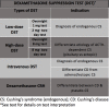
Figure
Dexamethasone Suppression Test - Types & Indications Contributed by Prerna Dogra, MD
References
- 1.
- Keevil BG. Improving the Dexamethasone Suppression Test. Clin Chem. 2021 Jul 06;67(7):929-931. [PubMed: 34125167]
- 2.
- LIDDLE GW. Tests of pituitary-adrenal suppressibility in the diagnosis of Cushing's syndrome. J Clin Endocrinol Metab. 1960 Dec;20:1539-60. [PubMed: 13761950]
- 3.
- Williams DM. Clinical Pharmacology of Corticosteroids. Respir Care. 2018 Jun;63(6):655-670. [PubMed: 29794202]
- 4.
- Tanaka K, Toriumi M, Itoh S, Ogino Y. [Dexamethasone suppression test]. Nihon Rinsho. 1997 Apr;55 Suppl 2:353-6. [PubMed: 9172546]
- 5.
- Nieman LK, Biller BM, Findling JW, Newell-Price J, Savage MO, Stewart PM, Montori VM. The diagnosis of Cushing's syndrome: an Endocrine Society Clinical Practice Guideline. J Clin Endocrinol Metab. 2008 May;93(5):1526-40. [PMC free article: PMC2386281] [PubMed: 18334580]
- 6.
- Fleseriu M, Auchus R, Bancos I, Ben-Shlomo A, Bertherat J, Biermasz NR, Boguszewski CL, Bronstein MD, Buchfelder M, Carmichael JD, Casanueva FF, Castinetti F, Chanson P, Findling J, Gadelha M, Geer EB, Giustina A, Grossman A, Gurnell M, Ho K, Ioachimescu AG, Kaiser UB, Karavitaki N, Katznelson L, Kelly DF, Lacroix A, McCormack A, Melmed S, Molitch M, Mortini P, Newell-Price J, Nieman L, Pereira AM, Petersenn S, Pivonello R, Raff H, Reincke M, Salvatori R, Scaroni C, Shimon I, Stratakis CA, Swearingen B, Tabarin A, Takahashi Y, Theodoropoulou M, Tsagarakis S, Valassi E, Varlamov EV, Vila G, Wass J, Webb SM, Zatelli MC, Biller BMK. Consensus on diagnosis and management of Cushing's disease: a guideline update. Lancet Diabetes Endocrinol. 2021 Dec;9(12):847-875. [PMC free article: PMC8743006] [PubMed: 34687601]
- 7.
- Spencer RL, Deak T. A users guide to HPA axis research. Physiol Behav. 2017 Sep 01;178:43-65. [PMC free article: PMC5451309] [PubMed: 27871862]
- 8.
- Aguilera G, Liu Y. The molecular physiology of CRH neurons. Front Neuroendocrinol. 2012 Jan;33(1):67-84. [PMC free article: PMC4341841] [PubMed: 21871477]
- 9.
- Brownstein MJ. Adrenocorticotropic hormone (ACTH) in the central nervous system. Adv Biochem Psychopharmacol. 1980;22:93-9. [PubMed: 6249088]
- 10.
- Tanaka K. [Cortisol]. Nihon Rinsho. 1995 Mar;53 Su Pt 2:437-40. [PubMed: 8753275]
- 11.
- Hodes A, Meyer J, Lodish MB, Stratakis CA, Zilbermint M. Mini-review of hair cortisol concentration for evaluation of Cushing syndrome. Expert Rev Endocrinol Metab. 2018 Sep;13(5):225-231. [PMC free article: PMC6378952] [PubMed: 30234410]
- 12.
- Sederberg-Olsen P, Binder C, Kehlet H. Urinary excretion of free cortisol in impaired renal function. Acta Endocrinol (Copenh). 1975 Jan;78(1):86-90. [PubMed: 1172895]
- 13.
- Billon-Rey S, Beylot M, Mathian B, Patricot MC, Berthezene F, Mornex R, Revol A. [Comparison between the value of urinary free cortisol and 17-hydroxycorticosteroids for the diagnosis of Cushing's syndrome]. Presse Med. 1986 May 24;15(21):965-8. [PubMed: 2942852]
- 14.
- Mengden T, Hubmann P, Müller J, Greminger P, Vetter W. Urinary free cortisol versus 17-hydroxycorticosteroids: a comparative study of their diagnostic value in Cushing's syndrome. Clin Investig. 1992 Jul;70(7):545-8. [PubMed: 1327327]
- 15.
- Lee DY, Kim E, Choi MH. Technical and clinical aspects of cortisol as a biochemical marker of chronic stress. BMB Rep. 2015 Apr;48(4):209-16. [PMC free article: PMC4436856] [PubMed: 25560699]
- 16.
- Lowrance SA, Ionadi A, McKay E, Douglas X, Johnson JD. Sympathetic nervous system contributes to enhanced corticosterone levels following chronic stress. Psychoneuroendocrinology. 2016 Jun;68:163-70. [PMC free article: PMC5656452] [PubMed: 26974501]
- 17.
- Maidana P, Bruno OD, Mesch V. [A critical analysis of cortisol measurements: an update]. Medicina (B Aires). 2013;73(6):579-84. [PubMed: 24356273]
- 18.
- Gatti R, Antonelli G, Prearo M, Spinella P, Cappellin E, De Palo EF. Cortisol assays and diagnostic laboratory procedures in human biological fluids. Clin Biochem. 2009 Aug;42(12):1205-17. [PubMed: 19414006]
- 19.
- El-Farhan N, Rees DA, Evans C. Measuring cortisol in serum, urine and saliva - are our assays good enough? Ann Clin Biochem. 2017 May;54(3):308-322. [PubMed: 28068807]
- 20.
- Rosmalen JG, Kema IP, Wüst S, van der Ley C, Visser ST, Snieder H, Bakker SJ. 24 h urinary free cortisol in large-scale epidemiological studies: short-term and long-term stability and sources of variability. Psychoneuroendocrinology. 2014 Sep;47:10-6. [PubMed: 25001951]
- 21.
- Salazar-García S, Lares-Villaseñor E, Bárcenas-Morales A, Vargas-Morales Juan M. Impact of chemical preservative in urine samples. EJIFCC. 2020 Mar;31(1):56-64. [PMC free article: PMC7109501] [PubMed: 32256289]
- 22.
- Poulsen CS, Kaas RS, Aarestrup FM, Pamp SJ. Standard Sample Storage Conditions Have an Impact on Inferred Microbiome Composition and Antimicrobial Resistance Patterns. Microbiol Spectr. 2021 Oct 31;9(2):e0138721. [PMC free article: PMC8510183] [PubMed: 34612701]
- 23.
- Turpeinen U, Hämäläinen E. Determination of cortisol in serum, saliva and urine. Best Pract Res Clin Endocrinol Metab. 2013 Dec;27(6):795-801. [PubMed: 24275191]
- 24.
- Casals G, Hanzu FA. Cortisol Measurements in Cushing's Syndrome: Immunoassay or Mass Spectrometry? Ann Lab Med. 2020 Jul;40(4):285-296. [PMC free article: PMC7054699] [PubMed: 32067427]
- 25.
- Alwani RA, Schmit Jongbloed LW, de Jong FH, van der Lely AJ, de Herder WW, Feelders RA. Differentiating between Cushing's disease and pseudo-Cushing's syndrome: comparison of four tests. Eur J Endocrinol. 2014 Apr;170(4):477-86. [PubMed: 24394725]
- 26.
- Mezzullo M, Fanelli F, Fazzini A, Gambineri A, Vicennati V, Di Dalmazi G, Pelusi C, Mazza R, Pagotto U, Pasquali R. Validation of an LC-MS/MS salivary assay for glucocorticoid status assessment: Evaluation of the diurnal fluctuation of cortisol and cortisone and of their association within and between serum and saliva. J Steroid Biochem Mol Biol. 2016 Oct;163:103-12. [PubMed: 27108942]
- 27.
- Hariharan M, Naga S, VanNoord T, Kindt EK. Simultaneous assay of corticosterone and cortisol in plasma by reversed-phase liquid chromatography. Clin Chem. 1992 Mar;38(3):346-52. [PubMed: 1547550]
- 28.
- Mezzullo M, Fazzini A, Gambineri A, Di Dalmazi G, Mazza R, Pelusi C, Vicennati V, Pasquali R, Pagotto U, Fanelli F. Parallel diurnal fluctuation of testosterone, androstenedione, dehydroepiandrosterone and 17OHprogesterone as assessed in serum and saliva: validation of a novel liquid chromatography-tandem mass spectrometry method for salivary steroid profiling. Clin Chem Lab Med. 2017 Aug 28;55(9):1315-1323. [PubMed: 28076306]
- 29.
- Wood PJ, Barth JH, Freedman DB, Perry L, Sheridan B. Evidence for the low dose dexamethasone suppression test to screen for Cushing's syndrome--recommendations for a protocol for biochemistry laboratories. Ann Clin Biochem. 1997 May;34 ( Pt 3):222-9. [PubMed: 9158818]
- 30.
- Cronin C, Igoe D, Duffy MJ, Cunningham SK, McKenna TJ. The overnight dexamethasone test is a worthwhile screening procedure. Clin Endocrinol (Oxf). 1990 Jul;33(1):27-33. [PubMed: 2401096]
- 31.
- NUGENT CA, NICHOLS T, TYLER FH. DIAGNOSIS OF CUSHING'S SYNDROME; SINGLE DOSE DEXAMETHASONE SUPPRESSION TEST. Arch Intern Med. 1965 Aug;116:172-6. [PubMed: 14315650]
- 32.
- Bruno OD, Rossi MA, Contreras LN, Gómez RM, Galparsoro G, Cazado E, Kral M, Leber B, Arias D. Nocturnal high-dose dexamethasone suppression test in the aetiological diagnosis of Cushing's syndrome. Acta Endocrinol (Copenh). 1985 Jun;109(2):158-62. [PubMed: 2990131]
- 33.
- Biemond P, de Jong FH, Lamberts SW. Continuous dexamethasone infusion for seven hours in patients with the Cushing syndrome. A superior differential diagnostic test. Ann Intern Med. 1990 May 15;112(10):738-42. [PubMed: 2158760]
- 34.
- Jung C, Alford FP, Topliss DJ, Burgess JR, Long F, Gome JJ, Stockigt JR, Inder WJ. The 4-mg intravenous dexamethasone suppression test in the diagnosis of Cushing's syndrome. Clin Endocrinol (Oxf). 2010 Jul;73(1):78-84. [PubMed: 20039897]
- 35.
- Tran HA, Petrovsky N. Dexamethasone infusion testing in the diagnosis of Cushing's syndrome. Endocr J. 2005 Feb;52(1):103-9. [PubMed: 15758565]
- 36.
- Yanovski JA, Cutler GB, Chrousos GP, Nieman LK. Corticotropin-releasing hormone stimulation following low-dose dexamethasone administration. A new test to distinguish Cushing's syndrome from pseudo-Cushing's states. JAMA. 1993 May 05;269(17):2232-8. [PubMed: 8386285]
- 37.
- Raff H, Carroll T. Cushing's syndrome: from physiological principles to diagnosis and clinical care. J Physiol. 2015 Feb 01;593(3):493-506. [PMC free article: PMC4324701] [PubMed: 25480800]
- 38.
- Bansal V, El Asmar N, Selman WR, Arafah BM. Pitfalls in the diagnosis and management of Cushing's syndrome. Neurosurg Focus. 2015 Feb;38(2):E4. [PubMed: 25639322]
- 39.
- Pektas SD, Dogan G, Cinar N. Iatrogenic Cushing's Syndrome with Subsequent Adrenal Insufficiency in a Patient with Psoriasis Vulgaris Using Topical Steroids. Case Rep Endocrinol. 2017;2017:8320254. [PMC free article: PMC5702924] [PubMed: 29259830]
- 40.
- Chabre O. The difficulties of pseudo-Cushing's syndrome (or "non-neoplastic hypercortisolism"). Ann Endocrinol (Paris). 2018 Jun;79(3):138-145. [PubMed: 29716734]
- 41.
- Stephens MA, Wand G. Stress and the HPA axis: role of glucocorticoids in alcohol dependence. Alcohol Res. 2012;34(4):468-83. [PMC free article: PMC3860380] [PubMed: 23584113]
- 42.
- Bae YJ, Kratzsch J. Corticosteroid-binding globulin: modulating mechanisms of bioavailability of cortisol and its clinical implications. Best Pract Res Clin Endocrinol Metab. 2015 Oct;29(5):761-72. [PubMed: 26522460]
- 43.
- Meikle AW. Dexamethasone suppression tests: usefulness of simultaneous measurement of plasma cortisol and dexamethasone. Clin Endocrinol (Oxf). 1982 Apr;16(4):401-8. [PubMed: 7094363]
- 44.
- Rush AJ, Schlesser MA, Giles DE, Crowley GT, Fairchild C, Altshuler KZ. The effect of dosage on the dexamethasone suppression test in normal controls. Psychiatry Res. 1982 Dec;7(3):277-85. [PubMed: 6962436]
- 45.
- Meinardi JR, Wolffenbuttel BH, Dullaart RP. Cyclic Cushing's syndrome: a clinical challenge. Eur J Endocrinol. 2007 Sep;157(3):245-54. [PubMed: 17766705]
- 46.
- Elamin MB, Murad MH, Mullan R, Erickson D, Harris K, Nadeem S, Ennis R, Erwin PJ, Montori VM. Accuracy of diagnostic tests for Cushing's syndrome: a systematic review and metaanalyses. J Clin Endocrinol Metab. 2008 May;93(5):1553-62. [PubMed: 18334594]
- 47.
- Flack MR, Oldfield EH, Cutler GB, Zweig MH, Malley JD, Chrousos GP, Loriaux DL, Nieman LK. Urine free cortisol in the high-dose dexamethasone suppression test for the differential diagnosis of the Cushing syndrome. Ann Intern Med. 1992 Feb 01;116(3):211-7. [PubMed: 1728204]
- 48.
- Aron DC, Raff H, Findling JW. Effectiveness versus efficacy: the limited value in clinical practice of high dose dexamethasone suppression testing in the differential diagnosis of adrenocorticotropin-dependent Cushing's syndrome. J Clin Endocrinol Metab. 1997 Jun;82(6):1780-5. [PubMed: 9177382]
- 49.
- Pecori Giraldi F, Pivonello R, Ambrogio AG, De Martino MC, De Martin M, Scacchi M, Colao A, Toja PM, Lombardi G, Cavagnini F. The dexamethasone-suppressed corticotropin-releasing hormone stimulation test and the desmopressin test to distinguish Cushing's syndrome from pseudo-Cushing's states. Clin Endocrinol (Oxf). 2007 Feb;66(2):251-7. [PubMed: 17223996]
- 50.
- Tirabassi G, Papa R, Faloia E, Boscaro M, Arnaldi G. Corticotrophin-releasing hormone and desmopressin tests in the differential diagnosis between Cushing's disease and pseudo-Cushing state: a comparative study. Clin Endocrinol (Oxf). 2011 Nov;75(5):666-72. [PubMed: 21554373]
- 51.
- Findling JW, Raff H. DIAGNOSIS OF ENDOCRINE DISEASE: Differentiation of pathologic/neoplastic hypercortisolism (Cushing's syndrome) from physiologic/non-neoplastic hypercortisolism (formerly known as pseudo-Cushing's syndrome). Eur J Endocrinol. 2017 May;176(5):R205-R216. [PubMed: 28179447]
- 52.
- Pivonello R, De Martino MC, De Leo M, Lombardi G, Colao A. Cushing's Syndrome. Endocrinol Metab Clin North Am. 2008 Mar;37(1):135-49, ix. [PubMed: 18226734]
- 53.
- Nieman LK, Ilias I. Evaluation and treatment of Cushing's syndrome. Am J Med. 2005 Dec;118(12):1340-6. [PubMed: 16378774]
- 54.
- Nieman LK. Diagnosis of Cushing's Syndrome in the Modern Era. Endocrinol Metab Clin North Am. 2018 Jun;47(2):259-273. [PubMed: 29754631]
- 55.
- Poh DKH, Lim CY, Tan RZ, Markus C, Loh TP. Internal quality control: Moving average algorithms outperform Westgard rules. Clin Biochem. 2021 Dec;98:63-69. [PubMed: 34534518]
- 56.
- Kearney E. Internal quality control. Methods Mol Biol. 2013;1065:277-89. [PubMed: 23996371]
- 57.
- Bayat H. Selecting multi-rule quality control procedures based on patient risk. Clin Chem Lab Med. 2017 Oct 26;55(11):1702-1708. [PubMed: 28236626]
- 58.
- Cornish NE, Anderson NL, Arambula DG, Arduino MJ, Bryan A, Burton NC, Chen B, Dickson BA, Giri JG, Griffith NK, Pentella MA, Salerno RM, Sandhu P, Snyder JW, Tormey CA, Wagar EA, Weirich EG, Campbell S. Clinical Laboratory Biosafety Gaps: Lessons Learned from Past Outbreaks Reveal a Path to a Safer Future. Clin Microbiol Rev. 2021 Jun 16;34(3):e0012618. [PMC free article: PMC8262806] [PubMed: 34105993]
- 59.
- Munson E, Bowles EJ, Dern R, Beck E, Podzorski RP, Bateman AC, Block TK, Kropp JL, Radke T, Siebers K, Simmons B, Smith MA, Spray-Larson F, Warshauer DM. Laboratory Focus on Improving the Culture of Biosafety: Statewide Risk Assessment of Clinical Laboratories That Process Specimens for Microbiologic Analysis. J Clin Microbiol. 2018 Jan;56(1) [PMC free article: PMC5744218] [PubMed: 29118166]
- 60.
- Ionescu G, Neguţ M, Combiescu AA. [Biosafety and biosecurity in the medical laboratory. Update and trends]. Bacteriol Virusol Parazitol Epidemiol. 2007 Jul-Dec;52(3-4):91-9. [PubMed: 19326721]
Disclosure: Prerna Dogra declares no relevant financial relationships with ineligible companies.
Disclosure: Narasimha Vijayashankar declares no relevant financial relationships with ineligible companies.
Publication Details
Author Information and Affiliations
Authors
Prerna Dogra1; Narasimha P. Vijayashankar2.Affiliations
Publication History
Last Update: April 23, 2023.
Copyright
This book is distributed under the terms of the Creative Commons Attribution-NonCommercial-NoDerivatives 4.0 International (CC BY-NC-ND 4.0) ( http://creativecommons.org/licenses/by-nc-nd/4.0/ ), which permits others to distribute the work, provided that the article is not altered or used commercially. You are not required to obtain permission to distribute this article, provided that you credit the author and journal.
Publisher
StatPearls Publishing, Treasure Island (FL)
NLM Citation
Dogra P, Vijayashankar NP. Dexamethasone Suppression Test. [Updated 2023 Apr 23]. In: StatPearls [Internet]. Treasure Island (FL): StatPearls Publishing; 2024 Jan-.
