Continuing Education Activity
Congenital vertical talus is a foot deformity characterized by dorsal dislocation of the navicular resulting in equinus and hindfoot valgus. It is a relatively rare condition and may be associated with other anomalies. More than 50% of the cases are idiopathic, and the remaining are mainly associated with genetic or neuromuscular disorders. It may result in significant disability due to multiple bony deformities with a rocker-bottom appearance, callosities formation, and difficulty in wearing shoes if it is not managed properly. The management starts with nonoperative treatment in the form of serial casting. Minimally invasive surgeries have also shown promising results in the treatment after serial casting. Postoperative rehabilitation plays a significant role in preventing relapse of this rare foot condition after correcting the deformity. This activity examines congenital vertical talus from a diagnostic and therapeutic standpoint and covers how the interprofessional healthcare team should address this condition in patients to derive optimal outcomes.
Objectives:
Describe the etiology of congenital vertical talus.
Outline the common presentation of a patient with congenital vertical talus.
Review the classification system of congenital vertical talus.
Summarize the various treatment options for patients with congenital vertical talus and the anticipated complications.
Access free multiple choice questions on this topic.
Introduction
Congenital vertical talus is characterized by equinus and hindfoot valgus. It is usually associated with multiple deformities of the foot, including abduction at the forefoot and dorsiflexion at the midfoot, primarily due to the fixed dorsal dislocation of the navicular bone over the talar head.[1][2]
In 1913, the Congenital vertical talus term was first described by Rocher as a foot in piole. It has also been known by a few other terms, such as reversed club foot, congenital valgus flat foot, pied plat valgus congenital, congenital convex pes valgus, and rocker bottom foot.[3] However, now, days Congenital convex pes valgus is most commonly used.[1]
Congenital vertical talus has also been associated with several clinical conditions, including cryptorchidism, microcephaly, and hypoxic birth injury.[4][5] If this condition is missed or not managed properly may result in significant disability, including the formation of painful callosities around the prominent talar head and severe ankle and foot pain.[3]
Traditionally, surgical management of congenital vertical talus was aggressive, with high chances of complications.[1] Nowadays minimally invasive approach has shown promising results in providing deformity correction and limited soft tissue release.[6]
Etiology
The exact etiology of congenital vertical talus is still debatable.[3] More than 50% of the cases are idiopathic, and the remaining are mainly associated with genetic or neuromuscular conditions. Congenital vertical talus can manifest itself in either systemic or isolated forms.[1] In rare instances, it can be associated with a nonsyndromic condition.[3]
Commonly associated conditions include Marfan syndrome, De Barsy Syndrome, chromosomal abnormalities of 13,15,18,30,31, Multiple pterygium syndrome, Cri du chat syndrome, and muscle abnormalities like ischiocalcaneal band, spinal abnormalities, migration anomalies, and fetal brain abnormalities.[2][5]
Other possible etiologies are increased intrauterine pressure, muscle imbalance, intrauterine growth retardation, and arrest in fetal development or the rotational development of the foot.[4]
Epidemiology
The estimated prevalence of congenital vertical talus is 1 in 10,000 live births, which qualifies it as a rare condition and contributes to it being difficult to diagnose in a newborn child.[4]
Pathophysiology
Congenital vertical talus is a condition of the foot in which there is plantar flexion of the talus and equinus of the calcaneum bone.[1] There is associated talonavicular dislocation, which is rigid and irreducible. It results in the prominence of the talar head over the medial aspect, and the appearance of rocker bottom on the sole of the foot is seen.[3]
The valgus and equinus deformity in congenital vertical talus occur mainly due to the contracture of the subtalar joint capsule, tendon Achilles tendon, and involvement of primarily the posterolateral part of the ankle.[3]
The dorsiflexion and abduction at the forefoot and midfoot occur due to the contracture of the peroneus tertius, extensor digitorum longus, tibialis anterior, extensor hallucis longus tendon, extensor hallucis brevis, and dorsal part of the talonavicular capsule.[4] The anterior subluxation of the peroneus longus and tibialis posterior tendon results in dorsiflexion of the ankle joint.[1][2]
All of this results in hypoplasia of the navicular bone, abnormal shape of the talus, and dorsolateral dislocation of the calcaneocuboid joint.[4]
History and Physical
Whenever a child is suspected of having a congenital vertical talus, a detailed history must be taken from the guardian, including prepartum, intrapartum, and postpartum.[3] It is usually characterized by valgus and equinus at the hindfoot, abduction at the forefoot, and forefoot dorsiflexion at the midtarsal joint.[1]
Due to the vertical positioning of the talus and weakening of the spring ligament, a convexity over the plantar aspect of the foot develops, which ultimately results in a rocker-bottom appearance.[2] Deep creases can be easily noticed over the dorsolateral aspect of the foot. The talar head can be palpated as a bony prominence over the medial and plantar aspect of the midfoot.
If this condition is left untreated, it may increase the rigidity and development of the adaptive changes in the tarsal bones. When a child starts weight-bearing, callosities develop around the talar head along the medial border of the foot.[4] The forefoot abduction and its rigidity restrict the heel from touching the ground. In all patients with congenital vertical talus, a thorough examination of the neuroaxis should be done to rule out any associated abnormalities.[6]
Evaluation
Anteroposterior and lateral roentgenogram in the neutral position of the foot should be done for infants and standing views for weight-bearing children.[2] Sometimes the diagnosis of congenital vertical talus at birth is challenging due to the lack of ossification of multiple bones in the foot.[3]
The metatarsals, calcaneus, tibia, and talus are usually ossified at the time of birth. The ossification of the cuboid bone occurs in the first month of life, whereas the ossification of the cuneiforms and the navicular bone usually happens around the ages of 2 and 3 years, respectively.[4]
As most cases of congenital vertical talus are presented in the newborn period, the relationship of the ossified talus and calcaneum relative to the tibia and the relationship of the metatarsals with regard to the hindfoot becomes important while evaluating the radiographs.
Classical radiological findings are increased in talocalcaneal angle. The long axis of the talus lies vertically and looks parallel on the lateral roentgenogram with the longitudinal axis of the tibia. To establish the diagnosis of vertical talus, forced dorsiflexion, and plantar flexion, roentgenograms are required, which is also helpful in opting out of the diagnosis of calcaneovalgus and oblique talus.[2]
On lateral views of the roentgenogram, forced dorsiflexion shows decreased tibiocalcaneal angle and malalignment of the long axis of the talus bone with respect to the navicular bone, whereas forced plantar flexion shows malalignment of the long axis of the talus with respect to the first metatarsal.
Treatment / Management
The primary aim of managing congenital vertical talus is to resume the normal functioning of the foot by achieving the correct anatomical alignment between the talus, calcaneum, and navicular bone. Non-invasive conservative management options include manipulation followed by serial casting.[4]
In cases of congenital vertical talus role of early casting is sporadic with unpredicted mixed outcomes, unlike in congenital talipes equino varus, where early casting is associated with successful outcomes.[7] In cases where conservative treatment fails, surgical intervention is necessary, which can be performed either in a single or multiple stages.[8] Many surgeons initially go for conservative treatment in the form of serial casting until the talonavicular joint is reduced and then plan for minimal surgical procedures.[9]
The preferable age group for surgical management is below two years.[6][7] Various surgeries are described in the literature for managing congenital vertical talus. The type of surgery mainly depends on the severity of the deformity, the age of the patient, and the surgeon's preference.
Children below the age of 2 years are usually managed by open reduction of the talonavicular joint, which can be performed using either single-stage or two-stage surgery. If a two-stage surgery is being performed, the first stage usually includes the reduction of the talonavicular joint and lengthening of the extensor tendons, the tibialis anterior tendon. The second stage of surgery includes the lengthening of the peroneal tendons and correction of the equinus contracture through the lengthening of an Achilles tendon and release of the posterior ankle and subtalar soft tissues.[6][9]
However, nowadays, most surgeons prefer a single-stage procedure due to fewer complications like avascular necrosis of the talus bone. There are basically three main components to the one-stage procedure. The initial first step is to achieve the reduction of the talonavicular joint. The second step is to lengthen the peroneals and toe extensors. The final third step is to correct the equinus contracture of the ankle by lengthening the Achilles tendon and releasing the subtalar and ankle joint capsules.[6][7]
Differential Diagnosis
Congenital vertical talus needs to be differentiated from various other deformities of the foot like congenital talipes equinovarus, calcaneovalgus foot, and posterior medial bowing of the tibia. In congenital vertical talus, displacement of the talonavicular joint is directed medially and downward, whereas, in congenital vertical talus, it is directed laterally and upward. In cases of calcaneovalgus foot, a gap that is palpated between the neck of the talus and navicular bone dorsally occurs primarily due to the forefoot dorsiflexion at the midtarsal joints, vanishes on doing plantar flexion.[3][4]
Staging
Coleman proposed that anatomical classification is widely used, subdividing the congenital vertical talus into two types. Type 1 deformity includes dorsal rigid dislocation of the talonavicular joint. Type II deformity includes the features of type 1 along with subluxation or dislocation of the calcaneocuboid joint. One of the milder forms of congenital vertical talus includes oblique talus, which is primarily diagnosed based on clinical and radiographic workup.[1]
This form shows a similar clinical picture as seen in the vertical talus, but the deformities are less rigid here. While performing the plantar flexion in such patients, the navicular bone will reduce, whereas, in patients with vertical talus, the navicular bone is rigidly dislocated.[3]
Ogata and Schoenecker classify it into three groups. Group 1 is idiopathic vertical talus without any associated diagnoses. Group 2 is a vertical talus patient with associated congenital anomalies but no neurological deficit. Group 3 is a vertical talus patient with an associated neurological disorder.[10]
Hamanishi divides the vertical talus into five groups based on its association with: (1) spinal anomalies or neural tube defects, (2) neuromuscular conditions, (3) malformation anomalies, (4) chromosomal aberrations, and (5) idiopathic.[10]
Prognosis
The prognosis of the condition varies, and it mainly depends on what age group the condition has been diagnosed with and what was the staging at the time of diagnosis.
Complications
If congenital vertical talus is left untreated, it will result in multiple bony deformities with a rocker-bottom appearance. It also results in callosities formation and difficulty in wearing shoes. Other complications are mainly due to associated disorders or surgery related if needed.
Postoperative and Rehabilitation Care
Once deformity is corrected by manipulation and serial casting, there is still a high chance of relapse. Hence postoperative rehabilitation plays a significant role in preventing this, which includes bracing and stretching. Parents must ensure that their child wears a brace for at least 23 hours a day for the initial three months and then a minimum of 12 to 14 hours a day for at least two years.[8]
Parents are also trained in stretching exercises for their child’s foot, which need to be performed every time when changing the diaper to maintain foot flexibility.[7]
Deterrence and Patient Education
Children with congenital vertical talus should be promptly evaluated and treated, as it can lead to significant long-term disability without proper management. The treatment options are aimed at resuming the normal functioning of the foot by restoring the anatomical alignment of the foot bones.
Enhancing Healthcare Team Outcomes
The management of congenital vertical talus begins with appropriate evaluation and staging. Proper radiographs should be performed in the assessment. As such, the radiology technicians should be well-trained in getting the appropriate view done so that the diagnosis is promptly made. Also, a consultation with a pediatrician should be sought to evaluate other associated anomalies.
A pediatric orthopedic specialist is an essential component of the multi-disciplinary and interprofessional team. This team approach will lead to optimal diagnosis and management of the condition regardless of what form the treatment takes and will result in the best patient outcomes. [Level 5]
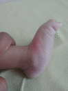
This clinical picture showing the rocket bottom appearance in a case of congenital vertical talus Contributed by Prateek Behera, MS
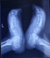
A radiograph of a child with congenital vertical talus Contributed by Prateek Behera, MS
References
- 1.
Pannier S. Congenital pseudarthrosis of the tibia.
Orthop Traumatol Surg Res. 2011 Nov;97(7):750-61. [
PubMed: 21996526]
- 2.
Mahnken AH, Staatz G, Hermanns B, Gunther RW, Weber M. Congenital pseudarthrosis of the tibia in pediatric patients: MR imaging.
AJR Am J Roentgenol. 2001 Nov;177(5):1025-9. [
PubMed: 11641162]
- 3.
Boyd HB. Pathology and natural history of congenital pseudarthrosis of the tibia.
Clin Orthop Relat Res. 1982 Jun;(166):5-13. [
PubMed: 7083685]
- 4.
Hefti F, Bollini G, Dungl P, Fixsen J, Grill F, Ippolito E, Romanus B, Tudisco C, Wientroub S. Congenital pseudarthrosis of the tibia: history, etiology, classification, and epidemiologic data.
J Pediatr Orthop B. 2000 Jan;9(1):11-5. [
PubMed: 10647103]
- 5.
McClure PK, Franzone JM, Herzenberg JE. Congenital Pseudarthrosis of the Tibia Associated With Cleidocranial Dysostosis: Case Report and Literature Review.
JBJS Case Connect. 2021 Nov 04;11(4) [
PubMed: 34735385]
- 6.
Ohnishi I, Sato W, Matsuyama J, Yajima H, Haga N, Kamegaya M, Minami A, Sato M, Yoshino S, Oki T, Nakamura K. Treatment of congenital pseudarthrosis of the tibia: a multicenter study in Japan.
J Pediatr Orthop. 2005 Mar-Apr;25(2):219-24. [
PubMed: 15718906]
- 7.
Khan T, Joseph B. Controversies in the management of congenital pseudarthrosis of the tibia and fibula.
Bone Joint J. 2013 Aug;95-B(8):1027-34. [
PubMed: 23908415]
- 8.
Dobbs MB, Rich MM, Gordon JE, Szymanski DA, Schoenecker PL. Use of an intramedullary rod for the treatment of congenital pseudarthrosis of the tibia. Surgical technique.
J Bone Joint Surg Am. 2005 Mar;87 Suppl 1(Pt 1):33-40. [
PubMed: 15743845]
- 9.
Paley D, Catagni M, Argnani F, Prevot J, Bell D, Armstrong P. Treatment of congenital pseudoarthrosis of the tibia using the Ilizarov technique.
Clin Orthop Relat Res. 1992 Jul;(280):81-93. [
PubMed: 1611768]
- 10.
El-Rosasy MA. Congenital pseudarthrosis of the tibia: the outcome of a pathology-oriented classification system and treatment protocol.
J Pediatr Orthop B. 2020 Jul;29(4):337-347. [
PubMed: 31503102]
Disclosure: Udit Agrawal declares no relevant financial relationships with ineligible companies.
Disclosure: Vivek Tiwari declares no relevant financial relationships with ineligible companies.