U.S. flag

An official website of the United States government

NCBI Bookshelf. A service of the National Library of Medicine, National Institutes of Health.

Arechavala-Gomeza V, Garanto A, editors. Antisense RNA Design, Delivery, and Analysis [Internet]. New York: Humana; 2022. doi: 10.1007/978-1-0716-2010-6_13

Cover of Antisense RNA Design, Delivery, and Analysis

Antisense RNA Design, Delivery, and Analysis [Internet].

Show details

Chapter 13Rapid Determination of MBNL1 Protein Levels by Quantitative Dot Blot for the Evaluation of Antisense Oligonucleotides in Myotonic Dystrophy Myoblasts

, , , and .

Author Information and Affiliations

Published online: February 25, 2022.

Western blot assays are not adequate for high-throughput screening of protein expression because it is an expensive and time-consuming technique. Here we demonstrate that quantitative dot blots in plate format are a better option to determine the absolute contents of a given protein in less than 48 h. The method was optimized for the detection of the Muscleblind-like 1 protein in patient-derived myoblasts treated with a collection of more than 100 experimental oligonucleotides.

Key words:

Myotonic dystrophy, Oligonucleotides, Quantitative dot blot, Muscleblind-like protein 1, DM1 myoblastsMyoblasts

1. Introduction

Myotonic dystrophy type 1 (DM1) is a degenerative genetic disease that is classified as rare because it affects less than 1 in 2000 people (1/3000 to 1/8000; [1]). DM1 originates from an expansion of the CTG trinucleotide repeat in the 3′-untranslated region (UTR) of the DMPK gene that, upon transcription, forms CUG hairpins that behave as toxic RNAs. CUG expansion RNA aberrantly binds and sequesters essential developmental proteins of the Muscleblind-like (MBNL) family, which are key regulators of alternative splicing. The depletion in MBNL protein function causes alterations in RNA metabolism that originate defined symptoms of the disease [2]. Studies in animal models have shown that the increase of MBNL in a genetic background of DM improves the pathological phenotypes and that the overexpression of MBNL1 in control mice is well tolerated [3].

Patients suffer from myotonia and muscle atrophy and weakness, which, in advanced stages of the disease, lead to respiratory distress and early death. Currently, there is no effective treatment for DM1, and management of symptoms is the only option to preserve the quality of life of people living with DM1. In its most common form, the onset of symptoms occurs during adolescence, and affected people have a significantly shortened lifespan of 48–55 years. Because of its prevalence and the severity of the clinical manifestations, finding a cure for DM1 is a social and medical need [4, 5].

The development of effective high-throughput tools in drug discovery research has increased the demand for complementary high capacity immunoblot methods in which to assess the consequences of drug candidates at protein level. One example is the need to quickly evaluate the levels of MBNL1 protein in patient-derived myoblasts [6] treated with hundreds of oligonucleotide variants to block repressive miRNAs miR-23b and miR-218, as a means to boost endogenous levels and compensate sequestration by CUG expansions in mutant DMPK [7]. To this end, we have generated a diversity (>100) of highly modified antisense oligonucleotides (AONs) to block miR-23b and miR-218. Examples of these modifications are the substitution of natural ribose rings by locked nucleic acid (LNAs), C2′ hydroxyl substitutions with a methoxy (2′OMe) or methoxyethyl (2′-MOE ), or the use of phosphorothioates to link two nucleotides, instead of natural phosphodiester bonds, to improve stability in vivo as they make these ASOs resistant to intracellular and extracellular nucleases. AON can also be made electrostatically neutral by using phosphorodiamidate morpholino oligomers (PMO) and can be conjugated to a cholesterol moiety to improve the diffusion through cell membranes and cell uptake [8, 9].

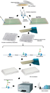
For this purpose, we propose the use of quantitative dot blot (QDB) analysis as an alternative to Western blot. For the development of the QDB assay, we have modified two previously published protocols [10, 11]. Dot blot was developed to simplify the process of Western blot analysis (Fig. 1) when the antibody is very specific for the detection of the protein of interest, and there is no need to determine its molecular weight, for example, when screening the effects of several molecules on the expression of a single protein. Specifically, QDB transforms traditional immunoblots into quantitative assays and allows expression analysis of a certain protein in your samples in 96-well format, being more efficient and faster than a Western blot.

Fig. 1

Fig. 1

Illustration of the entire QDB process for evaluation of antisense oligonucleotides in DM1 myoblast

2. Materials

2.1. Cell Culture and Transfection

1.

Standard tissue culture facilities.

2.

Six-well plates.

3.

PBS 1×.

4.

Opti-MEM Reduced Serum medium.

5.

Transfection Reagent (see Note 1).

6.

Complete medium (for 500 mL): 445 mL DMEM Dulbecco’s Modified Eagle Medium 4500 mg/glucose, 50 mL FBS, and 5 mL penicillin-streptomycin.

7.

Differentiation medium (for 100 mL): 1 mL penicillin-streptomycin, 2 mL Horse Serum, 1 mL apotransferrin, 100 μL insulin, 20 μL doxiciclin and 95.88 mL DMEM 4500 mg/glucose.

2.2. QDB Assay

1.

Protein quantification kit (see Note 2).

2.

QDB plates (Quanticision Diagnostics, Inc.).

3.

Primary and secondary antibodies:

(a)

Anti-MBNL1 (Abcam, ab77017).

(b)

Anti-GAPDH (Santa Cruz Biotechnology, G-9: sc-365062).

(c)

Anti-mouse POD.

4.

Pierce™ ECL Western blotting substrate.

5.

White 96-well plate, flat bottom.

6.

TECAN infinite M200 Pro.

7.

Orbital shaker.

8.

Transfer buffer: 14.4 g glycine, 3 g Tris base, 700 mL ddH2O, 200 mL methanol and fill to 1 L with ddH2O. Store at 4 °C.

9.

10× TBS: 24.2 g Tris base, 80 g NaCl, bring to 0.8 L with ddH2O, adjust the pH to 7.6 with HCl and fill to 1 L with ddH2O. Store at 4 °C.

10.

1× TBST: 100 mL 10× TBS, 5 mL Tween 20 and fill to 1 L with ddH2O. Store at 4 °C.

11.

Blocking buffer: 5 g milk powder and bring to 100 mL with TBST 1×. Store at 4 °C.

12.

4× Loading buffer: 0.8 mL 3 M Tris-HCl pH 6.8, 4 mL glycerol, 0.8 g SDS, 0.04 g bromophenol blue, 4 mL β-mercaptoethanol and fill to 10 mL with ddH2O. Store at −20 °C.

13.

RIPA solution: 10 mL RIPA and 1 tablet of protease inhibitor cocktail. Storage in 4 °C.

3. Methods

3.1. Cell Transfection

1.

Seed cells in 6-well plates at a density of 200,000 cells per well in complete medium and are incubated for 24 h at 37 °C 5% CO2.

2.

Prepare the mixes for transfection. Volumes in this example are calculated for 3 wells of a 6-well plate and in 5 different concentrations in the range between 10 and 5000 nM, diluted in Opti-MEM in a sterile tube in a final volume of 600 μL. For example, to transfect AO at 200 μM mix 594 μL of Opti-MEM and add 6 μL of AO at 100 μM. To test other concentrations, adapt the volumes accordingly. Pipet gently to mix.

3.

Add 3 μL of X-tremeGENE HP DNA transfection reagent to the diluted mix. Incubate for 30 min at RT while preparing the following step.

4.

Remove medium from wells and wash twice with PBS 1×.

5.

Add 200 μL of transfection mix to each well in a dropwise manner. Swirl the wells to ensure the distribution over the entire plate.

6.

Leave the cells in the incubator for 4 h.

7.

Add 2.3 mL of differentiation medium per well.

3.2. Sample Collection and Quantification

1.

After 72 h of incubation of the DM1 myoblasts with the test oligonucleotides, wash each well with PBS before protein collection.

2.

Collect cells in RIPA solution (120 μL/well).

3.

Lyse cells with an ultrasonic homogenizer such as the UP100H. Sonication is at an ultrasonic cycle mode of 30 s and an amplitude of 60%. The sample must be kept in ice during the entire procedure.

4.

Centrifuge the lysate at 16,000 × g for approximately 15 min after ultrasonic homogenization/extraction.

5.

After centrifuging, collect the supernatant and determine the protein concentration by a protein assay such as Pierce protein assay BCA.

3.3. Sample Preparation

1.

Calculate enough protein extract for ten replicate wells at 1 μg/well to account for pipetting errors, as they will be later loaded in quadruplicates in two different plates: one for detection of MBNL1 and the other for GAPDH, as an endogenous control (see Note 3).

2.

Add the protein extract at the indicated concentration, add 10.4 μL of loading buffer 4× and complete to a final volume of 50 μL of ddH2O (see Note 4).

3.

Boil the freshly prepared sample for 5 min in a water bath and after denaturation of the protein leave it on ice.

3.4. Sample Application and Transference

1.

Place the plates upside down to load the samples. Pipette 5 μL of the previously prepared protein sample on each membrane circle (Fig. 2).

2.

Dry loaded QDB plates for 30 min at room temperature in a well-ventilated space.

3.

After drying, dip the QDB plate in transfer buffer for 1 min. During this minute, shake it with an orbital movement until the blue color in the wells is eliminated.

4.

Rinse QDB plates gently with TBST in constant shaking for 1 min three times.

5.

Block QDB plates with blocking buffer for 1 h with constant shaking.

Fig. 2. A photograph of a QDB plate upside down.

Fig. 2

A photograph of a QDB plate upside down. The figure shows the different components of the plate and how to load the sample

3.5. Primary Antibody Incubation

1.

Dilute primary antibodies in blocking buffer at the appropriate concentrations (anti-MBNL1: 1/1000 and anti-GAPDH: 1/500) and aliquot into the wells of two 96-well plates, one for each antibody, at 100 μL/well (see Note 5).

2.

Insert each QDB plate into the 96-well plates and incubate overnight at 4 °C in constant shaking.

3.

Rinse the plates briefly with TBST and then wash with TBST in constant shaking for 5 min, three times.

3.6. Secondary Antibody Incubation

1.

Dilute the appropriate secondary antibody (anti-mouse POD) in blocking buffer at the final concentration of 1/2000 and aliquot it in the 96-well plates at 100 μL/well. QDB plates are inserted into the 96-well plates and incubated in constant shaking for 2 h.

2.

Rinse QDB plates gently three times with TBST and then wash with TBST in constant shaking for 5 min, three times.

3.7. Quantification

1.

Before the last washes, prepare the ECL substrate by following the manufacturer’s instructions.

2.

Aliquot 50 μL of ECL substrate into each well of a 96-well plate, and insert the QDB plate inside the 96-well plates for 1 min.

3.

After a minute, remove the QDB plate from the 96-well plate and shake it briefly to remove the remaining reagent and introduce it into a clean white 96-well plate.

4.

For data acquisition, use a TECAN infinite M200 Pro using an initial acquisition time of 1 s/well in the luminescence and select “plate with cover.”

3.8. Data Analysis

1.

First, look at the negative control values, which have to be below the rest of the samples. Pay also attention to the integrity of the plate since any alteration of the membrane in one of the wells may lead to artifactually low chemiluminescence values.

2.

Next, subtract the average value of the negative controls from all the samples, thus removing the contribution of the background to the quantification.

3.

Obtain the average of the quadruplicates for both the MBNL1 and the GAPDH plates. Divide the average of the replicates of the MBNL1 samples by those of GAPDH.

4.

GAPDH normalized values can be further normalized to values in mock-transfected DM1 cells to obtain fold changes.

5.

As a positive control, confirm in each plate that the difference between the average values of MBNL1 in control and untreated DM1 cells is around 1.6 times.

4. Notes

1.

Could be done with any transfection reagent, but we have tested only X-tremeGENE HP DNA Transfection Reagent.

2.

Could be done with any protein quantification kit, but we have tested only PierceTM BCA Protein Assay kit.

3.

Always include positive and negative controls in each of the plates. Our positive controls were healthy cell samples while the negative contained RIPA buffer only. Negative control wells provide the background luminescence reading, which must be significantly lower than our experimental data.

4.

It is very important to prepare the protein and load the samples in the gas hood since β-mercaptoethanol is toxic by inhalation.

5.

To ensure a valid result, the specificity of the antibody used needs to be verified through Western blot analysis .

Acknowledgments

The project leading to these results has received funding from “la Caixa” Banking Foundation under the project code HR17-00268 (TATAMI project to R.A.). I.G.-M. was funded by the Precipita Project titled “Desarrollo de una terapia innovadora contra la distrofia miotónica”; E.C.-H. was supported by the post-doctoral fellowship APOSTD/2019/142; and N.M.-C was supported by the pre-doctoral fellowship PRE2019-090622. Part of the equipment employed in this work has been funded by Generalitat Valenciana and cofinanced with ERDF funds (OP ERDF of Comunitat Valenciana 2014-2020).

References

1.
Ashizawa T, Gagnon C, Groh WJ, Gutmann L, Johnson NE, Meola G, Moxley R III, Pandya S, Rogers MT, Simpson E, Angeard N, Bassez G, Berggren KN, Bhakta D, Bozzali M, Broderick A, Byrne JLB, Campbell C, Cup E, Day JW, De Mattia E, Duboc D, Duong T, Eichinger K, Ekstrom AB, van Engelen B, Esparis B, Eymard B, Ferschl M, Gadalla SM, Gallais B, Goodglick T, Heatwole C, Hilbert J, Holland V, Kierkegaard M, Koopman WJ, Lane K, Maas D, Mankodi A, Mathews KD, Monckton DG, Moser D, Nazarian S, Nguyen L, Nopoulos P, Petty R, Phetteplace J, Puymirat J, Raman S, Richer L, Roma E, Sampson J, Sansone V, Schoser B, Sterling L, Statland J, Subramony SH, Tian C, Trujillo C, Tomaselli G, Turner C, Venance S, Verma A, White M, Winblad S (2018) Consensus-based care recommendations for adults with myotonic dystrophy type 1. Neurol Clin Pract 8(6):507–520. https://doi​.org/10.1212/CPJ​.0000000000000531. [PMC free article: PMC6294540] [PubMed: 30588381] [CrossRef]
2.
Jiang H, Mankodi A, Swanson MS, Moxley RT, Thornton CA (2004) Myotonic dystrophy type 1 is associated with nuclear foci of mutant RNA, sequestration of muscleblind proteins and deregulated alternative splicing in neurons. Hum Mol Genet 13(24):3079–3088. https://doi​.org/10.1093/hmg/ddh327. [PubMed: 15496431] [CrossRef]
3.
Chamberlain CM, Ranum LP (2012) Mouse model of muscleblind-like 1 overexpression: skeletal muscle effects and therapeutic promise. Hum Mol Genet 21(21):4645–4654. https://doi​.org/10.1093/hmg/dds306. [PMC free article: PMC3471398] [PubMed: 22846424] [CrossRef]
4.
Landfeldt E, Nikolenko N, Jimenez-Moreno C, Cumming S, Monckton DG, Gorman G, Turner C, Lochmuller H (2019) Disease burden of myotonic dystrophy type 1. J Neurol 266(4):998–1006. https://doi​.org/10.1007​/s00415-019-09228-w. [PMC free article: PMC6420885] [PubMed: 30788616] [CrossRef]
5.
Mathieu J, Allard P, Potvin L, Prevost C, Begin P (1999) A 10-year study of mortality in a cohort of patients with myotonic dystrophy. Neurology 52(8):1658–1662. [PubMed: 10331695] [CrossRef]
6.
Arandel L, Polay Espinoza M, Matloka M, Bazinet A, De Dea DD, Naouar N, Rau F, Jollet A, Edom-Vovard F, Mamchaoui K, Tarnopolsky M, Puymirat J, Battail C, Boland A, Deleuze JF, Mouly V, Klein AF, Furling D (2017) Immortalized human myotonic dystrophy muscle cell lines to assess therapeutic compounds. Dis Model Mech 10(4):487–497. https://doi​.org/10.1242/dmm.027367. [PMC free article: PMC5399563] [PubMed: 28188264] [CrossRef]
7.
Cerro-Herreros E, Sabater-Arcis M, Fernandez-Costa JM, Moreno N, Perez-Alonso M, Llamusi B, Artero R (2018) miR-23b and miR-218 silencing increase Muscleblind-like expression and alleviate myotonic dystrophy phenotypes in mammalian models. Nat Commun 9(1):2482. https://doi​.org/10.1038​/s41467-018-04892-4. [PMC free article: PMC6018771] [PubMed: 29946070] [CrossRef]
8.
Crooke ST, Witztum JL, Bennett CF, Baker BF (2019) RNA-targeted therapeutics. Cell Metab 29(2):501. https://doi​.org/10.1016/j​.cmet.2019.01.001. [PubMed: 30726759] [CrossRef]
9.
Smith CIE, Zain R (2019) Therapeutic oligonucleotides: state of the art. Annu Rev Pharmacol Toxicol 59:605–630. https://doi​.org/10.1146​/annurev-pharmtox-010818-021050. [PubMed: 30285540] [CrossRef]
10.
Tian G, Tang F, Yang C, Zhang W, Bergquist J, Wang B, Mi J, Zhang J (2017) Quantitative dot blot analysis (QDB), a versatile high throughput immunoblot method. Oncotarget 8(35):58553–58562. https://doi​.org/10.18632/oncotarget​.17236. [PMC free article: PMC5601674] [PubMed: 28938578] [CrossRef]
11.
Qi X, Zhang Y, Zhang Y, Ni T, Zhang W, Yang C, Mi J, Zhang J, Tian G (2018) High throughput, absolute determination of the content of a selected protein at tissue levels using quantitative dot blot analysis (QDB). J Vis Exp (138):56885. https://doi​.org/10.3791/56885. [PMC free article: PMC6231693] [PubMed: 30199009]

Nerea Moreno and Irene González-Martínez contributed equally to this work.

Copyright 2022, The Author(s)

Open Access This chapter is licensed under the terms of the Creative Commons Attribution 4.0 International License (http://creativecommons.org/licenses/by/4.0/), which permits use, sharing, adaptation, distribution and reproduction in any medium or format, as long as you give appropriate credit to the original author(s) and the source, provide a link to the Creative Commons license and indicate if changes were made.

The images or other third party material in this chapter are included in the chapter's Creative Commons license, unless indicated otherwise in a credit line to the material. If material is not included in the chapter's Creative Commons license and your intended use is not permitted by statutory regulation or exceeds the permitted use, you will need to obtain permission directly from the copyright holder.


Bookshelf ID: NBK584210DOI: 10.1007/978-1-0716-2010-6_13

Views

  • PubReader
  • Print View
  • Cite this Page
  • PDF version of this page (619K)

Related information

  • PMC
    PubMed Central citations
  • PubMed
    Links to PubMed

Similar articles in PubMed

See reviews...See all...

Recent Activity

Your browsing activity is empty.

Activity recording is turned off.

Turn recording back on

See more...