The desmoplastic reaction is a prominent pathological characteristic of pancreatic cancer. Desmoplasia is marked by a dramatic increase in the proliferation of alpha-smooth muscle actin- positive fibroblasts and is also accompanied by increased deposition of many extracellular matrix components. Changes in stromal cell proliferation and the deposition of extracellular matrix components result in dramatic changes in overall tissue heterogeneity and elasticity, as well as accompanying interstitial fluid pressure. These changes have been suggested to contribute to chemoresistance in cancer. Chemoresistance brought about by desmoplasia has both biological and physiological causes and consequences. In this chapter, we discuss some of the origins of desmoplasia in pancreatic cancer, and how it might be contributing to resistance to current chemotherapeutic interventions.
Introduction
Pancreatic ductal adenocarcinoma (PDAC) stands as the fourth leading cause of cancer-related death in the United States, with a 5-year survival rate of less than 5% (1). Factors contributing to this poor prognosis include both intrinsic and acquired chemoresistance to the current first-line therapy of choice, gemcitabine. There are multiple factors which contribute to chemoresistance observed in pancreatic cancer. Among them, desmoplasia and the tumor microenvironment (TME) are increasingly seen as major contributors to chemoresistance in PDAC. Since Paget’s seminal ‘seed’ and ‘soil’ hypothesis was put forward in order to reconcile measureable differences in the frequency of distal sites of metastasis, the capacity of the ‘soil,’ or tumor microenvironment, to promote tumor cell growth has been increasingly recognized (2). The tumor microenvironment, or stroma, is home to many components, both cellular and molecular. Unfortunately, the contribution of the stroma to disease in patients is not fully understood in pancreatic cancer (3). Desmoplasia, which is a result of the proliferation of alpha-smooth muscle actin-positive fibroblasts (also known as the myofibroblast, or activated pancreatic stellate cell (PSC)) and increased deposition of extracellular matrix (ECM) components, leads to reduced elasticity of tumor tissue with a concomitant increase in tumor interstitial fluid pressure (IFP). Increased IFP results in a decreased rate of perfusion of therapeutic agents and consequently decreased efficacy (4). This physiological chemoresistance has been shown to be a major contributor to the reduced efficacy of chemotherapeutics in multiple tumor types (4). In addition, desmoplasia can result in multiple signaling cascades that increase biological chemoresistance to therapeutic agents. Thus, targeting components of the tumor stroma that contribute to desmoplasia, in combination with agents directly targeting the tumor cells, is gaining traction as a potential approach for overcoming resistance and improving efficacy (5). In this chapter, we will review some of the general concepts of desmoplasia and chemoresistance, as well as how desmoplasia might be contributing to chemoresistance in pancreatic cancer.
Desmoplasia
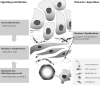
Desmoplasia, which is also referred to as the desmoplastic reaction, is a fundamental characteristic of PDAC. Originating from the Greek words desmos meaning “band” or “fastening” and plassein which is to “form” or “mold,” desmoplasia as we know clinically manifests itself in two ways: (1) significant overproduction of extracellular matrix proteins, and (2) extensive proliferation of myofibroblast-like cells (6). The resulting dense and fibrous connective tissue therefore is comprised of both cellular and non-cellular components (Figure 1). The cellular components of desmoplasia can include, among others, stellate cell-derived myofibroblast-like cells and an assortment of infiltrating immune cell types. Of the non-cellular components, multiple extracellular matrix (ECM) proteins, including collagen types I, III, and IV, fibronectin, laminin, hyaluronan, as well as the glycoprotein osteonectin (also secreted protein, acidic and rich in cysteine, or SPARC), have been identified (7, 8). Desmoplasia results from multiple intercellular and intracellular biological signaling events (see Figure 1). Reports have demonstrated that transforming growth factor β (TGFβ), basic fibroblast growth factor (FGF2) connective tissue growth factor (CTGF), and interleukin-1β (IL-1β) each stimulate ECM production. Platelet-derived growth factor (PDGF), however, has been shown to stimulate proliferation of the myofibroblast-like cell population (3). Importantly, both the cellular and non-cellular components of desmoplasia contribute to the pathogenesis of pancreatic cancer.
Cellular components
Among the multitude of cell types found within the tumor stroma, the pancreatic stellate cell is the most proliferative and is the primary synthetic source of many of the extracellular matrix components in the tumor microenvironment (6, 9). As this topic was also discussed in Chapter 1, the role of the PSC in desmoplasia will only be covered briefly here. In its quiescent state, the pancreatic stellate cell is positive for vitamin A-containing lipid droplets and serves as a reservoir for vitamin A in the normal pancreas. Its activation from a quiescent to an activated state, including changes to its proliferation rate, morphology, and sensitivity to mitogenic factors, are all primary features of PDAC. While the activation process is not yet fully understood, many of the signaling events leading to stellate cell activation have been identified. Following activation, the PSC loses its lipid droplets, undergoes changes to its morphology, and upregulates alpha smooth muscle actin (3). Intercellular signaling originating from multiple cell types, including tumor cells and immune cells, contribute to this increased activation and proliferation of PSCs in the desmoplastic reaction.
Tumor-associated macrophages
Rudolf Virchow first observed in 1863 that leukocytes could infiltrate tumor tissues, suggesting a possible connection between inflammation and cancer (10). In his study, he suggested that chronic inflammation may in fact lead to the development of cancer. In diseased tissue, the immune response leading to inflammation can stall and become locked in a chronic state that fosters fibrotic conditions which may stimulate tumorigenesis and tumor progression, including increased genomic instability and increased secretion of growth factors. It is generally accepted that the reactive oxygen species (ROS) generated by phagocytic cells, including both neutrophils and macrophages, can contribute to multiple types of genotoxicity, including DNA strand breaks or DNA base modifications. Many of the inflammatory cells that infiltrate tumors often localize primarily to the tumor stroma, including tumor-associated macrophages (TAM), neutrophils, and regulatory T cells (Treg) (11). TAMs are among the most populous immune cell type to infiltrate into the tumor microenvironment (12). TAMs are recruited to tumor sites by chemoattractants, including the CC chemokines CCL2, 3, 4, 5, and 8. Also, TAMs can be recruited via VEGF, colony stimulating factor 1 (CSF-1), stromal-derived factor 1 (SDF-1), and thrombospondin-1 (TSP-1) (13-15). Once guided to the tumor and activated, macrophages secrete various growth factors and cytokines into the immediate tumor microenvironment, indirectly contributing to an altered stromal environment and enhanced desmoplasia (8). For example, once activated, TAMs are known to express the most potent mediator of PSC proliferation, PDGF, as well as to release ROS (16). In addition, macrophages also have the capacity to stimulate the synthesis of collagen type I and fibronectin in PSC via the release of TGFβ (17, 18). Ultimately, infiltration and localization of leukocytes to the stromal compartment vastly alters its properties, and leads to increased desmoplasia.
Neutrophils and regulatory T cells
Two additional cell types thought to contribute to desmoplasia in pancreatic cancer include the neutrophils and regulatory T (Treg) cells. While the neutrophils, along with the TAMs can function as phagocytes at sites of inflammation, they act as the first responders in acute inflammatory responses with the release of toxic granules that are a significant source of ROS. ROS are a significant source of genomic instability and indeed have been shown to promote carcinogenesis in pancreatic ductal epithelial cells (19, 20). However, ROS can also directly result in the activation of PSCs. Furthermore, ROS can stimulate the spontaneous, non-enzymatic activation of TGFβ in vivo (21). By release of ROS, therefore, neutrophils may be contributing to the mobilization of TGFβ signaling. In addition, Nozawa et al. suggest that infiltrating neutrophils mediate the angiogenic switch and ECM remodeling through their expression of MMP-9 (22). In this way, neutrophils may contribute to an altered stromal compartment supportive of tumor progression. Treg cells play a significant role in the immune response to cancer. While Treg cells do not necessarily induce carcinogenesis or mutagenesis, they play an important role in suppressing an effective response that the immune system may attempt to mount. Reports have demonstrated that the prevalence of Treg cells is increased in patients with PDAC, suggesting that they may play a significant role in suppressing an immune response in PDAC (23, 24). In addition, Treg cells express and release multiple factors, including TGFβ, into the tumor microenvironment and have been shown to exhibit potent anti-inflammatory effects and participate in suppressing auto-immunity (25-28). The Treg cell population, which is defined as CD4+CD25+Foxp3+, expresses both active and latent TGFβ at it its cell surface that is absent in CD4+CD25- cells (31). Though it does not appear that TGFβ expression is entirely necessary for its immunosuppressive effects, the release of Treg-derived TGFβ may further contribute to desmoplasia in PDAC following receptor binding on the PSC (18, 29).
The cellular components of desmoplasia include the PSCs and infiltrating immune cells. In its normal quiescent state, the pancreatic stellate cell functions in the metabolism of retinoids, the development of the pancreas, and in immune cell regulation by means of its antigen presentation abilities (30). In PDAC, however, proliferating stromal cells prevent the normal function of the pancreas. Utilizing various signaling mechanisms, both tumor and immune cells contribute to the dramatic proliferation of the stromal cells. In contrast, non-cellular components of desmoplasia arise from increased deposition of extracellular matrix components.
Non-cellular components
While desmoplasia is characterized by a marked increase in the proliferation of the myofibroblasts and the infiltration of multiple immune cell types, the deposition of ECM components contributes significantly to the pathogenic potential of the desmoplastic reaction. The non-cellular components of desmoplasia consist largely of the extracellular matrix components: the fibrous proteins (e.g. collagen) and the polysaccharide chain glycosaminoglycans (GAGs, including hyaluronan) (Table 1) (31-41). In the normal pancreas, the GAGs function structurally to support compressive forces on the tissue. Conversely, the fibrous proteins serve to support any tensile forces on the tissue (33). The various ECM components also serve to allow for proper diffusion of various molecules, including nutrients and hormones, at the bloodstream:tissue interface. In the diseased pancreas, the significant over-production of ECM components can be described as the failed resolution of a healing wound, which leads to fibrosis. TGFβ-mediated cell signaling has been recognized as the primary signaling mechanism for fibrogenesis (42). Here we will discuss some of the signaling mechanisms contributing to the increased deposition of the ECM components of desmoplasia and how they might contribute to the pathogenesis of PDAC.
Table 1.
Extracellular matrix components and their contribution to chemoresistance.
Immunohistochemical analyses of PDAC have reported increased expression of collagen, including types I, III, and IV, localizing to the extracellular space (43-45). Collagen type I is composed of two alpha1 and one alpha2 protein subunits which are encoded by the COL1A1 and COL1A2 genes, respectively (46). Following translation of pro-collagen peptide chains, the peptides are processed in the endoplasmic reticulum before being secreted via the Golgi apparatus. Subsequent processing by extracellular proteases allows the collagen to coalesce into fibrils (on the order of 1μm in diameter) and collagen fibers (up to 10μm in diameter) (33). The natural arrangement of crosslinking within the collagen fibrils makes the collagen fibers highly resistant to tensile forces, and capable of dissipating significant deformation energy (47). Collagen expression is upregulated in response to TGFβ/Smad signaling and is a product of activated fibroblasts (3, 46). Importantly, collagen fibers can function in concert with the fibroblast-mediated tissue contraction to help in providing structural support in closing and healing wounds (42). In PDAC, this same function of collagen decreases tissue elasticity and increases interstitial fluid pressure, resulting in reduced drug perfusion.
Hyaluronan, a protein-free GAG, is also an important component of the ECM and contributes to tissue rigidity and decreased elasticity (34, 48). Hyaluronan (HA) accumulation within diseased tissue is the product of increased synthesis and is secreted by activated fibroblasts in pancreatic cancer (49). Although increased expression of hyaluronan synthases in tumor cells has been observed, the overall regulation of hyaluronan synthesis is not fully understood (50). HA maintains significant interaction with water molecules, and functions to preserve tissue hydration in the normal pancreas. However, in diseased tissue and with increased deposition of HA, interstitial edema can ensue. Consequently, interstitial fluid pressure increases, and the ability of the interstitia to conduct fluid is decreased (51). The lack of a functional lymphatic system in tumor tissue combines with this increased interstitial edema brought about by HA to significantly reduce the exchange of various molecules at the bloodstream:tissue interface, including chemotherapeutics. The significant accumulation of HA in PDAC is one characteristic feature of pancreatic cancer that contributes to the observed desmoplastic reaction, and a decrease of chemotherapeutic penetration.
The deposition of multiple ECM components in the desmoplastic reaction in PDAC is a characteristic that contributes significantly to the pathogenesis of tumors and ultimately to chemoresistance. Both the proliferation of the fibroblast cell compartment and the increased deposition of ECM components contribute to the desmoplastic reaction and subsequent chemoresistance. Importantly, these cellular and non-cellular components of desmoplasia contribute to biological and physiological forms of chemoresistance in PDAC.
Desmoplasia induced chemoresistance
Chemoresistance can occur by multiple mechanisms in cancer. It can arise from physiological barriers to drug absorption or penetration into target tissues, or from biological mechanisms within individual tumor cells which reduce the effectiveness at their intended site of action. Biological chemoresistance appears to arise as the result of three different general types of mechanisms: first, target cells can develop resistance to drug uptake; second, target cells can develop altered sensitivity to drugs at their intended targets through, for example, increased expression of anti-apoptotic proteins; and third, target cells can develop increased efflux of drugs, preventing the drugs from reaching their intended site of action (52). Physiological chemoresistance can include poor tissue vasculature or perfusion, which may result from increased interstitial fluid pressure and increased production of extracellular matrix proteins that arises from the desmoplastic reaction (53). Chemoresistance is a problem in pancreatic cancer due to the presence of both biological and physiological mechanisms. In this section, we will review some of these mechanisms, how desmoplasia can contribute to these forms of chemoresistance, and approaches to overcoming chemoresistance therapeutically.
Biological chemoresistance
Biological chemoresistance has been well documented in multiple tumor types. Reduced uptake of drugs is one mechanism by which cells resist chemotherapeutic activity. A well documented example of such an effect is seen in the resistance to such cancer chemotherapeutics as the cytotoxic folate analogs, including methotrexate. Mutations arising in either the folate binding protein, or the reduced folate transporter give way to decreased transport of methotrexate and subsequent chemoresistance (54). In pancreatic cancer, epigenetic changes in the epithelial tumor cells, resulting in reduced expression of the nucleoside transporter required for gemcitabine uptake, hENT1, has also been reported to contribute to chemoresistance (55). Increased efflux of drugs by target cells, including the expression of the ATP-binding cassette (ABC) efflux transporter family of proteins, is one way in which cells can exhibit chemoresistance to a number of therapeutics. Indeed, expression of ABC transporters such as MDR1 or MRP1-6 can evoke resistance to numerous hydrophobic natural-products, such as doxorubicin, vinblastine, and paclitaxel, and to the nucleoside analogs, respectively (56-58), by reducing intracellular drug concentrations. Both of these proteins are widely expressed in human cancers. Drug resistance has also been observed resulting from altered cellular sensitivity to drugs’ mechanisms of action. For example, increased expression of anti-apoptotic proteins such as the Bcl-2 family of proteins confers a growth advantage to tumor cells but also confers drug resistance (59). Furthermore, increased drug metabolism, increased repair of DNA damage, altered intracellular drug targets, and altered cell cycle checkpoints have all been identified as potential mechanisms of biological chemoresistance (57). The non-cellular components of desmoplasia contribute to the biological chemoresistance through multiple mechanisms, including stimulating the expression of ABC multidrug resistance transporters in tumor cells. One report demonstrated that the binding of HA to its receptor, CD44, resulted in a Stat-3-mediated increase in the expression of MDR1 in breast and ovarian tumor cell lines (60). Importantly, similar observations of induced chemoresistance by CD44 activity have also been reported in pancreatic cancer cell lines (61). In addition, HA interaction with CD44 has also been shown to activate the phosphoinositide 3-kinase/AKT signaling pathway. Activation of AKT signaling is upregulated in many cancers, resulting in phosphorylation of Bad, and downregulation of apoptosis (62-64). In this sense, desmoplasia and chemoresistance are tightly regulated and interwoven. Factors stimulating desmoplasia in fact result in chemoresistance on multiple levels, both biologically and physiologically.
Physiological chemoresistance
Physiological chemoresistance has also been well documented in cancer, though studies of its clinical relevance are made difficult by the lack of good models to study their effects. Physiological chemoresistance, also referred to as “host factors” by Gottesman (57), can develop following biological changes in tumor cells but arises largely as a consequence of extracellular changes that occur in the stromal compartment. Physiological chemoresistance can arise from, among others, increased interstitial fluid pressure (as a consequence of the desmoplastic reaction), increased extracellular matrix protein deposition afforded by the expansion of the stromal compartment, or from the development of a disorganized tumor vasculature that gives rise to poor tumor perfusion. The mechanisms that lead to some of these effects, including increases in IFP, are not well understood.
Tumor extracellular matrix components contribute significantly to physiological chemoresistance in cancer. Collagens I, III, and IV are secreted at high levels in PDAC. Tumor collagen content, specifically, has been shown to maintain an inverse relationship to that of macromolecule penetration. In one report it was demonstrated that a U87 (human glioblastoma) xenograft tumor containing high collagen content displayed a significantly decreased diffusion coefficient (0.87 versus 1.89 × 10⁻⁷ cm2 s-1) of IgG molecules compared to a LS174T (human colon adenocarcinoma) xenograft tumor containing little collagen (65). Further confirming the specific contribution of collagen, it was noted that collagenase treated U87 tumors showed diffusion coefficients similar to the untreated LS174T tumors with low collagen content. Similar effects have also been observed in pancreatic cancer. Diop-Frimpong and colleagues demonstrated that the collagen-reducing effects of the anti-hypertensive agent, losartan, resulted in a significant increase in the penetration of nanoparticles (66). Using an orthotopic xenograft mouse model, they demonstrated that the increased collagen content, and decreased penetration of nanoparticles, in their L3.6pl tumors corresponded to a significantly decreased response to doxorubicin treatment. Taken together, these results suggest that collagen content may be sufficient to predict the delivery of drugs to tumor, and to predict physiological resistance to therapy.
Hyaluronan has also been shown to contribute to physiological chemoresistance in cancer. As a component of the ECM, HA contributes to the structural rigidity of the network of ECM proteins and may act as a molecular sieve to molecules extravasating from the vasculature (67). In acting as a molecular sieve, HA is thought to reduce the penetration of chemotherapeutics in PDAC. There is, however, some debate as to the extent or role that HA may play in tissue diffusion. Indeed, it has been shown that tissue elasticity and hydraulic conductivity do not correlate with total tissue hyaluronan content (65). Further, it has been shown that depletion of tissue HA by hyaluronidase decreased the diffusion of large molecules, namely FITC-dextran (500kDa molecular weight) (34). However, additional studies have demonstrated that the molecular selectivity of the ECM is tightly regulated through variation of the ECM composition, and that this affects molecule penetration based on charge and size (68). Furthermore, it has been demonstrated that depletion of HA in a PC3 human prostate xenograft model both decreased tumor IFP and increased vascular area (69). These studies suggest that HA can indeed increase IFP in tumors, but also that its ability to elicit a decrease in the penetration of chemotherapeutics will vary depending on the charge and size characteristics of the drug used.
In their paper on the potential efficacy of hedgehog inhibitors in pancreatic cancer, Olive et al outlined several careful experiments demonstrating in a transgenic mouse model for PDAC that tumors arising spontaneously within the pancreas are in fact poorly vascularized, and show poor drug perfusion relative to transplanted xenograft tumors or normal pancreatic tissue (70). Known most widely for its role in embryonic development, the hedgehog pathway has also been shown to play important roles in angiogenesis. This angiogenic potential of the hedgehog pathway has been reported in ischemic limbs and in the cornea of adult mice, or in rat models of diabetic neuropathy (71, 72). However, as shown by Olive and colleagues, the clinical efficacy of the hedgehog inhibitors in treating PDAC and in increasing overall survival is working through a mechanism that is not anti-angiogenic, but rather potently pro-perfusion. The researchers demonstrated that the hedgehog signaling inhibition increased endothelial cell proliferation and decreased fibroblast proliferation. The results presented in this study indicate that increased perfusion and decreased fibrosis are potentially important in treating PDAC. Certainly, however, more thorough studies characterizing the role of hedgehog inhibition in both pancreatic tumor and stromal cells will be necessary and will help translate the findings into the clinic.
Mechanisms of chemoresistance arise from both physiological barriers and biological adaptations of the tumor epithelial cells. Considering the number of obstacles to effective and penetrating treatment, we propose that future regimens consider a multipronged approach to targeting pancreatic cancer. By targeting the pathways involved in desmoplasia, in combination with cytotoxic agents to inhibit tumor cell growth, we would anticipate increased efficacy resulting in greater survival in pancreatic cancer patients.
Potential therapeutic approaches for reducing desmoplasia induced chemoresistance
Many approaches have been pursued in hopes of increasing the efficacy of chemotherapeutics against tumor epithelial cells in pancreatic cancer. Unfortunately, the desmoplastic reaction may abrogate much of the therapeutic activity of test agents. Indeed, we have observed significant chemoresistance to many therapeutics by primary human fibroblasts isolated from pancreatic tumors, even in the absence of a supportive microenvironment and collagen or hyaluronan network (unpublished data). Therapeutics reducing the contribution of the desmoplastic reaction to chemoresistance are being actively pursued as a potential therapeutic approach (5).
Members of the TGFβ family of mediators have been implicated in the activation of the PSC. TGFβ proteins are multifunctional cytokines known to be involved in a host of cellular functions, including regulation of immune cell function, cell growth and differentiation, and in ECM production (73). The canonical TGFβ signaling involves the binding to TGFβ receptors (TGFβRI/II) and subsequent activation of downstream Smad-mediated transcription. Since Smad4/DPC4 is reportedly mutated or deleted in approximately 55% of pancreatic tumors, it is unclear mechanistically how TGFβ antagonists may offer clinical benefit (74). Indeed, studies have demonstrated the complex nature of TGFβ signaling, demonstrating both tumor-suppressive and tumor-promoting features of TGFβ signaling (75). Despite the complexity and multifunctional nature of the signaling pathways, recent studies have indicated that interventions aiming at TGFβ signaling can have therapeutic benefit, without the danger of the potential side-effects that may result from inhibition of its tumor suppressive activity (reviewed in 76). Indeed, both the TGFβ antagonist, SR2F, and a TGFβ-neutralizing antibody were reportedly administered in animals with few side effects (77, 78). These studies have demonstrated that TGFβ antagonism can prevent metastases, as well as prevent cerulein-induced fibrosis and TGFβ signaling (77, 79, 80). As it appears that TGFβR1 haploinsufficiency can itself significantly inhibit the development of fibrosis and progression of precancerous lesions in a mouse model for PDAC, efforts have been made to examine the effects of TGFβ inhibition on the secretion of ECM components in fibroblast cells (80, 81). Multiple studies taking different approaches have observed decreased ECM (fibronectin, collagen, and others) deposition by fibroblasts following inhibition of TGFβ-mediated signaling (80, 82, 83). While it is important to note that the authors commented on the potential importance of the TGFβR1/TGFβR2 ratio on downstream signaling and outcome, inhibition of TGFβ-mediated signaling may yet be a viable pathway for therapeutic intervention if, as Yingling and colleagues suggested, “patient populations in which the tumor-promoting role of TGFβ signaling predominates” are identified (76, 81). Furthermore, due to the important nature of TGFβ in the perpetuation of PSC activation, recent studies have focused on utilizing TGFβ antagonists in targeting pancreatic fibrosis as a result of chronic pancreatitis (84). The translation of these findings to the clinic could potentially make a significant impact on the treatment of patients with PDAC. As reduction in PSC load and fibrotic buildup comes as a result of TGFβ antagonism, we would hope to see significant increases in the penetration of drugs targeting tumor cells.
In addition to the TGFβ antagonists, the peroxisome proliferator- activated receptor γ (PPARγ) agonists are also being investigated for its anti- desmoplasia activity. PPARγ has been studied in a variety of conditions, but is most recognized in its ability to increase insulin sensitivity (85, 86). Currently, the PPARγ ligand troglitazone (also known as Rezulin) is used clinically as an oral antihyperglycemic agent for the management of type II diabetes. Other ligands for PPARγ include lysophosphatidic acid, a few specific prostaglandins, and the thiazolidinediones (87-89). Besides insulin biology, several groups have shown that PPARγ is also involved in, for example, cell differentiation, inflammation regulation, and fibrosis in the lung (90-92). Following activation, its anti-fibrotic capabilities appear to be mediated by its ability to inhibit TGFβ signaling, as is especially noted in its ability to inhibit differentiation of fibroblasts to myofibroblasts (93). By activating PPARγ via a natural or synthetic ligand, fibrosis can be reduced and clinical benefit achieved through inhibition of the stromal contribution made by myofibroblasts such as their increased collagen production and increased matrix metalloproteinase expression. Indeed, improved organ function and decreased fibrosis has been reported in steatohepatitis patients being treated with pioglitazone, a PPARγ agonist, for diabetes (94).
The target in the aforementioned approaches is clearly the stromal compartment, specifically the myofibroblast, and would function as part of a larger therapeutic regimen. In addition to these approaches, enzymatic degradation of ECM components has also been proposed. Though the collagenases have been shown to enhance the penetration of macromolecules, sensitivity to and stability in physiological pH may be a therapeutic hindrance (34). To our knowledge, no collagenase is yet clinically available. In contrast, hyaluronidase has been used clinically in multiple trials, and has been shown to enhance tumor sensitivity to chemotherapeutics, even in tumors deemed chemoresistant (95). Importantly, hyaluronidase treatment of cultured tumor cells has been shown to enhance the penetration of drug into cultured spheroid cells (96). Early clinical trials utilizing hyaluronidase employed a bovine form of the enzyme from which allergic reactions had been reported. Currently, a recombinant human hyaluronidase has been made available for early pilot clinical studies and has been shown not to induce significant allergic reactions (97). The availability of the human recombinant hyaluronidase is particularly exciting as many of the early clinical studies demonstrating increased chemotherapeutic efficacy of anti-tumor agents administered in combination with bovine hyaluronidase may be expanded upon. It is our hope that such agents will effect greater therapeutic outcomes for patients.
Conclusions
Desmoplasia is indeed a fundamental characteristic of pancreatic cancer that contributes significantly to its chemoresistance. Deposition of ECM proteins such as collagen and hyaluronan contributes physiologically to chemoresistance by mediating a decrease in macromolecular diffusion in pancreatic tissues. Their deposition also mediates biological signaling cascades that result in, for example, decreased apoptotic signaling by Bad. Therefore, targeting desmoplasia, which is the response of the tumor microenvironment to molecular cues from the epithelial cell compartment, constitutes a viable, combinatorial therapeutic strategy in pancreatic cancer. Whether through enzymatic digestion of ECM components, or inhibitors of myofibroblast activation, targeting desmoplasia in pancreatic cancer will likely improve outcomes for patients with pancreatic cancer where chemoresistance remains a significant hurdle to effective systemic treatment.
Acknowledgements
Research in the authors’ laboratories is supported by grants from the NIH/NCI (CA109552, CA140924, and CA095031), Stand Up to Cancer (SU2C), and the Katz Family Foundation.
References
- 1.
- Jemal A, Siegel R, Xu J, Ward E. CA Cancer J Clin. 2010;60:277. [PubMed: 20610543]
- 2.
- Paget S. Lancet. 1889;133:571.
- 3.
- Shimizu K. J Gastroenterol. 2008;43:823. [PubMed: 19012035]
- 4.
- Heldin CH, Rubin K, Pietras K, Ostman A. Nat Rev Cancer. 2004;4:806. [PubMed: 15510161]
- 5.
- Garber K. J Natl Cancer Inst. 2010;102:448. [PubMed: 20339135]
- 6.
- Yen TW, Aardal NP, Bronner MP, et al. Surgery. 2002;131:129. [PubMed: 11854689]
- 7.
- Bachem MG, Schneider E, Gross H, et al. Gastroenterology. 1998;115:421. [PubMed: 9679048]
- 8.
- Apte MV, Haber PS, Darby SJ, et al. Gut. 1999;44:534. [PMC free article: PMC1727467] [PubMed: 10075961]
- 9.
- Faouzi S, Le Bail B, Neaud V, et al. J Hepatol. 1999;30:275. [PubMed: 10068108]
- 10.
- Balkwill F, Mantovani A. Lancet. 2001;357:539. [PubMed: 11229684]
- 11.
- Negus RP, Stamp GW, Hadley J, Balkwill FR. Am J Pathol. 1997;150:1723. [PMC free article: PMC1858213] [PubMed: 9137096]
- 12.
- Solinas G, Germano G, Mantovani A, Allavena P. J Leukoc Biol. 2009;86:1065. [PubMed: 19741157]
- 13.
- Murdoch C, Giannoudis A, Lewis CE. Blood. 2004;104:2224. [PubMed: 15231578]
- 14.
- Coffelt SB, Hughes R, Lewis CE. Biochim Biophys Acta. 2009;1796:11. [PubMed: 19269310]
- 15.
- Martin-Manso G, Galli S, Ridnour LA, Tsokos M, Wink DA, Roberts DD. Cancer Res. 2008;68:7090. [PMC free article: PMC2562557] [PubMed: 18757424]
- 16.
- Ross R. Lancet. 1989;1:1179. [PubMed: 2566744]
- 17.
- Schmid-Kotsas A, Gross HJ, Menke A, et al. Am J Pathol. 1999;155:1749. [PMC free article: PMC1866993] [PubMed: 10550331]
- 18.
- Aoyagi Y, Oda T, Kinoshita T, et al. Br J Cancer. 2004;91:1316. [PMC free article: PMC2409911] [PubMed: 15365564]
- 19.
- Toyokuni S, Okamoto K, Yodoi J, Hiai H. FEBS Lett. 1995;358:1. [PubMed: 7821417]
- 20.
- Vaquero EC, Edderkaoui M, Pandol SJ, Gukovsky I, Gukovskaya AS. J Biol Chem. 2004;279:34643. [PubMed: 15155719]
- 21.
- Koli K, Myllarniemi M, Keski-Oja J, Kinnula VL. Antioxid Redox Signal. 2008;10:333. [PubMed: 17961070]
- 22.
- Nozawa H, Chiu C, Hanahan D. Proc Natl Acad Sci U S A. 2006;103:12493. [PMC free article: PMC1531646] [PubMed: 16891410]
- 23.
- Liyanage UK, Moore TT, Joo HG, et al. J Immunol. 2002;169:2756. [PubMed: 12193750]
- 24.
- Liyanage UK, Goedegebuure PS, Moore TT, et al. J Immunother. 2006;29:416. [PubMed: 16799337]
- 25.
- Li MO, Sanjabi S, Flavell RA. Immunity. 2006;25:455. [PubMed: 16973386]
- 26.
- Li MO, Wan YY, Sanjabi S, Robertson AK, Flavell RA. Annu Rev Immunol. 2006;24:99. [PubMed: 16551245]
- 27.
- Moore KW, de Waal Malefyt R, Coffman RL, O'Garra A. Annu Rev Immunol. 2001;19:683. [PubMed: 11244051]
- 28.
- Miyara M, Sakaguchi S. Trends Mol Med. 2007;13:108. [PubMed: 17257897]
- 29.
- Kullberg MC, Hay V, Cheever AW, et al. Eur J Immunol. 2005;35:2886. [PubMed: 16180248]
- 30.
- 31.
- Miyamoto H, Murakami T, Tsuchida K, Sugino H, Miyake H, Tashiro S. Pancreas. 2004;28:38. [PubMed: 14707728]
- 32.
- McKee TD, Grandi P, Mok W, et al. Cancer Res. 2006;66:2509. [PubMed: 16510565]
- 33.
- Alberts B, Bray D, Lewis J, Raff M, Roberts K, Watson J. Extracellular matrix of animals. Molecular Biology of the Cell. 3rd Ed. ed. New York: Garland Publishing; 1994. p. 971.
- 34.
- Magzoub M, Jin S, Verkman AS. FASEB J. 2008;22:276. [PubMed: 17761521]
- 35.
- Stern R. Semin Cancer Biol. 2008;18:275. [PubMed: 18485730]
- 36.
- Ricciardelli C, Sakko AJ, Ween MP, Russell DL, Horsfall DJ. Cancer Metastasis Rev. 2009;28:233. [PubMed: 19160015]
- 37.
- Marastoni S, Ligresti G, Lorenzon E, Colombatti A, Mongiat M. Connect Tissue Res. 2008;49:203. [PubMed: 18661343]
- 38.
- Kaspar M, Zardi L, Neri D. Int J Cancer. 2006;118:1331. [PubMed: 16381025]
- 39.
- Sethi T, Rintoul RC, Moore SM, et al. Nat Med. 1999;5:662. [PubMed: 10371505]
- 40.
- Mahadevan D, Von Hoff DD. Mol Cancer Ther. 2007;6:1186. [PubMed: 17406031]
- 41.
- Desai N, Trieu V, Damascelli B, Soon-Shiong P. Transl Oncol. 2009;2:59. [PMC free article: PMC2670572] [PubMed: 19412420]
- 42.
- Raghow R. Chest. 1991;99:61S. [PubMed: 1997279]
- 43.
- Imamura T, Iguchi H, Manabe T, et al. Pancreas. 1995;11:357. [PubMed: 8532652]
- 44.
- Mollenhauer J, Roether I, Kern HF. Pancreas. 1987;2:14. [PubMed: 3554225]
- 45.
- Linder S, Castanos-Velez E, von Rosen A, Biberfeld P. Hepatogastroenterology. 2001;48:1321. [PubMed: 11677955]
- 46.
- Verrecchia F, Mauviel A. Cell Signal. 2004;16:873. [PubMed: 15157666]
- 47.
- Buehler MJ. Proc Natl Acad Sci U S A. 2006;103:12285. [PMC free article: PMC1567872] [PubMed: 16895989]
- 48.
- Laurent TC, Fraser JR. FASEB J. 1992;6:2397. [PubMed: 1563592]
- 49.
- Hiltunen EL, Anttila M, Kultti A, et al. Cancer Res. 2002;62:6410. [PubMed: 12438225]
- 50.
- Itano N, Sawai T, Atsumi F, et al. J Biol Chem. 2004;279:18679. [PubMed: 14724275]
- 51.
- Levick JR. Q J Exp Physiol. 1987;72:409. [PubMed: 3321140]
- 52.
- Szakacs G, Paterson JK, Ludwig JA, Booth-Genthe C, Gottesman MM. Nat Rev Drug Discov. 2006;5:219. [PubMed: 16518375]
- 53.
- Minchinton AI, Tannock IF. Nat Rev Cancer. 2006;6:583. [PubMed: 16862189]
- 54.
- Longo-Sorbello GS, Bertino JR. Haematologica. 2001;86:121. [PubMed: 11224479]
- 55.
- Giovannetti E, Del Tacca M, Mey V, et al. Cancer Res. 2006;66:3928. [PubMed: 16585222]
- 56.
- Borst P, Evers R, Kool M, Wijnholds J. Biochim Biophys Acta. 1999;1461:347. [PubMed: 10581366]
- 57.
- Gottesman MM. Annu Rev Med. 2002;53:615. [PubMed: 11818492]
- 58.
- Higgins CF. Annu Rev Cell Biol. 1992;8:67. [PubMed: 1282354]
- 59.
- Hamacher R, Schmid RM, Saur D, Schneider G. Mol Cancer. 2008;7:64. [PMC free article: PMC2515336] [PubMed: 18652674]
- 60.
- Bourguignon LY, Peyrollier K, Xia W, Gilad E. J Biol Chem. 2008;283:17635. [PMC free article: PMC2427357] [PubMed: 18441325]
- 61.
- Hong SP, Wen J, Bang S, Park S, Song SY. Int J Cancer. 2009;125:2323. [PubMed: 19598259]
- 62.
- Fujita Y, Kitagawa M, Nakamura S, et al. FEBS Lett. 2002;528:101. [PubMed: 12297287]
- 63.
- Benitez A, Yates TJ, Lopez LE, Cerwinka WH, Bakkar AA, Lokeshwar VB. Cancer Res. 2011;71:4085. [PMC free article: PMC3117105] [PubMed: 21555367]
- 64.
- Mitsiades CS, Mitsiades N, Koutsilieris M. Curr Cancer Drug Targets. 2004;4:235. [PubMed: 15134532]
- 65.
- Netti PA, Berk DA, Swartz MA, Grodzinsky AJ, Jain RK. Cancer Res. 2000;60:2497. [PubMed: 10811131]
- 66.
- Diop-Frimpong B, Chauhan VP, Krane S, Boucher Y, Jain RK. Proc Natl Acad Sci U S A. 2011;108:2909. [PMC free article: PMC3041115] [PubMed: 21282607]
- 67.
- Stern R. Eur J Cell Biol. 2004;83:317. [PubMed: 15503855]
- 68.
- Lieleg O, Baumgartel RM, Bausch AR. Biophys J. 2009;97:1569. [PMC free article: PMC2749787] [PubMed: 19751661]
- 69.
- Thompson CB, Shepard HM, O'Connor PM, et al. Mol Cancer Ther. 2010;9:3052. [PubMed: 20978165]
- 70.
- Olive KP, Jacobetz MA, Davidson CJ, et al. Science. 2009;324:1457. [PMC free article: PMC2998180] [PubMed: 19460966]
- 71.
- Pola R, Ling LE, Silver M, et al. Nat Med. 2001;7:706. [PubMed: 11385508]
- 72.
- Kusano KF, Allendoerfer KL, Munger W, et al. Arterioscler Thromb Vasc Biol. 2004;24:2102. [PubMed: 15358602]
- 73.
- Naber HP, ten Dijke P, Pardali E. Curr Cancer Drug Targets. 2008;8:466. [PubMed: 18781893]
- 74.
- Hahn SA, Schutte M, Hoque AT, et al. Science. 1996;271:350. [PubMed: 8553070]
- 75.
- Wakefield LM, Roberts AB. Curr Opin Genet Dev. 2002;12:22. [PubMed: 11790550]
- 76.
- Yingling JM, Blanchard KL, Sawyer JS. Nat Rev Drug Discov. 2004;3:1011. [PubMed: 15573100]
- 77.
- Yang YA, Dukhanina O, Tang B, et al. J Clin Invest. 2002;109:1607. [PMC free article: PMC151015] [PubMed: 12070308]
- 78.
- Ruzek MC, Hawes M, Pratt B, et al. Immunopharmacol Immunotoxicol. 2003;25:235. [PubMed: 12784916]
- 79.
- Muraoka RS, Dumont N, Ritter CA, et al. J Clin Invest. 2002;109:1551. [PMC free article: PMC151012] [PubMed: 12070302]
- 80.
- Zion O, Genin O, Kawada N, et al. Pancreas. 2009;38:427. [PubMed: 19188864]
- 81.
- Adrian K, Strouch MJ, Zeng Q, et al. Cancer Res. 2009;69:9169. [PubMed: 19951995]
- 82.
- Mulsow JJ, Watson RW, Fitzpatrick JM, O'Connell PR. Ann Surg. 2005;242:880. [PMC free article: PMC1409881] [PubMed: 16327498]
- 83.
- Fu K, Corbley MJ, Sun L, et al. Arterioscler Thromb Vasc Biol. 2008;28:665. [PubMed: 18202322]
- 84.
- Nagashio Y, Ueno H, Imamura M, et al. Lab Invest. 2004;84:1610. [PubMed: 15502860]
- 85.
- Kobayashi M, Iwanishi M, Egawa K, Shigeta Y. Diabetes. 1992;41:476. [PubMed: 1318856]
- 86.
- Kellerer M, Kroder G, Tippmer S, et al. Diabetes. 1994;43:447. [PubMed: 7508875]
- 87.
- McIntyre TM, Pontsler AV, Silva AR, et al. Proc Natl Acad Sci U S A. 2003;100:131. [PMC free article: PMC140905] [PubMed: 12502787]
- 88.
- Forman BM, Tontonoz P, Chen J, Brun RP, Spiegelman BM, Evans RM. Cell. 1995;83:803. [PubMed: 8521497]
- 89.
- Shimizu K, Shiratori K, Hayashi N, Kobayashi M, Fujiwara T, Horikoshi H. Pancreas. 2002;24:184. [PubMed: 11854624]
- 90.
- Burgess HA, Daugherty LE, Thatcher TH, et al. Am J Physiol Lung Cell Mol Physiol. 2005;288:L1146. [PubMed: 15734787]
- 91.
- Buckingham RE. Hepatol Res. 2005;33:167. [PubMed: 16198619]
- 92.
- Hummasti S, Tontonoz P. Mol Endocrinol. 2006;20:1261. [PubMed: 16556736]
- 93.
- Sime PJ. J Investig Med. 2008;56:534. [PubMed: 18317437]
- 94.
- Belfort R, Harrison SA, Brown K, et al. N Engl J Med. 2006;355:2297. [PubMed: 17135584]
- 95.
- Baumgartner G, Gomar-Hoss C, Sakr L, Ulsperger E, Wogritsch C. Cancer Lett. 1998;131:85. [PubMed: 9839623]
- 96.
- Kohno N, Ohnuma T, Truog P. J Cancer Res Clin Oncol. 1994;120:293. [PubMed: 8126058]
- 97.
- Yocum RC, Kennard D, Heiner LS. J Infus Nurs. 2007;30:293. [PubMed: 17895809]
Publication Details
Author Information and Affiliations
Authors
Clifford J. Whatcott, 1 Richard G. Posner,1 Daniel D. Von Hoff,1 and Haiyong Han1.
Affiliations
Correspondence/Reprint request: Dr. Clifford J. Whatcott, Clinical Translational Research Division, The Translational Genomics Research Institute, Scottsdale, Arizona, 85259, USA. E-mail: gro.negt@ttoctahwcCopyright
Publisher
Transworld Research Network, Trivandrum (India)
NLM Citation
Whatcott CJ, Posner RG, Von Hoff DD, et al. Desmoplasia and chemoresistance in pancreatic cancer. In: Grippo PJ, Munshi HG, editors. Pancreatic Cancer and Tumor Microenvironment. Trivandrum (India): Transworld Research Network; 2012. Chapter 8.
