From: Intracellular Control of Cell-Cycle Events

NCBI Bookshelf. A service of the National Library of Medicine, National Institutes of Health.

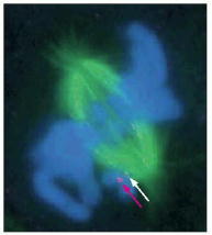
This fluorescence micrograph shows a mammalian cell in prometaphase, with the mitotic spindle in green and the sister chromatids in blue. One sister chromatid pair is not yet attached to the spindle. The presence of Mad2 on the kinetochore of the unattached chromosome is revealed by the binding of anti-Mad2 antibodies (red dot, indicated by red arrow). Another chromosome has just attached to the spindle, and its kinetochore has a low level of Mad2 still associated with it (pale dot, indicated by white arrow). (From J.C. Waters et al., J. Cell Biol. 141:1181–1191, 1998. © The Rockefeller University Press.)
From: Intracellular Control of Cell-Cycle Events

NCBI Bookshelf. A service of the National Library of Medicine, National Institutes of Health.