

NCBI Bookshelf. A service of the National Library of Medicine, National Institutes of Health.
Varki A, Cummings RD, Esko JD, et al., editors. Essentials of Glycobiology [Internet]. 3rd edition. Cold Spring Harbor (NY): Cold Spring Harbor Laboratory Press; 2015-2017. doi: 10.1101/glycobiology.3e.032


Essentials of Glycobiology [Internet]. 3rd edition.
Show details
The L-type lectins occur in the seeds of leguminous plants, and they have structural motifs that are present in a variety of glycan-binding proteins (GBPs) from other eukaryotic organisms. The structures of many of these lectins have been characterized, and many L-type lectins are used in a wide range of biomedical and analytical procedures. This chapter discusses the structure–function relationships of these lectins and the various biological roles they have in different organisms.
HISTORICAL BACKGROUND
The L-type lectins have a rich history going back to the end of the 19th century, when it was found that extracts from the seeds of leguminous plants (Fabaceae family but also called the Leguminosae family) could agglutinate red blood cells. These agglutinins (later named lectins) were found to be soluble proteins that are abundant in the seeds of leguminous plants. Different species of legumes were found to differ in hemagglutination specificity. Much work on these proteins was performed in the early part of the 20th century, including the crystallization of concanavalin A (ConA; the hemagglutinin [HA] from jack beans), which was first isolated by James Sumner in 1919 and crystallized in 1936 and was the first commercially available lectin. The legume lectins were also found to have hemagglutinating properties because of their ability to bind glycans on the cell surface.
The abundance of these proteins in the soluble extracts of legume seeds (up to ∼5%–10% total protein) enabled many lectins to be isolated and characterized. Such seed lectins have considerable amino acid sequence homology, and the variety of carbohydrate-binding specificities found among these lectins made them useful tools in a wide variety of analytical and biomedical procedures.
The crystal structures of a number of legume seed lectins allowed the identification of the carbohydrate-binding sites. Structural similarities in the tertiary structures were identified among these lectins and several other lectins, including the galectins (see Chapter 36), as will be discussed in this chapter. For this reason, the term “L-type lectins” has recently been designated as a classification for all proteins with this legume seed lectin-like protein structure.
COMMON FEATURES OF L-TYPE LECTINS
The L-type lectins are distinguished from other lectins primarily on the basis of tertiary structure and not the primary sequence. In general, either the entire lectin monomer or the carbohydrate-recognition domains (CRDs) of the more complex lectins is composed of antiparallel β-sheets connected by short loops and β-bends, and they usually lack any α-helices. These sheets form a dome-like structure related to the “jelly-roll fold,” and it is often called a “lectin fold.” The carbohydrate-binding site is generally localized toward the apex of this dome. The tertiary structure of the monomer of ConA, the lectin from the seeds of the legume Canavalia ensiformis, is shown in Figure 32.1. The crystal structures of at least 20 other legume L-type lectin monomers have been determined by high-resolution X-ray crystallography and are almost superimposable on this structure. Thus, it is not surprising that the primary amino acid sequences of legume lectins show remarkable homology with one another, as well as with the sequences of many other legume seed lectins sequenced but not yet crystallized. Fewer, but significant, homologies in primary structure have been found between legume L-type lectins compared with some L-type lectins from far distant sources, such as ERGIC-53 and VIP36. Yet, in other L-type lectins, no homology is found with the seed lectins, although they contain similar lectin folds. For example, a comparison of the tertiary structure of the legume soybean lectin with the structure of human galectin-3 shows that both proteins contain the typical L-type lectin fold, but no amino acid sequence homology exists between the two lectins (Figure 32.2).
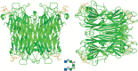
FIGURE 32.1.
Structure of concanavalin A (ConA), a legume seed lectin in complex with a branched pentasaccharide GlcNAcβ1-2Manα1-3(GlcNAcαβ1-2Manα1-6)Man to 2.7 Å. The pentasaccharide is depicted in the center. The tetrameric (more...)
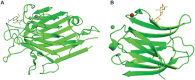
FIGURE 32.2.
Comparison of the subunit structures of soybean agglutinin (A) complexed with a pentasaccharide containing Galβ1-4GlcNAc-R) and human galectin-3 at 1.4 Å (B) complexed with Galβ1-4GlcNAc. Both lectins display a related β-barrel (more...)
Relationships between sequences of L-type lectins from legumes compared with the phylogeny of the various species within the Fabaceae family of plants suggest that these lectins most probably arose from divergent evolution. It remains an open possibility that the tertiary structures of some of the other members of the “L-type lectin family” arose by convergent evolution. It must also be noted that for a protein to be firmly placed in the L-type lectin category it must have the lectin fold and glycan-binding activity.
All soluble L-type lectins found to date are multimeric proteins, although all do not have the same quaternary structure. Thus, these lectins are multivalent with more than one glycan-binding site per lectin molecule. The same multivalent principle applies to the membrane bound L-type lectins because the presence of two or more molecules on a membrane surface essentially presents a multivalent situation. In addition to increasing the avidity of the lectins for branched and/or cell-surface glycans, this multivalence can have great biological significance. Binding of the lectins to the cell surface can lead to aggregation of specific glycan receptors, which can promote a variety of biological responses such as mitogenesis and various signal transduction processes.
PLANT L-TYPE LECTINS
Distribution and Localization
Plant L-type lectins are primarily found in the seeds of leguminous plants and are synthesized during seed development several weeks after flowering; they are transported to the vacuole where they become condensed into specialized vesicles called protein bodies. They are stable during desiccation of the seeds and can remain in that state indefinitely until the seeds germinate. They represent one of several classes of proteins stored in high concentrations in the seeds and are often called storage proteins. During seed germination, the storage bodies become the vacuoles of the cotyledons, which appear as the first leafy appendages of the plant. During the first week of development, these cotyledons provide food for the plant and eventually shrivel up and disappear. L-type lectins have also been found in the bark of some leguminous trees, and very low amounts of these lectins are also found in other vegetative tissues of legumes. In some cases, these latter lectins have been found to be encoded by separate but very similar genes. More than 100 of the seed legume L-type lectins have been characterized, and are the most extensively studied proteins of this class.
Structure
A common feature of the legume L-type lectins is their oligomeric structure. The structures of the monomers consist of three antiparallel β-sheets: a flat six-stranded “back” sheet, a concave seven-stranded “front” sheet, and a short “top” sheet that keeps the two major sheets together (Figure 32.1A,B). All of these lectins require Ca++ and a transition metal ion (usually Mn++) for their carbohydrate-binding activity. The glycan-binding and metal-binding sites are localized in close proximity to each other at the top of the “front” sheet.
The glycan-binding site is composed of four loops: A, B, C, and D (Figure 32.3, top). These loops contain four invariant amino acids that are essential for carbohydrate binding (Figure 32.3, bottom). Loop A contains an invariant aspartate, which forms hydrogen bonds between its side chain and the glycan ligand. This amino acid is linked to its preceding amino acid (usually alanine) by a rare cis-peptide bond, which is stabilized by the metal ions and is necessary for the proper orientation of the aspartate in the combining site. Loop B contains an invariant glycine, which also forms hydrogen bonds with the ligand. An exception to this case is found in two lectins (ConA and the closely related Dioclea grandiflora lectin) where the glycine is replaced with an arginine. Both the glycine and arginine form hydrogen bonds with the ligand via their main-chain amides. Loop C contains an invariant asparagine, which forms a hydrogen bond with the ligand via its side chain. This loop also contains an invariant hydrophobic amino acid.
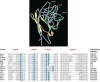
FIGURE 32.3.
(Top) Three-dimensional structure of a peanut agglutinin (PNA) monomer showing the four loops involved in sugar binding: loops A, B, C, and D. The bound sugar (lactose) is shown as a “ball-and-stick” model. Calcium and manganese ions are (more...)
The legume L-type lectins are generally classified into groups based on their carbohydrate specificities as often identified by the ability of monosaccharides to inhibit their agglutinating activity. These differences in specificities are brought about by variability in the conformation and size of the D loop and to some extent by the C loop. Although the main specificity regions of the legume lectins are determined by the loops, there are sites other than these that contribute to lectin specificity. There are several additional modes of refining these specificities, such as interaction with water, posttranslational modifications, and state of oligomerization.
Within these oligomeric proteins, the back β-sheet is involved in formation of the oligomers, which are mostly dimeric or tetrameric in nature. A variety of quaternary structures are found among the lectins. The dimeric and tetrameric structures of ConA are shown in Figure 32.1C,D. Although some of the other lectins occur as dimeric and tetrameric structures, several other different orientations of the β-sheets account for the variability in dimeric and tetrameric structures of other lectins in this class. Interestingly, some legume lectins have a hydrophobic binding site that binds adenine and adenine-derived plant hormones with micromolar affinity; this is two to three orders of magnitude higher than their affinity for monosaccharides. Three of these lectins (the soybean agglutinin, phytohemagglutinin L [PHAL], and Dolichos biflorus lectin) have been crystallized and found to have a unique tetrameric structure, in that the dimer–dimer interface creates a channel running through the center of the tetramer. Two identical adenine-binding sites are found at opposite ends of this channel.
Another common feature of the legume lectins is that they are secretory proteins and undergo cotranslational signal peptide removal, which accompanies their entry into the secretory system. All but peanut agglutinin (PNA) are N-glycosylated as precursors; the N-glycans undergo normal posttranslational modification that occur as they transit the Golgi apparatus. The lectins vary from one another as to whether the mature proteins contain oligomannose-type, complex-type, or a mixture of both types of N-glycans. The lectins may also undergo a variety of proteolytic modifications as they transit through the secretory system. Some of the lectins are cleaved to generate a β-chain, corresponding to the amino terminus and an α-chain corresponding to the carboxyl terminus. For example, the pea lectin and favin (the lectin from Vicia faba) are tetrameric glycoproteins that contain two types of subunits, α and β, which are ∼5 kDa and ∼21 kDa, respectively. These two lectins are each synthesized as single polypeptide precursors that contain the sequences of both chains in the following orientation: β-chain–α-chain. The chains associate to form dimers; they are then proteolytically processed in the protein bodies to form tetramers containing two separate α- and β-chains. Other lectins may undergo carboxy-terminal trimming of only some of their subunits. For example, the soybean agglutinin, phytohemagglutinin E (E-PHA), and D. biflorus lectins are tetramers of equimolar mixtures of intact and trimmed subunits.
The most intriguing proteolytic modification occurs in the case of ConA. A small segment is removed from the interior of the protein and the original amino terminus is ligated with the original carboxyl terminus. This forms what is termed a circularly permuted protein. The N-glycosylated segment of the protein is removed during this transpeptidation process; thus, the mature ConA is not a glycoprotein, in contrast to most seed lectins in the Fabaceae family, which have N-glycans.
Thus, the protein sequence of isolated ConA aligns with other seed lectins, whereas the alignment of the DNA encoding the protein with other lectin genes suggested that the gene is circularly permuted.
Glycan-Binding Activities and Functions
Despite years of research, the intrinsic biological roles of L-type lectins are poorly understood. The glycan-binding specificity can vary significantly; ConA binds mannose and glucose-containing glycans, whereas the lectins from the leguminous trees Maackia amurensis and Sambucus nigra bind to sialylated glycans (see Chapters 15 and 29). There is evidence from many approaches that the legume lectins are insecticidal, antifungal, and antimicrobial and can be toxic to animals that eat raw seeds. Thus, the seed lectins may function in plant immunity as a type of pattern-recognition receptor to protect the offspring of the plant. As highly abundant seed constituents, the lectins could also serve as a storage protein for the plant development. L-type lectins are involved in the symbiosis of plants with nitrogen-fixing bacteria, such as in Rhizobium-legume symbiosis, but the precise function of lectins in this regard is unclear. A recent study has shown that the D. biflorus seed lectin is also a lipoxygenase. It will be of interest to see how many other L-type lectins have this activity, which is necessary to initiate the wound-induced defense pathway in plants.
L-TYPE LECTINS IN PROTEIN QUALITY CONTROL AND SORTING
Calnexin/Calreticulin
Calnexin (CNX) and calreticulin (CRT) are homologous protein chaperones that mediate quality control of proteins in the endoplasmic reticulum (ER) (see Chapter 39). Although CRT is a soluble ER luminal component, CNX is membrane-bound and is perhaps closely associated with the protein-translocating channel that imports nascent proteins into the ER. Both CNX and CRT bind to monoglucosylated, high-mannose-type glycans and prevent their exit from the ER until they are properly folded and assembled into correct quaternary structures (Chapter 39). During the binding and dissociation from CRT or CNX, if the glycoprotein folds correctly, then glucose removal by glucosidase-II allows its passage out of the ER. In the event that a glycoprotein misfolds or aggregates, it is reglucosylated by UDP-Glc:glycoprotein glucosyltransferase (UGGT); this enzyme only recognizes misfolded or aggregated glycoproteins. Following reglucosylation, the monoglucosylated protein binds again to CRT or CNX. Thus, there is a cycle of glucose removal and addition by the alternating actions of glucosidase-II and UGGT and interactions with CNX/CRT.
Both CRT and CNX are Ca++-binding proteins, and their carbohydrate-binding activity is sensitive to changes in Ca++ concentration. CNX is a type I membrane protein with its carboxy-terminal end in the cytoplasm. The lumenal portion of the protein is divided into three domains: a Ca++-binding domain (which is adjacent to the transmembrane domain), a proline-rich long hairpin loop called the P domain, and the amino-terminal L-type lectin domain. CRT has a similar structure, but it is missing the cytoplasmic and transmembrane regions; it is retained in the ER through its KDEL-retrieval signal at the carboxyl terminus (Figure 32.4).
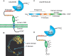
FIGURE 32.4.
Schematic representation of calnexin (CNX) showing the lectin domain, the P domain (containing the proline repeats), and the calcium-binding domain (A). Structure of CNX based on crystallographic data (B). (Adapted, with permission of Elsevier, from Schrag (more...)
ERGIC-53 and Its Related Proteins ERGL, VIP36, and VIPL
ERGIC-53 (human gene LMAN1) and its sequence-related proteins ERGL and VIP36 are type I membrane proteins that participate in vesicular protein transport in the secretory system (see Chapter 39). All share an L-type lectin folding motif. Orthologs of ERGIC-53 are found in plants and all animals and have been independently identified as being important in production of infectious viruses (e.g., coronavirus and filovirus). ERGIC-53 occurs in the ER–Golgi intermediate compartment (ERGIC), and its cytoplasmic carboxyl terminus contains the dilysine/diphenylalanine KKFF retention/retrieval motif. The dilysine is recognized by the COPI coatomer complex; this binding enables the coated vesicles to be recycled from the ERGIC back to the ER. The diphenylalanine helps to direct the COPII-coated vesicles to ER export sites by binding to the COPII coatomer. The location of VIP36 (vesicular integral membrane protein 36) is uncertain; overexpressed protein has been found in both the ER and ERGIC, as well as cis-Golgi. ERGIC 53 can associate with the soluble ER partner MCFD2 (multiple coagulation factor deficiency protein 2), which forms a cargo receptor complex important in factor V and VIII biosynthesis. Human mutations in ERGIC-53 are associated with deficiencies of circulating blood clotting factors V and VIII, which are glycoproteins with multiple N-glycans. LMAN1-deficient mice show FV and FVIII deficiencies and liver ER distension, with accumulation of α1-antitrypsin and GRP78. Partially penetrant, perinatal lethality occurs, dependent on inbred genetic background, suggesting potential roles for as yet unidentified LMAN1-dependent cargo proteins (PMID: 21795745).
Both ERGIC-53 and VIP36 bind to oligomannose-type glycans and require Ca++ for their carbohydrate-binding activity. These two proteins were the first animal lectins that were found to share some sequence and structural homology with the legume seed lectins. Although the overall sequence identity of these proteins to the seed lectins is only ∼19%–24%, those amino acids important for metal and carbohydrate binding in the seed lectins are conserved, including the invariant aspartate, glycine, and asparagine. The invariant aspartate also participates in a cis-peptide bond with its preceding amino acid; this is similar to the case of the legume seed lectins discussed in the above section. The crystal structure of the CRD of ERGIC-53 has been determined and confirms the structural similarity of these lectins. ERGL (ERGIC-53-like) lacks some of the key sugar-binding residues and its function is unclear. VIPL (VIP36-like) is an ER resident protein and also functionally able to bind oligomannose-type glycans, as well as associate with ERGIC-53, which may be important in regulating ERGIC-53 localization.
OTHER L-TYPE LECTINS
A variety of other proteins have been described that have carbohydrate-binding domains with tertiary structures similar to the L-type lectin fold and may be considered as members of this family. Members of the galectin family of lectins also fit into this category and are the subject of a separate chapter in this volume (see Chapter 36). Other GBPs that may fit into this category are briefly discussed below.
Pentraxins and Related Proteins
The pentraxins are a superfamily of plasma proteins that are involved in innate immunity in invertebrates and vertebrates and are designated as pattern-recognition receptors (PRRs). They contain L-type lectin folds and require Ca++ ions for ligand binding. Their name is based on the pentameric arrangement of their subunits. The short pentraxins, C-reactive protein (CRP), which binds phosphocholine residues on polysaccharides and on phospholipids, and the serum amyloid P (SAP) component, which also binds carbohydrate derivatives on bacterial polysaccharides and to amyloid fibrils, are acute-phase proteins in humans and mice, respectively. This family also contains long pentraxins that have an unrelated long amino-terminal domain coupled to the pentraxin domain. PTX3 is one of these long pentraxins; in addition to its role in innate immunity, PTX3 may help in the assembly of a hyaluronan-rich extracellular matrix (ECM), which may involve binding to the hyaluronan–heavy chain-1 complex (inter-α-trypsin inhibitor or IαI or ITI) and regulation by TNF-stimulated gene-6 (TSG-6).
Laminin G domain-like (LG) modules are made of 180–200 amino acid residues and were first identified in laminins. The carboxyl terminus of the laminin α-chain has five tandem lamnin G domains, which are important in heparin and sulfatide binding and cell/basement membrane adhesion, such as to the novel glycans of α-dystroglycan (α-DG). Some LG modules share binding properties for cellular receptors and carbohydrate ligands, indicating that the LG fold may have evolved from the L-type lectin fold for participation in related functions.
VP4 and VP8 in Rotaviruses
VP4 is the surface spike protein in rotaviruses and is proteolytically cleaved to generate VP8, which is a glycan-binding protein, and VP5, a hydrophobic region required for membrane entry. VP8 has an L-type lectin fold and structural features in common with galectins. This domain is required for infectivity of most animal rotaviruses. In some rotavirus strains, VP8 binds sialylated glycans, whereas most rotavirus VP8 binds type 1 and/or type 2 N- or O-glycans lacking sialic acid (Sia) and can especially bind to human milk oligosaccharides with appropriate sequences.
OTHER PROTEINS WITH JELLY-ROLL MOTIFS AND L-TYPE LECTIN DOMAINS
A number of proteins are known to contain the jelly-roll motif found in L-type lectins. These include the binding domain of Clostridium neurotoxins, which can bind to gangliosides (e.g., GT1b and GD1b); Pseudomonas aeruginosa exotoxin A in which one of its three domains, the amino-terminal domain Ia, displays an L-type lectin structure; Vibrio cholerae sialidase, a three-domain protein with a six-bladed β-propeller neuraminidase domain flanked by two L-type lectin domains; and the Leech intramolecular trans-sialidase (TS), which has a multidomain architecture with a lectin-like domain II and an irregular β-stranded domain III that is built around a canonical catalytic domain C.
Although the L-type lectin domain occurs in several proteins, in many cases evidence for carbohydrate binding and/or specificity is lacking. Examples include the L-type lectin receptor kinases (LecRKs) in plants, which is a large family comprised of dozens of membrane receptors that are key to plant development, immunity, and adaptive responses to stimuli; and mammalian thrombospondins, such as TSP-1, which has a carboxy-terminal domain that shows the typical β-sandwich of two curved antiparallel β-sheets, a feature of the jelly-roll topology, in which the L-type lectin domain is compactly assembled with three calcium-binding type 3 (T3) repeats.
This domain II may be involved in carbohydrate recognition through sugar ring and aromatic side-chain interactions, as observed in many lectins.
ACKNOWLEDGMENTS
The authors acknowledge helpful comments and suggestions from Oliver Pearce.
FURTHER READING
- Lis H, Sharon N. 1998. Lectins: Carbohydrate-specific proteins that mediate cellular recognition. Chem Rev 98: 637–674. [PubMed: 11848911]
- Hamelryck TW, Loris R, Bouckaert J, Dao-Thi M-H, Strecker G, Imberty A, Fernandez E, Wyns L, Etzler ME. 1999. Carbohydrate binding, quaternary structure and a novel hydrophobic binding site in two legume lectin oligomers from Dolichos biflorus. J Mol Biol 286: 1161–1177. [PubMed: 10047489]
- Loris R, Bouckaert J, Hamelryck T, Wynn L. 1999. Legume lectin structure. Biochim Biophys Acta 1383: 9–36. [PubMed: 9546043]
- Srinivas VR, Reddy GB, Ahmad N, Swaminathan CP, Mitra N, Surolia A. 2007. Legume lectin family, the “natural mutants of the quaternary state”, provide insights into the relationship between protein stability and oligomerization. Biochim Biophys Acta 1527: 102–111. [PubMed: 11479026]
- Bouwmeester K, Govers F. 2009. Arabidopsis L-type lectin receptor kinases: Phylogeny, classification, and expression profiles. J Exp Bot 60: 4383–4396. [PubMed: 19773388]
- Kouno T, Watanabe N, Sakai N, Nakamura T, Nabeshima Y, Morita M, Mizuguchi M, Aizawa T, Demura M, Imanaka T, Tanaka I, Kawano K. 2011. The structure of Physarum polycephalum hemagglutinin I suggests a minimal carbohydrate recognition domain of legume lectin fold. J Mol Biol 405: 560–569. [PubMed: 21094650]
- Klaus JP, Eisenhauer P, Russo J, Mason AB, Do D, King B, Taatjes D, Cornillez-Ty C, Boyson JE, Thali M, et al. 2013. The intracellular cargo receptor ERGIC-53 is require for the production of infectious arenavirus, coronavirus and filovirus particles. Cell Host Microbe 14: 522–534. [PMC free article: PMC3999090] [PubMed: 24237698]
- Satoh T, Suzuki K, Yamaguchi T, Kato K. 2014. Structural basis for disparate sugar-binding specificities in the homologous cargo receptors ERGIC-53 and VIP36. PLoS ONE 9: e87963. [PMC free article: PMC3912170] [PubMed: 24498414]
- Grandhi NJ, Mamidi AS, Surolia A. 2015. Pattern recognition in legume lectins to extrapolate amino acid variability to sugar specificity. Adv Exp Med Biol 842: 199–215. [PubMed: 25408345]
- Kim DJ, Christofidou ED, Keene DR, Hassan Milde M, Adams JC. 2015. Intermolecular inteactions of thrombospondins drive their accumulation in extracellular matrix. Mol Biol Cell 26: 2640–2654. [PMC free article: PMC4501361] [PubMed: 25995382]
- Wang Y, Weide R, Govers F, Bouwmeester K. 2015. L-type lectin receptor kinases in Nicotiana benthamiana and tomato and their role in Phytophthora resistance. J Exp Bot 66: 6731–6743. [PMC free article: PMC4623685] [PubMed: 26248665]
- Doni A, D'Amico G, Morone D, Mantovani A, Garlanda C. 2017. Humoral innate immunity at the crossroad between microbe and matrix recognition: The role of PTX3 in tissue damage. Semin Cell Dev Biol 61: 31–40. [PMC free article: PMC5419421] [PubMed: 27476448]
- Lamriben L, Graham JB, Adams BM, Hebert DN. 2017. N-glycan-based ER molecular chaperone and protein quality control system: The calnexin binding cycle. Traffic 17: 308–326. [PMC free article: PMC4805476] [PubMed: 26676362]
- Review L-Type Lectins.[Essentials of Glycobiology. 2022]Review L-Type Lectins.Cummings RD, Etzler ME, Ramya TNC, Kato K, Rabinovich GA, Surolia A. Essentials of Glycobiology. 2022
- Review L-type Lectins.[Essentials of Glycobiology. 2009]Review L-type Lectins.Etzler ME, Surolia A, Cummings RD. Essentials of Glycobiology. 2009
- Identifying glycan motifs using a novel subtree mining approach.[BMC Bioinformatics. 2020]Identifying glycan motifs using a novel subtree mining approach.Coff L, Chan J, Ramsland PA, Guy AJ. BMC Bioinformatics. 2020 Feb 4; 21(1):42. Epub 2020 Feb 4.
- Overall strategy for functional analysis of animal lectins.[Methods Mol Biol. 2014]Overall strategy for functional analysis of animal lectins.Kawasaki N. Methods Mol Biol. 2014; 1200:337-51.
- Fluorescence-based solid-phase assays to study glycan-binding protein interactions with glycoconjugates.[Methods Enzymol. 2010]Fluorescence-based solid-phase assays to study glycan-binding protein interactions with glycoconjugates.Leppänen A, Cummings RD. Methods Enzymol. 2010; 478:241-64.
- L-Type Lectins - Essentials of GlycobiologyL-Type Lectins - Essentials of Glycobiology
Your browsing activity is empty.
Activity recording is turned off.
See more...