A new version of this title is available
See the updated version of this chapter
Fungi are a fascinating group of predominantly multicellular organisms. Fungal species, such as Saccharomyces cerevisiae, have been instrumental in defining the fundamental processes of glycosylation, but their glycobiology is significantly different from animal or plant systems. This chapter describes the glycan structures that compose the fungal cell wall, offers some insights into novel glycobiology revealed through studying fungal systems, addresses the use of fungi as experimental and synthetic systems, and delineates the relationships of several important glycoconjugates to fungal biology and pathogenesis.
FUNGAL DIVERSITY
More than 70,000 species of fungi have been described, and it is estimated that more than 5,000,000 fungal species exist. The major fungal phyla are the Chytridiomycota (zoosporic fungi), the Zygomycota, the Glomerulomycota (abuscular mycorrhizal fungi), the Ascomycota (sac fungi, e.g., Saccharomyces, Candida, Aspergillus, Neurospora, and morel mushrooms), and the Basidiomycota (e.g., mushrooms, rot fungi, and puffballs). Most fungi are primarily made of hyphae (branching filaments) that form the mycelium and multicellular structures such as fruiting bodies, whereas the alternative fungal life-form is growth as unicellular yeast. The extracellular matrix of all fungi, the cell wall, comprises complex polysaccharides including mannans, galactans, glucans, and chitin and represents a major target of fungicides.
FUNGI AS MODEL SYSTEMS FOR GENETICS, BIOCHEMISTRY, AND GLYCOBIOLOGY
Historical Perspective
More than 100 years ago, Louis Pasteur discovered that fermentation requires a viable organism; since then yeast have been used as a model system to study cellular metabolism. In fact, Pasteur coined the word “ferment” during his work on alcohol production by yeast. S. cerevisiae, or baker's yeast, has been a wonderful resource for biologists and glycobiologists, especially because many of the fundamental enzymes in aerobic and anerobic metabolism (terms also invented by Pasteur) are shared between yeast and animals. Breakthroughs in enzymology occurred following the 1897 discovery by the Buchner brothers that extracts of yeast could make ethanol and carbon dioxide from glucose, just like intact cells. Mannose is a major component of the yeast cell wall; it was discovered by Emil Fischer in 1888, and the mannose-rich glycans in yeast, historically called yeast gum, have been known since the 1890s. The discovery that the yeast cell wall was composed of D-mannose and work elucidating the chemical structures of other carbohydrates (and vitamin C) led to Sir Walter Norman Haworth's 1937 Nobel Prize in Chemistry. Luis Leloir subsequently discovered the activated precursors required for carbohydrate synthesis, identifying UDP-glucose, GDP-mannose, and other nucleotide sugars from yeast extracts. He was awarded the 1970 Nobel Prize in Chemistry for this work. The discovery of hetherothallic yeast strains and the subsequent development of an elaborate genetic system led to multiple ground-breaking discoveries. For example, genetic studies initiated by the laboratory of Phil Robbins led to the molecular characterization of the conserved N- and O-glycosylation pathway in the endoplasmic reticulum (ER) and the biosynthesis of glycosylphosphatidylinositol (GPI)-anchored proteins. Yeast secretory (sec) mutants helped define the protein secretory pathway, by which polypeptides travel from the ER through the Golgi apparatus to the cell surface or surrounding milieu, becoming glycosylated en route. This foundational work in cell biology was recognized by the Nobel Prize in Physiology or Medicine awarded to Randy Schekman in 2013.
The Fungal Cell Wall
The fungal cell wall, like the plant cell wall, is composed of highly cross-linked glycan polymers (Figure 23.1), which adapt to growth conditions in a dynamic and flexible way and provide high mechanical stability. In contrast to the plant cell wall, it is directly connected to the plasma membrane and the specific cell-wall polysaccharides differ from those of plants. The cell wall polymers of yeast cells or hyphae are composed of N-acetylglucosamine, glucose, mannose, galactose, and/or rhamnose residues that are incorporated into complex polysaccharides such as chitin, glucans, mannans, galactomannans, glucomannans, rhamnomannans, and phosphomannans.
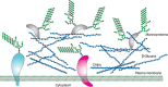
FIGURE 23.1.
Illustration of the cell wall of fungi, showing glycan polymers and mannoproteins. The presence and abundance of different glucans and chitin varies between different fungal species.
Chitin is a polymer of β1-4-linked GlcNAc, which occurs in chains that typically exceed 1000 residues. These chains self-associate to form microfibrils and are deposited primarily at the bud neck of yeast or at septa in filamentous fungi. Chitin synthesis is highly regulated by the coordinated action of multiple chitin synthases, so that deposition occurs at the specific sites and times required for normal cell growth and division. In S. cerevisiae, three chitin synthases (Chs1p, Chs2p, and Chs3p) have been described, along with multiple additional gene products that participate in the regulation and localization of chitin synthesis. Chitin may also be deacetylated to form the cationic polymer, chitosan.
β1-3 glucan, synthesized from UDP-glucose at the plasma membrane, is the major polysaccharide of the fungal cell wall. It is cross-linked at the nonreducing end to chitin and β1-6 glucan, another UDP-glucose-derived polysaccharide, but branched and water-soluble. In contrast to β1-3 glucan and chitin, which are generated at the cell surface, β1-6 glucan is generated in the Golgi. In filamentous ascomycetes, α1-3 and β1-4 glucans are found as well. The β1-6 glucan chains act as an attachment point for an external glycoprotein layer. The majority of these cell wall proteins are GPI-dependent and carry highly mannosylated N- and O-linked glycans (mannans). Cell-wall mannoproteins have the conserved N-glycan core structure linked to Asn residues, but this structure is further elaborated with an extensive repeating α1-6-linked mannose chain. The repeating α1-6-linked mannose backbone is usually branched by short chains of α1-2- and α1-3-linked mannose structures; some of these side chains may be in phosphodiester linkage (Figure 23.2). The mannans are highly heterogeneous in length and branching. In addition to large N-glycans rich in mannose, yeast cell walls contain proteins bearing Ser/Thr-linked O-mannose glycans, although these structures are of modest size compared with the mannans on N-glycans (Figure 23.3A). During cell wall assembly, these cell wall mannoproteins are linked via the oligosaccharide part of the GPI anchor to the β1-6 glucan. Cell wall construction is temporally and spatially controlled during the cell cycle, determining cellular shape (hyphae vs. yeast) and function. The relative abundance of the cell wall polymers varies between fungal species, and some include additional polymers, such as galactomannans or rhamnomannans.
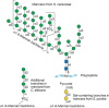
FIGURE 23.2.
Structures of selected yeast mannans. Note that a single pyruvate is (R)4,6 acetyl-(ketal)-linked to the terminal galactose residue in the pyruvylated structure.
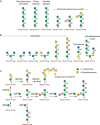
FIGURE 23.3.
Structures of selected O-linked glycans in fungi: (A) yeast, (B) Aspergillus, and (C) Cryptococcus.
Protein Glycosylation
Yeast proteins are abundantly glycosylated, often bearing extensive N- and O-glycans, as well as GPI anchors. As outlined in Chapter 9, N-glycan synthesis begins with synthesis of the conserved lipid-linked core glycan donor, Glc3Man9GlcNAc2-P-P-Dol, which is transferred to nascent polypeptides in the ER. Following core N-glycosylation, the Glc3Man9GlcNAc2Asn-R is processed in mammals and yeast, with removal of glucose residues by α-glucosidases I and II to generate Man9GlcNAc2Asn-R. In mammals and S. cerevisiae, the Man9GlcNAc2Asn-R is further trimmed to Man8GlcNAc2 Asn-R by an ER-mannosidase. Schizosaccharomyces pombe, however, lacks this enzyme and stops processing at Man9GlcNAc2Asn-R. Man8GlcNAc2Asn-R in mammals and Man9GlcNAc2Asn-R in S. pombe are then substrates for the UDP-Glc: glycoprotein glucosyltransferase (UGT) that generates Glc1Man8GlcNAc2Asn and Glc1Man9GlcNAc2Asn in mammals and S. pombe, respectively. This reglucosylation is part of the quality control system for protein folding in the ER (Chapter 39). The monoglucosylated structure is a ligand for the chaperone lectins calnexin and calreticulin in mammalian cells. S. cerevisiae lacks this glucosyltransferase, whereas both S. cerevisiae and S. pombe express calnexin but lack a calreticulin homolog. Specific trimming of the N-linked glycan regulates ER-associated degradation (ERAD) of improperly folded proteins or unassembled protein complex units. In S. cerevisiae, trimming of the N-linked glycan to Man7GlcNAc2 structure by the mannosidase Htm1p generates the degradation signal that is interpreted by the lectin Yos9p and leads to export of the glycoprotein to the cytoplasm and subsequent degradation. Notably, the quality control and ERAD processes differ between fungal species; in some cases, components of the complete pathway are absent. Reglucosylation of N-linked glycans by UGT is absent in S. cerevisiae, for example, and trimming of the Man9GlcNAc2 structure by ER-localized mannosidases is not observed in S. pombe.
Fungal proteins are rich in O-linked mannose. This protein modification is initiated by ER protein mannosyltransferases (PMTs) that use Dol-P-Man as a mannose donor. There are several hetero- or homodimeric PMTs in S. cerevisiae and each may have a different substrate specificity and glycoprotein preference. The Dol-P-Man for the yeast PMTs is synthesized in the cytosol and then flipped into the secretory organelle lumen (Figure 23.4, top right) to be used for both N- and O-glycosylation pathways. Subsequent additions of mannose residues to growing chains occur in the Golgi apparatus, where GDP-Man serves as the donor for reactions catalyzed by Mn++-dependent mannosyltransferases. This is interesting topologically, because GDP-Man is used to generate both the biosynthetic Man5GlcNAc2-P-P-Dol precursor, which is then “flipped” into the ER, and the Dol-P-Man donor of additional mannosylation reactions (Figure 23.4). Thus, GDP-Man does not appear to be transported into the ER, although it is transported into the Golgi apparatus.
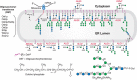
FIGURE 23.4.
Biosynthesis of N-glycans and addition of N-glycans to -Asn-X-Ser/Thr- residues in newly synthesized glycoproteins in the yeast endoplasmic reticulum (ER). Individual steps in the biosynthesis pathway from dolichol phosphate (simplest structure at top (more...)
Mannan extensions are generated in the Golgi apparatus by mannosyltransferases that use GDP-Man as the donor, with one or more specific glycosyltransferases to catalyze synthesis of each linkage and branch. Phosphomannose addition occurs by transfer of Man-1-P from GDP-Man donors in the Golgi apparatus.
Fungi express abundant GPI-anchored glycoproteins. As in other systems (Chapter 12), GPI synthesis begins with construction of a glycolipid precursor; this is then incorporated near the carboxyl terminus of nascent glycoproteins in the ER by a GPI transamidase, which is directed to the site of addition by a signal sequence that is concomitantly cleaved. Yeast GPIs include the conserved trimannose core linked to glucosamine-inositol-phosphatide, but this may bear modifications including additional mannose residues or ethanolamine phosphodiesters (Figure 23.5). In an interesting divergence from “higher” eukaryotes, the GPI anchor serves as a substrate for transglycosylation reactions in cell wall assembly, leading to covalent linkage of glycoproteins to the glucan matrix of the cell wall.
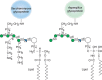
FIGURE 23.5.
Structures of two yeast glycosylphosphatidylinositol (GPI) anchors. Hexagon indicates myo-D-inositol.
Glycolipids
Yeast express a relatively simple array of glycolipids, although Candida albicans is notable for its large lipid-linked mannans. Many fungi make short-chain glycolipids, commonly containing myo-inositol phosphate linkers to mannose that may be modified by galactofuranose (as in Histoplasma capsulatum) or an additional mannose residue. S. cerevisiae generates forms with a single residue of mannose, whereas some longer galactose- and mannose-containing glycolipids are found in Aspergillus niger. Short-chain glycosylceramides such as Glc-Cer and Gal-Cer are also found in the fungi Schizophyllum commune and Aspergillus fumigatus, respectively.
MODEL FUNGI
Saccharomyces cerevisiae as an Experimental System
Yeasts have been valued for baking and brewing for thousands of years, but in the last century or so scientific attention has particularly focused on S. cerevisiae, an oval budding yeast 5–10 µm across. The rapid growth of this simple eukaryote, combined with its inexpensive culture and genetic tractability, has made it a powerful and popular model system. Studies of S. cerevisiae have enormously influenced the fields of eukaryotic cell biology and genetics, in addition to their impact on basic metabolism and enzymology noted above.
S. cerevisiae also contributed to defining the enzymology of GPI lipid precursor biosynthesis (Chapter 12). This complex process, involving more than 20 genes, presented a significant biochemical challenge to researchers in the field. However, as many of the steps are conserved from yeast to mammals, analysis of S. cerevisiae mutants offered a complementary and powerful approach to its dissection. Mutants have also been useful for dissecting yeast-specific processes, such as mannan synthesis; this was elucidated by identifying mnn mutants, which displayed aberrant antibody or dye binding.
Despite the tremendous value of S. cerevisiae as a model, it does have certain limitations. These cells do not synthesize complex N-glycans, mucins or mucin-type O-glycans, O-linked N-acetylglucosamine (O-GlcNAc), sialic acids, or glycosaminoglycans (GAGs) of the types found in vertebrates. However, recent glycomic and site mapping studies suggest that S. cervisiae cells use O-mannose on their nucleocytoplasmic proteins in a manner analogous to O-GlcNAc in plants and animals. Like most other fungi, S. cerevisiae also lacks long-chain glycolipids (apart from those participating in the GPI synthesis) and does not synthesize sialic acid or complex glycosphingolipids or gangliosides like those found in mammals (although it is still valuable for studying sphingosine and sphingolipid metabolism). S. cerevisiae expresses limited glycan diversity even compared with other fungi, with no galactose, xylose, or glucuronic acid reported in its glycans. This must be kept in mind when generalizing from this model to other organisms.
Schizosaccharomyces pombe, a Model for Ultrastructure
S. pombe is a rod-shaped yeast, ∼3–4 µm in diameter and 7–14 µm long. Rather than budding, this organism grows by elongation and fission to give equal-sized daughter cells. Like S. cerevisiae, it has a relatively small genome of ∼14 million base pairs. S. pombe has been useful as a genetically manipulatable model organism for studying the cell cycle. Because it has well-defined organelle structures compared with other yeast, it is a popular choice for studies of intracellular structure. S. pombe also synthesizes mannoproteins and mannans, some containing galactose (Figure 23.3A); this may occur as α1-2-linked caps that may also be pyruvylated. The galactose residues are important in lectin recognition in nonsexual flocculation (clumping) of S. pombe, as evidenced by inhibition of this process by free galactose. In contrast, flocculation in S. cerevisiae is mannose-dependent and inhibited by free mannose. The newly synthesized N-glycans in both S. cerevisiae and S. pombe have nine mannose residues (Man9GlcNAc2Asn; Figure 23.4). In S. cerevisiae, these structures are trimmed in the ER to form Man8GlcNAc2Asn (as discussed above), but this does not occur in S. pombe.
HARNESSING YEAST FOR PRODUCTION
Pichia pastoris and Its Advantages for Expression
Pichia pastoris is a methylotrophic, nonpathogenic organism that was discovered in 1969 in a screen for yeast capable of using methanol. Methanol is oxidized to formaldehyde and hydrogen peroxide by alcohol oxidase (AOX) in the peroxisome. The formaldehyde exits the peroxisome and is oxidized to formate and carbon dioxide in the cytoplasm for the purpose of energy production. Any remaining formaldehyde is assimilated into glyceraldehyde-3-phosphate and dihydroxyacetone by condensation with xylulose-5-monophosphate, in a reaction catalyzed by the peroxisomal enzyme dihydroxyacetone synthase. P. pastoris has become popular as a model system for making recombinant proteins because it is easy to manipulate genetically and can be grown to very high densities. The promoter for AOX is methanol inducible, and transcripts driven by this promoter may comprise up to 5% of the total poly(A)+ RNA in induced cells.
P. pastoris has several advantages over Escherichia coli as an expression system, in that it does not produce inclusion bodies and it promotes the correct folding of eukaryotic proteins. It also has certain advantages over typical model yeast. First, although its basic N-glycosylation pathway is similar to that of S. cerevisiae and yields glycoproteins with oligomannose-type N-glycans, these structures in P. pastoris have only five to 15 mannose residues (typically Man9GlcNAc2Asn and Man8GlcNAc2Asn) compared with the 50 to 150 mannose residues found in glycoproteins from S. cerevisiae (see Figure 23.2). Hyperglycosylation in S. cerevisiae can interfere with protein folding, requiring use of mnn mutants to limit hyperglycosylation and avoid this problem. Also, P. pastoris does not add outer α1-3-linked mannose residues to its N-glycans. These structures are highly antigenic to humans, making proteins expressed in S. cerevisiae unsuitable for human pharmaceutical use. P. pastoris synthesizes O-glycans with an O-linked mannose core attached to Ser/Thr residues; most of these are short α1-2-linked mannose structures (see Figure 23.3A). Genetic tools have been used to manipulate the machinery for glycoprotein assembly of both S. cerevisiae and P. pastoris. Deletion of ER- and Golgi-specific functionalities in combination with the introduction of heterologous hydrolases and glycosyltransferases has generated “humanized” yeasts and filamentous fungi for the production of therapeutic glycoproteins (Chapter 56).
Kluyveromyces lactis in Industry
Kluyveromyces lactis metabolizes lactose to lactic acid and, along with A. niger and E. coli, is grown to produce rennet for making cheese and other products. K. lactis is also a rich source of β-galactosidase, which hydrolyzes lactose. K. lactis synthesizes mannans similar to those in S. cerevisiae, but they lack mannose phosphate modifications, and some side chains are capped with a residue of N-acetylglucosamine. A K. lactis mutant that lacks these N-acetylglucosamine residues because of a deficiency of the Golgi UDP-GlcNAc nucleotide sugar transporter has been productively exploited in studies of heterologous transporters.
BASIDIOMYCETE DIVERSITY
To convey the enormous diversity of fungi we will consider the basidiomycete phylum, which encompasses fungi that produce spores from a pedestal-like structure called the basidium. Basidiomycetes range from fungi with gills or pores, such as common mushrooms and bracket fungi, to budding yeast that are deadly human pathogens.
Lifestyle and Polysaccharides
The basidiomycete Dictyonema glabratum illustrates a distinct fungal lifestyle. It lives in symbiosis with cyanobacteria Scytonema sp. forming a lichen, which is notable for its many unusual glycans. For example, although the β-glucans of most lichens are linear, in D. glabratum they are branched with β1-3 and β1-6 linkages. The mannans of D. glabratum also have an α1-3-linked backbone, rather than the typical α1-6 linkages found in other lichens, along with branches at the 2 and 4 positions. Finally, the xylans of this organism are linear β1-4-linked polymers of xylose, more typical of those found in “higher” plants and algae than in fungi. D. glabratum also synthesizes some unusual short glycolipids, including glycosyldiacylglycerolipids, which are similar to plant glycolipids and contain monosaccharides, disaccharides, and linear trisaccharides of α1-6-linked galactopyranose. This fungus thus highlights the extensive glycan diversity of the fungal kingdom.
O-Glycans
Another example of glycan diversity is offered by Cryptococcus laurentii, which has the unusual property of producing toxins that kill a pathogenic yeast, C. albicans (see below). The O-glycans of C. laurentii are unusual in that they contain mannose, xylose, and galactose (Figure 23.3C); these glycans are synthesized by a unique set of mannosyl-, xylosyl-, and galactosyltransferases that are not homologous to human enzymes. This diversity would not have been predicted by studies of model yeast alone, emphasizing the importance of examining glycans in a wide range of fungal species.
N-Glycan Diversity
Coprinopsis cinerea is a mushroom species that has gained prominence as a model for studies of diverse topics including mating, sexual development, meiosis, and the evolution of multicellularity. Its N-glycans typically are high mannose type with five to nine mannoses, but may also have a bisecting α1-4GlcNAc at the β-mannose.
PATHOGENIC FUNGI
Pathogenic fungi are a significant cause of plant and animal disease, responsible for devastation of crops (e.g., Phytophthora infestans, cause of the Irish potato famine), decimation of animal populations (e.g., certain bat, amphibian, and bee species), and serious human disease that kills an estimated one million people each year. These organisms display diverse glycans, which typically differ from those of the host and have been implicated in multiple pathogenic processes.
Candida albicans Glycans Are Central in Host Interactions
C. albicans (an ascomycete) is a normal commensal organism that can cause illness ranging from irritations of mucosal surfaces to life-threatening systemic infections. The C. albicans cell wall contains β1-3- and β1-6-linked glucans and chitin, similar to the S. cerevisiae wall, and mannans that are termed phosphopeptidomannans. It also produces unusual short β1-2-linked mannose chains (Figure 23.2) that are highly antigenic and are also expressed on phospholipomannan (PLM) antigens. PLM antigens contain phytoceramide derivatives of myo-inositol phosphate to which mannose and the β1-2-linked polysaccharides are linked. The abundant GPI-anchored proteins of C. albicans have been implicated in fungal adherence to host tissues.
The O-glycans of C. albicans are short chains of α1-2-linked mannose (Figure 23.3A), which lack the α1-3-linked mannose caps found in S. cerevisiae. As in S. cerevisiae, deficiencies of O-mannose addition generated through genetic deletions are lethal, indicating that O-mannosylation is essential in this yeast. C. albicans mannans are also important in its interactions with host cells, including macrophages and dendritic cells. In particular, these structures are recognized by the mannose receptor and by dectin-2. These are C-type lectins expressed by immune cells that are important in both innate and adaptive immune responses (see Chapter 34). PLM antigens may be shed by C. albicans and, through interactions with Toll-like receptors (TLR-2), they can induce nuclear factor-κB (NF-κB) activation and cytokine responses such as tumor necrosis factor-α (TNF-α) secretion. Galectin-3, a ubiquitous member of the galectin family of lectins that is highly expressed in macrophages, also appears to recognize C. albicans expressing β1-2-linked mannose residues, resulting in opsonization of the yeast.
Aspergillus fumigatus
A. fumigatus is an environmental mold that spreads by airborne particles. It causes serious disease in immunocompromised people that is difficult to treat, leading to high mortality rates. As with other fungal pathogens, the surface glycans of A. fumigatus are critical for interactions with the host. Presumably to alter surface properties and mask these structures from recognition by host immune receptors, the cell wall of infectious forms of this fungus is covered with specific proteins and melanin. The wall itself has a core layer of branched β1-3-glucan covalently linked to other glucan components, chitin, and galactomannan, which consists of a mannose backbone with short galactofuranose side chains. The core is surrounded by a less structured layer that mainly consists of α1-3-glucan. A. fumigatus also produces an extracellular matrix composed of monosaccharides, α1-3-glucan, galactomannan, and a galactosaminogalactan composed of variable galactopyranose repeats linked to N-acetylgalactosamine. This structure has been implicated in fungal virulence. N-glycans of A. fumigatus are generally high mannose structures without extensive elaboration, although they do generate polymannans in some conditions. Their O-glycans, which may include terminal glucose and galactofuranose modifications, are shown in Figure 23.3.
Cryptococcus neoformans and Its Capsule
Cryptococcus neoformans is a ubiquitous environmental basidiomycete yeast that causes severe disease in immunocompromised individuals, leading to roughly half a million deaths per year worldwide. It is unique among pathogenic fungi in having an extensive polysaccharide capsule that is required for virulence (Figure 23.6). The capsule is a dynamic structure that changes in thickness and composition depending on the environment and growth conditions. It is particularly large in the context of mammalian infection, in which it impedes host immune responses. It is composed of two large (millions of Da) polysaccharides named for their monosaccharide components: glucuronoxylomannan (GXM) and glucuronoxylomannogalactan (GXMGal). GXM is an extended α1-3 mannan substituted with β1-2Xyl, β1-4Xyl, and β1-2GlcA (Figure 23.7); a subset of the mannose residues are 6-O-acetylated (not shown). The second polymer, GXMGal, is based on α1-6 galactan, with side chains of galactose, glucuronic acid, mannose, and xylose (Figure 23.7); the backbone is also modified with small amounts of β1-2-linked galactofuranose (not shown). Association of the capsule with the cell surface relies on a cell-wall component, α1-3 glucan. Although α1-3 glucan is not present in the cell walls of S. cerevisiae or C. albicans, it is common in other fungi. N-glycans of C. neoformans are generally high mannose with modest outer chain extensions and may include xylose β1-2-linked to the trimannosyl core. Both N- and O-glycans may also be modified with xylose and xylose phosphate.
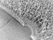
FIGURE 23.6.
A quick-freeze deep-etch image of the edge of a Cryptococcus neoformans cell. The polysaccharide capsule (open meshwork at right) is linked to the cell wall (central structure dividing the image from upper left to lower right) via α1-3 glucan. (more...)
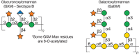
FIGURE 23.7.
Structures of capsular polysaccharides in Cryptococcus neoformans.
Fungal Glycans as Drug Targets
In the context of fungal diseases, the similarity of fungi to their eukaryote hosts becomes a liability, because it is challenging to develop antifungal drugs that are not hampered by toxicity. The unique features of fungal glycans may suggest drug targets to help improve this picture, and decrease the roughly one million deaths each year due to fungal infections.
The major success story using this approach is the development of echinocandin drugs. These antifungal lipopeptides inhibit β1-3-glucan synthesis in fungi including Candida and Aspergillus, leading to cell wall compromise, and are used clinically to treat invasive fungal infections. Despite this recent advance, however, not all fungal pathogens are susceptible to the echinocandins, and resistance to these compounds is a concern. Continued efforts to target novel aspects of fungal glycobiology may advance the ongoing search for new therapies.
ACKNOWLEDGMENTS
The authors appreciate helpful comments and suggestions from Robyn Peterson.
FURTHER READING
- Ballou CE, Lipke PN, Raschke WC. 1974. Structure and immunochemistry of the cell wall mannans from Saccharomyces chevalieri, Saccharomyces italicus, Saccharomyces diastaticus, and Saccharomyces carlsbergensis. J Bacteriol 117: 461–467. [PMC free article: PMC285535] [PubMed: 4590470]
- Huffaker TC, Robbins PW. 1983. Yeast mutants deficient in protein glycosylation. Proc Natl Acad Sci 80: 7466–7470. [PMC free article: PMC389972] [PubMed: 6369318]
- Dickson RC, Lester RL. 1999. Yeast sphingolipids. Biochim Biophys Acta 1426: 347–357. [PubMed: 9878820]
- Poulain D, Jouault T. 2004. Candida albicans cell wall glycans, host receptors and responses: Elements for a decisive crosstalk. Curr Opin Microbiol 7: 342–349. [PubMed: 15358252]
- Daly R, Hearn MT. 2005. Expression of heterologous proteins in Pichia pastoris: A useful experimental tool in protein engineering and production. J Mol Recognit 18: 119–138. [PubMed: 15565717]
- Klutts JS, Yoneda A, Reilly MC, Bose I, Doering TL. 2006. Glycosyltransferases and their products: Cryptococcal variations on fungal themes. FEMS Yeast Res 6: 499–512. [PubMed: 16696646]
- Klis FM, Ram AF, De Groot PW. 2007. A molecular and genomic view of the fungal cell wall. In Biology of the fungal cell (ed. Howard RJ, Gow NAR.), 2nd ed, The Mycota VIII, pp. 97–120. Springer-Verlag, Berlin.
- Deshpande N, Wilkins MR, Packer N, Nevalainen H. 2008. Protein glycosylation pathways in filamentous fungi. Glycobiology 18: 626–637. [PubMed: 18504293]
- De Pourcq K, De Schutter K, Callewaert N. 2010. Engineering of glycosylation in yeast and other fungi: Current state and perspectives. Appl Microbiol Biotechnol 87: 1617–1631. [PubMed: 20585772]
- Everest-Dass AV, Jin D, Thaysen-Andersen M, Nevalainen H, Kolarich D, Packer NH. 2012. Comparative structural analysis of the glycosylation of salivary and buccal cell proteins: Innate protection against infection by Candida albicans. Glycobiology 22: 1465–1479. [PubMed: 22833316]
Publication Details
Author Information and Affiliations
Authors
Tamara L. Doering, Richard D. Cummings, and Markus Aebi.Publication History
Published online: 2017.
Copyright
PDF files are not available for download.
Publisher
Cold Spring Harbor Laboratory Press, Cold Spring Harbor (NY)
NLM Citation
Doering TL, Cummings RD, Aebi M. Fungi. 2017. In: Varki A, Cummings RD, Esko JD, et al., editors. Essentials of Glycobiology [Internet]. 3rd edition. Cold Spring Harbor (NY): Cold Spring Harbor Laboratory Press; 2015-2017. Chapter 23. doi: 10.1101/glycobiology.3e.023


