U.S. flag

An official website of the United States government

NCBI Bookshelf. A service of the National Library of Medicine, National Institutes of Health.

Adam MP, Feldman J, Mirzaa GM, et al., editors. GeneReviews® [Internet]. Seattle (WA): University of Washington, Seattle; 1993-2024.

Cover of GeneReviews®

GeneReviews® [Internet].

Show details

Williams Syndrome

Synonym: Williams-Beuren Syndrome

, MD, FACMG, FAAP.

Author Information and Affiliations

Initial Posting: ; Last Update: April 13, 2023.

Estimated reading time: 48 minutes

Summary

Clinical characteristics.

Williams syndrome (WS) is characterized by developmental delay, intellectual disability (usually mild), a specific cognitive profile, unique personality characteristics, cardiovascular disease (supravalvar aortic stenosis, peripheral pulmonary stenosis, hypertension), connective tissue abnormalities, growth deficiency, endocrine abnormalities (early puberty, hypercalcemia, hypercalciuria, hypothyroidism), and distinctive facies. Hypotonia and hyperextensible joints can result in delayed attainment of motor milestones. Feeding difficulties often lead to poor weight gain in infancy.

Diagnosis/testing.

The diagnosis of WS is established by identification of a heterozygous 1.5- to 1.8-Mb deletion of the Williams-Beuren syndrome critical region (WBSCR) on chromosome 7q11.23.

Management.

Treatment of manifestations: Infants with feeding issues may benefit from feeding therapy. Early intervention programs, special education programs, and vocational training address developmental disabilities; programs include speech-language, physical, occupational, feeding, and sensory integration therapies as well as hippotherapy; phonics methods are recommended to teach reading. Psychological and psychiatric evaluation and treatment provide individualized behavioral counseling and medications, especially for attention-deficit/hyperactivity disorder and anxiety. Surgery may be required for supravalvar aortic or pulmonary artery stenosis, mitral valve insufficiency, and/or renal artery stenosis. Anesthesia consultation and electrocardiogram is recommended prior to sedation and surgical procedures. Orthodontic referral should be considered for malocclusion. Constipation should be aggressively managed at all ages. The lower urinary tract should be evaluated in those with febrile urinary tract infections; refer to a nephrologist for management of nephrocalcinosis, persistent hypercalcemia, and/or hypercalciuria. Range of motion exercises are recommended to prevent or ameliorate joint contractures. Treatment of hypercalcemia may include diet modification, oral corticosteroids, and/or intravenous pamidronate. Early puberty may be treated with a gonadotropin-releasing hormone agonist. Treatment of hypertension, sleep disorders, ocular manifestations, recurrent otitis media, hearing loss, dental issues, hypothyroidism, and insulin resistance does not differ from that in the general population.

Surveillance: Children younger than age two years should have serum calcium studies every four to six months. Thyroid function should be checked yearly until age three years and every two years thereafter. Medical evaluation, vision screening, hearing evaluation, measurement of blood pressure in both arms, calcium-to-creatinine ratio in spot urine, and urinalysis should be performed annually. Additional periodic evaluations for all individuals include: measurement of serum concentration of calcium every two years; cardiology evaluation for elastin arteriopathy at least annually until age five years and every two to three years thereafter; and renal and bladder ultrasound examination every ten years. Oral glucose tolerance tests in adults should start at age 20 years.

Agents/circumstances to avoid: Multivitamins for children, because all pediatric multivitamin preparations contain vitamin D.

Genetic counseling.

WS is an autosomal dominant disorder. Most individuals diagnosed with WS have the disorder as the result of a de novo 1.5- to 1.8-Mb 7q11.23 deletion; rarely, an individual with WS has an affected parent. Recommendations for the parents of a proband with WS include obtaining a medical history to determine if signs or symptoms of WS are present. In the absence of clinical findings of WS in the parents, testing of the parents for the 7q11.23 deletion identified in the proband is not warranted. Each child of an individual with WS has a 50% chance of inheriting the 7q11.23 deletion and being affected. Once the WS-causing 1.5- to 1.8-Mb 7q11.23 deletion has been identified in an affected family member, prenatal and preimplantation genetic testing are possible.

Diagnosis

Suggestive Findings

Williams syndrome (WS) should be suspected in individuals with the following findings:

  • Intellectual disability affects most individuals and is typically mild.
  • Specific cognitive profile includes strengths in verbal short-term memory and language and extreme weakness in visuospatial construction.
  • Unique personality includes overfriendliness, empathy, generalized anxiety, specific phobias, and attention-deficit/hyperactivity disorder.
  • Cardiovascular disease (elastin arteriopathy). Supravalvar aortic stenosis is the most common. Peripheral pulmonic stenosis is common in infancy. Any artery may be narrowed.
  • Distinctive facies. Broad forehead, bitemporal narrowing, periorbital fullness, a stellate/lacy iris pattern (see Figure 1), strabismus, short nose, broad nasal tip, malar flattening, long philtrum, thick vermilion of the upper and lower lips, wide mouth, malocclusion, small jaw, and large ear lobes are observed at all ages (see Figure 2). Young children have epicanthal folds, full cheeks, and small, widely spaced teeth (see Figure 3); adults have a long face and neck and sloping shoulders (see Figure 4).
  • Connective tissue abnormalities include hoarse voice, inguinal/umbilical hernia, bowel/bladder diverticulae, rectal prolapse, joint limitation or laxity, and soft, lax skin.
  • Growth abnormalities include prenatal growth deficiency, postnatal poor weight gain, and decreased linear growth.
  • Endocrine abnormalities include early puberty, hypercalcemia, hypercalciuria, hypothyroidism, and diabetes mellitus.
Figure 1.

Figure 1.

Note the stellate iris pattern in an individual with Williams syndrome

Figure 2. . A broad forehead, bitemporal narrowing, periorbital fullness, strabismus, short nose, broad nasal tip, malar flattening, long philtrum, thick vermilion of the upper and lower lips, wide mouth, malocclusion, small jaw, and large earlobes are observed at all ages and in all ethnic groups.

Figure 2.

A broad forehead, bitemporal narrowing, periorbital fullness, strabismus, short nose, broad nasal tip, malar flattening, long philtrum, thick vermilion of the upper and lower lips, wide mouth, malocclusion, small jaw, and large earlobes are observed at (more...)

Figure 3.

Figure 3.

Young children with Williams syndrome typically have epicanthal folds, full cheeks, and small, widely spaced teeth, as seen in these children at the following ages: A. Newborn

Figure 4. . Adults typically have a long face and neck, accentuated by sloping shoulders, resulting in a gaunt appearance, as seen in this affected individual, age 43 years.

Figure 4.

Adults typically have a long face and neck, accentuated by sloping shoulders, resulting in a gaunt appearance, as seen in this affected individual, age 43 years.

Note: See the National Human Genome Research Institute (NHGRI) Atlas of Human Malformation Syndromes (scroll to ATLAS IMAGES) for photographs of individuals with WS from diverse ethnic backgrounds.

Establishing the Diagnosis

The diagnosis of WS is established by identification of a heterozygous 1.5-to 1.8-Mb deletion at chromosome 7q11.23. For this GeneReview, WS is defined as the presence of a recurrent 1.5- to 1.8-Mb deletion at the approximate position of chr7:73,330,452-74,728,172 in the reference genome (NCBI Build 38).

Note: The phenotype of significantly larger or smaller deletions within this region may be clinically distinct from WS (see Genetically Related Disorders).

Although several genes of interest (e.g., ELN) are within the 1.5- to 1.8-Mb recurrent deletion, no single gene in which pathogenic variants are causative of WS has been identified (see Molecular Genetics for genes of interest in the deleted region).

Genomic testing methods that determine the copy number of sequences can include chromosomal microarray (CMA) or targeted deletion analysis by fluorescence in situ hybridization (FISH). Note: The 7q11.23 recurrent deletion cannot be identified by routine analysis of G-banded chromosomes or other conventional cytogenetic banding techniques.

  • Chromosomal microarray (CMA) using oligonucleotide arrays or SNP genotyping arrays can detect the recurrent deletion in a proband. The ability to size the deletion depends on the type of microarray used and the density of probes in the 7q11.23 region.
    Note: (1) Most individuals with WS are identified by CMA performed in the context of evaluation for developmental delay, intellectual disability, or autism spectrum disorders. (2) The recurrent deletion was detected by early arrays (e.g., BAC arrays).
  • Targeted deletion analysis. A FISH probe targeted to the 7q11.23 region can be reliably used for diagnosis in situations where CMA is not available. FISH analysis may be used to test at-risk relatives of a proband known to have WS.
    Note: (1) Targeted deletion testing by FISH is not appropriate for the relative of an individual suspected of having WS in whom a deletion was not detected by FISH or by CMA designed to target 7q11.23. By definition, such individuals do not have WS. (2) It is not possible to routinely size the deletion by use of FISH.

Table 1.

Genomic Testing Used in Williams Syndrome

Deletion 1MethodSensitivity
ProbandAt-risk family members
1.5- to 1.8-Mb heterozygous deletion at 7q11.23
ISCN: seq[GRCh38] del(7)(q11.23) chr7:73,330,452-74,728,172 2
ISCA-37392
CMA 3100%100%
FISH100%100% 4
1.

See Molecular Genetics for details of the deletion and genes of interest.

2.

Standardized ISCN annotation and interpretation for genomic variants from the Clinical Genome Resource (ClinGen) project (formerly the International Standards for Cytogenomic Arrays [ISCA] Consortium). Genomic coordinates represent the minimum deletion size associated with the 7q11.23 recurrent deletion as designated by ClinGen. Deletion coordinates may vary slightly based on array design used by the testing laboratory. Note: The size of the deletion as calculated from these genomic positions may differ from the expected deletion size due to the presence of segmental duplications near breakpoints. The phenotype of significantly larger or smaller deletions within this region may be clinically distinct from the recurrent 7q11.23 deletion (see Genetically Related Disorders).

3.

Chromosomal microarray analysis (CMA) using oligonucleotide arrays or SNP arrays. CMA designs in current clinical use target the 7q11.23 region.

4.

FISH is not appropriate as a diagnostic method for the relative of an affected individual in whom Williams syndrome was not detected by FISH or by CMA designed to target this region.

Clinical Characteristics

Clinical Description

Williams syndrome (WS) is a multisystem disorder characterized by cardiovascular disease (elastin arteriopathy, most often manifesting as supravalvar aortic stenosis), developmental delay, usually mild intellectual disability, a specific cognitive profile, unique personality characteristics, connective tissue abnormalities, growth abnormalities, other endocrine manifestations, and distinctive facies. Additional manifestations can include sleep problems, ocular issues, hearing loss, dental problems, gastrointestinal difficulties, urinary tract abnormalities, and musculoskeletal issues.

Table 2.

Williams Syndrome: Frequency of Select Features

SystemFeature% of Persons w/FeatureComment
Neurodevelopment Developmental delay100%Average age for walking & talking is 21 mos.
Hypotonia80%
Intellectual disability75%Typically mild
Characteristic cognitive profile95%Severe impairment of visuospatial construction, relative strength in language
Neurobehavioral Unique personality95%Overfriendliness, difficulty w/emotional regulation
Anxiety (non-social)80%Specific phobia most common
ADHD65%
Sleep disorders65%
Autism spectrum disorder10%-20%
Cardiovascular Any arterial stenosis80%Elastin arteriopathy
SVAS75%
PPS40%-60%Typically improves w/time
Hypertension50%Renal artery stenosis may contribute
Mitral valve prolapse15%
QTc prolongation13%
Ocular/visual Esotropia50%
Hyperopia60%
Auditory Recurrent otitis media50%
Hypersensitivity to sound90%
Sensorineural hearing loss>60%Present in >90% of adults
Dental Microdontia95%
Malocclusion85%
Gastrointestinal Feeding difficulties70%
Constipation50%
Umbilical hernia50%
Inguinal hernia40%
Colon diverticula30%
Rectal prolapse15%
Urinary tract Urinary frequency70%
Enuresis50%
Bladder diverticula50%
Musculoskeletal Joint hypermobility90%Most common in young children
Joint contractures50%
Scoliosis20%-35%
Integument /
connective tissue
Hoarse voice90%
Soft, lax skin90%
Endocrine Short stature50%Mean adult height is <3rd centile
Early puberty50%
Hypercalcemia20%-40%Clinically significant hypercalcemia typically occurs at age <2 yrs
Hypercalciuria30%Nephrocalcinosis in 5%
Hypothyroidism10%Subclinical in 30%
Diabetes mellitus20%More common in adults
Craniofacial Characteristic facial features100%See Figures 1, 2, 3, and 4.
Microcephaly30%

ADHD = attention-deficit/hyperactivity disorder; PPS = peripheral pulmonic stenosis; SVAS = supravalvar aortic stenosis

Infancy. Infants with WS are often born post-term, and 50% of infants are small for gestational age [Li et al 2022]. Feeding difficulties leading to poor weight gain are common, including gastroesophageal reflux, disordered suck and swallow, textural aversion, and vomiting. Prolonged colic (lasting longer than four months) may be related to gastroesophageal reflux, chronic constipation, and/or idiopathic hypercalcemia.

Developmental delay. Infants with WS are hypotonic and typically have hyperextensible joints, resulting in delayed attainment of motor milestones [Morris & Mervis 2021]. Walking usually occurs by age 24 months. Speech is also delayed but later becomes a relative strength [Kozel et al 2021]. Fine motor difficulties are present at all ages.

Cognitive abilities. Intellectual disability, usually mild, occurs in 75% of individuals with WS. The cognitive profile is distinctive, consisting of strengths in verbal short-term memory and language but extreme weakness in visuospatial constructive cognition. As a result, children with WS usually score higher on verbal subtests than on tests measuring visuospatial construction [Mervis & Greiner de Magalhães 2022]. No difference in IQ between males and females is reported, and IQ is stable throughout childhood [Mervis et al 2012].

Academically, individuals with WS perform relatively well in reading, and adults may read at a high school level, though the range of achievement is wide. Reading skills correlate with method of reading instruction rather than IQ, with the highest reading skills following systemic phonics instruction [Mervis & Greiner de Magalhães 2022]. Difficulty with writing, drawing, and mathematics is significant, although many adults with WS are able to perform simple addition.

Unique personality/behavior. The characteristic personality profile of WS includes overfriendliness, social disinhibition, excessive empathy, attention problems, and non-social anxiety. Other common behavior problems include difficulty with sensory modulation/processing, emotional regulation, and perseveration. Some individuals have overlapping symptoms with autism spectrum disorder, such as restricted interests and repetitive behavior. Children with WS who meet diagnostic criteria for autism spectrum disorder (ASD; 10%-20%) fit in the active-but-odd subtype rather than the aloof subtype of ASD [Klein-Tasman et al 2018]. In children, attention-deficit/hyperactivity disorder occurs in 65% of individuals and anxiety disorder in 57% (usually specific phobias) [Leyfer et al 2006]. Anxiety is common across the life span; longitudinal studies of anxiety indicate a prevalence of 80% [Woodruff-Borden et al 2010]. Pharmacotherapy for psychiatric disorders in WS should take into account the medical comorbidities associated with WS [Thom et al 2021]. For instance, use of stimulants (which can increase blood pressure and heart rate) to treat ADHD should be discussed with the individual's cardiologist; atomoxetine is a non-stimulant alternative [Thom et al 2021].

Adaptive behavior is less than expected for IQ in both children and adults [Howlin & Udwin 2006, Mervis & Pitts 2015], and adversely affects the ability of adults with WS to function independently (less than 10% live independently). There is typically a relative strength in socialization and communication skills, with significant weakness in motor skills and daily living skills. Executive function deficits occur in 70% of children and adolescents with WS. Difficulties with behavioral and emotional regulation are associated with behavior issues and poor adaptive behavior; difficulty with inhibition is associated with attention problems; and difficulty with flexibility in shifting focus is associated with anxiety [Greiner de Magalhães et al 2022].

Sleep disorders occur in 65% of affected individuals and include increased sleep latency and decreased sleep efficiency [Goldman et al 2009, Mason et al 2011]. Abnormal or absent nocturnal melatonin peak has been documented [Sniecinska-Cooper et al 2015, Santoro et al 2016]; melatonin treatment may be helpful [Martens et al 2017, Morris & Mervis 2021]. Sleep-related breathing disorder (reported in 15%) and excessive daytime sleepiness (reported in 30%) were associated with more externalizing behavior problems in toddlers with WS at age two years [Greiner de Magalhães et al 2020].

Cardiovascular disease occurs in 80% of individuals with WS [Collins 2018, Honjo et al 2022]. Elastin arteriopathy is the major cause of morbidity and mortality. Any artery may be narrowed, but supravalvar aortic stenosis (SVAS) is most common and may worsen over time, especially in the first five years of life [Collins et al 2010b]. SVAS can be either a discrete hourglass stenosis or diffuse aortic stenosis. If untreated, the resultant increase in arterial resistance leads to elevated left heart pressure, cardiac hypertrophy, and cardiac failure. Progression is more likely if the stenosis is moderate or severe and presents in infancy or early childhood. Mild SVAS is less likely to progress. Approximately one third of children with SVAS will require surgical correction.

Peripheral pulmonic stenosis (PPS) is common in infancy but usually improves over time without intervention. However, individuals with combined SVAS and PPS (biventricular outflow tract obstruction) may develop biventricular hypertrophy and hypertension, increasing the risk for myocardial ischemia, dysrhythmias, and sudden death [Pham et al 2009]. The overall 30-year survival rate for children undergoing interventions for obstructive lesions is 90% in North America [Zinyandu et al 2023]. Individuals with either isolated left heart obstructive lesions or isolated right heart obstructive lesions have better survival rates than those with combined disease (96% vs 83%). In-hospital mortality was 2.5%, and 9% required reoperation (mostly for recurrent SVAS) [Zinyandu et al 2023].

Coronary artery stenosis has been implicated as a cause of sudden death in individuals with WS [Bird et al 1996]. Coronary artery stenosis or dilatation are found in 10%-30% of individuals with WS, and coronary blood flow may be restricted at the sinuses of Valsalva in the setting of narrowed sinotubular junction [Brown et al 2018]. The incidence of sudden death in one cohort of 293 individuals with WS was one in 1,000 patient years, which is 25 to 100 times higher than the age-matched population [Wessel et al 2004].

The prevalence of hypertension in individuals with WS is 40%-50%. Hypertension may present at any age [Bouchireb et al 2010] and may be secondary to renal artery stenosis in some instances [Honjo et al 2022]. Increased vascular stiffness has been documented in individuals with WS and responds to antihypertensive medication [Kozel et al 2014].

Mitral valve prolapse and aortic insufficiency have been reported in adults [Morris et al 1990, Collins et al 2010a].

Prolonged QTc has been reported in 13.6% of individuals with WS; screening for repolarization abnormalities is recommended [Collins et al 2010a, Brink et al 2022].

Anesthesia and sedation are associated with an increased risk for adverse events, including cardiac arrest, in individuals with WS [Olsen et al 2014]. Sedation and anesthesia risk assessment and management guidelines have been developed [Schmidt et al 2021, Staudt & Eagle 2021] with the goal of maintaining intravascular volume and a stable blood pressure to optimize coronary perfusion [Schmidt et al 2021]. In centers using these strategies specifically for individuals with WS, morbidity and mortality is decreased. Brown et al [2018] reported no adverse events in 90% of individuals undergoing cardiac procedures, and no adverse events in 95% of individuals undergoing noncardiac procedures. Schmidt et al [2021] compared a historical group with an intervention group after new anesthesia guidelines were adopted for WS and found that incidence of adverse cardiac events in the intervention group was 2% compared to 6% in the historical group.

Stenosis of additional arteries has been reported. Neurovascular abnormalities are rarely reported but may result in stroke [Cherniske et al 2004]. Stenosis of the mesenteric arteries may contribute to abdominal pain.

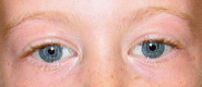
Ocular manifestations. Lacrimal duct obstruction, hyperopia (67%), and esotropia (50%) are common in individuals with WS [Weber et al 2014]. Cataracts have been reported in adults [Cherniske et al 2004].

Ears and hearing. Chronic otitis media is seen in 50% of affected individuals. Increased sensitivity to sound is common (90%), and individuals with WS report discomfort at 20 decibels lower than controls [Gothelf et al 2006]. Many affected individuals report specific phobias for certain sounds [Levitin et al 2005]. Hyperacusis occurs in 35% and is associated with absence of contralateral acoustic reflexes [Silva et al 2021].

Progressive sensorineural hearing loss has been observed; mild-to-moderate hearing loss is detected in 63% of children and 92% of adults [Gothelf et al 2006, Marler et al 2010]. Mild-to-moderate high-frequency sensorineural hearing loss is common in adults, as is excessive buildup of ear wax [Cherniske et al 2004].

Dental findings include generalized diastemas (70%), hypodontia (50%), microdontia, enamel hypoplasia, and malocclusion (angle class II and III) [Castro et al 2019].

Gastrointestinal manifestations. Individuals with WS have sensory defensiveness; difficulty with food textures leads to problems in transitioning from breast milk or formula to solid foods in infancy.

Chronic abdominal pain is common in children and adults with WS; possible causes include gastroesophageal reflux, hiatal hernia, peptic ulcer disease, cholelithiasis, diverticulitis, ischemic bowel disease, chronic constipation, and somatization of anxiety. The prevalence of diverticulitis is increased in adolescents [Stagi et al 2010] and adults with WS [Partsch et al 2005]. Complications of constipation may include rectal prolapse, hemorrhoids, or intestinal perforation.

Hypercalcemia may contribute to irritability, vomiting, constipation, and muscle cramps. Hypercalcemia is more common in infancy but may recur in adults [Morris et al 1990, Pober et al 1993].

Urinary tract abnormalities. Urinary frequency and enuresis are common in children with WS. Renal artery stenosis is found in 50% of individuals with WS, structural abnormalities of the urinary tract in 10%, bladder diverticulae in 50%, and nephrocalcinosis in fewer than 5% [Pober et al 1993, Pankau et al 1996, Sforzini et al 2002, Sammour et al 2006, Sammour et al 2014]. Bladder capacity is reduced, and detrusor overactivity is observed in 60% of affected individuals [Sammour et al 2006]. Average age of daytime urinary continence is four years; nocturnal continence occurs in 50% by age ten years. Nocturnal enuresis occurs in an estimated 3% of adults [von Gontard et al 2016].

Musculoskeletal and additional neurologic manifestations. The hypotonia and lax joints of the young child lead to abnormal compensatory postures to achieve stability. Older children and adults with WS typically have hypertonia and hyperactive deep-tendon reflexes. Gradual tightening of the heel cords and hamstrings occurs, resulting in a stiff and awkward gait, kyphosis, and lordosis by adolescence [Morris et al 1988, Kaplan et al 1989, Copes et al 2016]. Scoliosis is common [Damasceno et al 2014]. Ten percent of individuals with WS have radioulnar synostosis [Morris & Carey 1990]. Fine motor function is impaired, leading to difficulty with tool use and handwriting at all ages. Cerebellar signs in adults include ataxia, dysmetria, and tremor [Pober & Morris 2007].

Neuroimaging. Reduced brain size, reduced gray matter volume (especially in the parietal and occipital regions), and increased gyral complexity are seen on brain MRI [Jackowski et al 2009, Eisenberg et al 2010]. Reduced posterior fossa size coupled with preserved cerebellar size may contribute to Chiari I malformation found in some affected individuals [Pober & Filiano 1995, Mercuri et al 1997].

Integument and additional connective tissue manifestations. Most individuals have a hoarse or low-pitched voice; vocal cord abnormalities secondary to elastin deficiency are likely causative [Vaux et al 2003]. Soft, lax skin is typical. The hair grays prematurely [Morris et al 1988].

Growth deficiency. Poor weight gain is observed in 70% of infants. The growth pattern is characterized by prenatal growth deficiency, poor weight gain, and poor linear growth in the first four years, a rate of linear growth that is 75% of normal in childhood, and a brief pubertal growth spurt. Individuals with WS are shorter than predicted by midparental height [Levy-Shraga et al 2018]. The mean adult height is below the third centile. Specific growth curves for WS are available [Saul et al 1988, Martin et al 2007, Morris et al 2020]. For a systematic review of growth studies in WS cohorts, see de Sousa Lima Strafacci et al [2020]. Obesity or overweight is seen in 50% of older children and adults [Cherniske et al 2004, Stanley et al 2021]. A lipedema phenotype of the lower extremities in seen in 25% of adults [Shaikh et al 2018].

Puberty may occur early, and central precocious puberty is present in 18% of individuals with WS [Partsch et al 2002, Kim et al 2016]. Hormonal suppression with gonadotropin-releasing hormone agonist is well tolerated by girls with either early or precocious puberty, and treated girls are taller than WS controls [Spielmann et al 2015].

Hypercalcemia is most often symptomatic (irritability, vomiting, constipation) in the first two years of life [Martin et al 1984, Morris et al 1988, Kim et al 2016]. Hypercalcemia is associated with dehydration, hypercalciuria, and nephrocalcinosis. Compared to controls, higher median serum calcium levels are found in all age groups [Sindhar et al 2016]. The etiology of hypercalcemia in WS is unknown [Stagi et al 2016].

Additional endocrine problems. Endocrine abnormalities include hypothyroidism, including subclinical hypothyroidism (thyroid-stimulating hormone elevation with normal T3/T4 levels) [Palacios-Verdú et al 2015]. Prevalence of impaired glucose tolerance is 42%, and incidence of diabetes mellitus is increased in adolescents and adults with WS [Stanley et al 2021]. Low bone mineral density has been reported in 50% of adults with WS [Cherniske et al 2004] and is associated with low serum phosphate [Palmieri et al 2019].

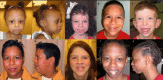
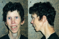
Distinctive facial features including broad forehead, bitemporal narrowing, periorbital fullness, a stellate/lacy iris pattern (see Figure 1), strabismus, short nose, broad nasal tip, malar flattening, long philtrum, thick vermilion of the upper and lower lips, wide mouth, malocclusion, small jaw, and large ear lobes are observed at all ages (see Figure 2). Young children have epicanthal folds, full cheeks, and small, widely spaced teeth (see Figure 3), while adults typically have a long face and neck, accentuated by sloping shoulders (see Figure 4).

Genotype-Phenotype Correlations

The 7q11.23 recurrent deletions of the Williams-Beuren syndrome critical region (WBSCR) comprise either 1.55 Mb (90%-95% of individuals with WS) or 1.84 Mb (5%-10% of individuals with WS) [Palacios-Verdú et al 2015].

Deletions within the WBSCR smaller than the recurrent deletions associated with WS are rare and are associated with a variable phenotype. Genotype-phenotype correlations are limited for genes other than ELN [Kozel et al 2021, Serrano-Juárez et al 2022].

  • Individuals with partial WBSCR deletions that include the usual telomeric breakpoint (including GTF2I) have classic manifestations of WS, including intellectual disability [Botta et al 1999, Heller et al 2003]. Individuals with deletions of the WBSCR that include distal genes in the region (especially GTF2IRD1 and GTF2I) are more likely to have intellectual disability and facial features characteristic of WS [Alesi et al 2021]. One individual with a partial deletion of the WBSCR that included GTF2I had borderline adaptive and language development [Zhou et al 2022].
  • Individuals with partial deletions that include ELN have SVAS.
  • Individuals with partial WBSCR deletions that include ELN but do not include deletion of GTF2I do not have intellectual disability but often demonstrate the WS cognitive profile [Morris et al 2003]. In two families, deletion of ELN and LIMK1 was associated with the WS cognitive profile but not with intellectual disability or other characteristics of WS [Frangiskakis et al 1996]. Another family with a similar deletion did not have the WS cognitive profile [Tassabehji et al 2005].

Deletions within the WBSCR may be of maternal or paternal origin [Ewart et al 1993, Urbán et al 1996]. No phenotypic differences have been related to the parent of origin in some series [Wu et al 1998], while microcephaly has been correlated with maternal origin of the WBSCR deletion in others [Del Campo et al 2006].

Penetrance

Penetrance is 100%.

Nomenclature

The first descriptions of WS were incomplete in that they reflected the chief complaint of the individual or the medical specialty of the observer. Thus, nephrologists and endocrinologists described "idiopathic infantile hypercalcemia" and cardiologists reported "supravalvular aortic stenosis syndrome."

Early reports also noted dysmorphic facial features that were thought to resemble elves of legend: for a time, the term "Williams elfin facies syndrome" was used.

After the reports of Williams et al [1961] and Beuren et al [1962], the condition was called Williams syndrome in the US and Williams-Beuren syndrome in Europe.

Prevalence

A study of WS in Norway reported a prevalence of 1:7,500 [Strømme et al 2002].

Differential Diagnosis

Williams syndrome (WS) should be distinguished from other chromosomal (see Table 4a) and single-gene disorders (see Table 4b) characterized by developmental delay, attention-deficit/hyperactivity disorder, short stature, distinctive facies, and/or congenital heart disease.

Table 4a.

Selected Chromosomal Disorders in the Differential Diagnosis of Williams Syndrome

Genetic MechanismDisorderClinical CharacteristicsComment
2.54-Mb heterozygous deletion at 22q11.2 22q11.2 deletion syndrome Wide range of highly variable features. Major clinical manifestations incl CHD (particularly conotruncal malformations), palatal abnormalities, immune deficiency, characteristic facial features (e.g., hooded eyelids, ear anomalies, prominent nasal bridge, bulbous nose, asymmetric crying facies), learning difficulties, & hearing loss.CHD in WS is typically SVAS.
17p11.2 deletion or intragenic RAI1 pathogenic variantSmith-Magenis syndrome (SMS)Distinctive physical features, DD, ID, behavioral abnormalities, sleep disturbance, & childhood-onset abdominal obesity. Behavioral phenotype, incl significant sleep disturbance, stereotypies, & maladaptive & self-injurious behaviors, is generally not recognized until age ≥18 mos & continues to change until adulthood. Significant anxiety & problems w/executive functioning are common.Phenotypic overlap between SMS & WS incl short stature, hoarse voice, & CHD, w/SVAS being rarely reported in SMS but common in WS. The facial gestalt is quite different.

CHD = congenital heart disease; DD = developmental delay; ID = intellectual disability; SVAS = supravalvar aortic stenosis

Table 4b.

Selected Single-Gene Disorders with Short Stature and Congenital Heart Disease in the Differential Diagnosis of Williams Syndrome

Gene(s)DisorderMOIClinical CharacteristicsComment
BRAF
KRAS
LZTR1
MAP2K1
MRAS
NRAS
PTPN11
RAF1
RASA2
RIT1
RRAS2
SOS1
SOS2
Noonan syndrome (NS)AD
AR 1
Characteristic facies, short stature, DD of variable degree, & CHD (most commonly pulmonary valve stenosis, often w/dysplasia). Hypertrophic cardiomyopathy may be present at birth or develop in infancy or childhood.The facial features of NS & WS are similar in infancy but are more easily distinguished in older children & adults. CHD in WS is typically supravalvar or peripheral pulmonic stenosis & in NS is commonly valvar stenosis.
KDM6A
KMT2D
Kabuki syndrome (KS)AD
XL 2
Typical facial features, minor skeletal anomalies, persistence of fetal fingertip pads, mild-to-moderate ID, & postnatal growth deficiency. ~70% of affected persons have CHD (most commonly left-sided obstructive lesions, esp coarctation of aorta).KS is assoc w/long palpebral fissures w/everted lower lid, leading to different facial gestalt than WS. The left-sided cardiac lesion in KS is typically aortic coarctation, while SVAS is most common CHD in WS.

AD = autosomal dominant; AR = autosomal recessive; CHD = congenital heart disease; DD = developmental delay; ID = intellectual disability; MOI = mode of inheritance; SVAS = supravalvar aortic stenosis; WS = Williams syndrome; XL = X-linked

1.

Noonan syndrome (NS) is most often inherited in an autosomal dominant manner. NS caused by pathogenic variants in LZTR1 can be inherited in either an autosomal dominant or an autosomal recessive manner.

2.

KMT2D-related Kabuki syndrome is inherited in an autosomal dominant manner; KDM6A-related Kabuki syndrome is inherited in an X-linked manner.

Fetal alcohol spectrum disorders associated with characteristic facial features (short palpebral fissures, flat philtrum, thin vermilion of the upper lip), growth deficiency, cognitive impairment, and disinhibited behavior can also be considered in the differential diagnosis of WS. Septal defects are the most common congenital heart defect in infants with fetal alcohol spectrum disorders, whereas supravalvar aortic stenosis is most common in WS.

Management

Clinical practice guidelines for Williams syndrome (WS) have been published [Morris et al 2020].

Evaluations Following Initial Diagnosis

To establish the extent of disease and needs in an individual diagnosed with WS, and to guide medical management, the evaluations summarized in Table 5 (if not performed as part of the evaluation that led to diagnosis) are recommended [Morris et al 2020].

Table 5.

Recommended Evaluations Following Initial Diagnosis in Individuals with Williams Syndrome

System/ConcernEvaluationComment
General
  • Plotting of growth parameters on WS growth charts
  • Complete physical & neurologic exam
WS growth charts are available. 1
Development Developmental assessment
  • To incl motor, adaptive, cognitive, & speech-language eval
  • Eval for early intervention / special education
Psychiatric/
Behavioral
Neuropsychiatric evalFor persons age >12 mos: screening for behavior concerns incl attention, anxiety, adaptive skills, & sleep disturbances
Cardiovascular
  • Eval by cardiologist w/experience in WS
  • Measurement of blood pressure in all four limbs
  • Echocardiogram, incl doppler flow studies
  • Electrocardiogram
Additional cardiovascular imaging studies (CT, MRA, or cardiac catheterization) may be required in persons w/diminished pulses, bruits, or signs of diffuse thoracic aortic stenosis.
Eyes Ophthalmologic evalTo assess for reduced vision, abnormal ocular movement, best corrected visual acuity, refractive errors, strabismus, & lacrimal duct stenosis
Hearing Audiologic evalTo assess for hearing loss
Dental Dental evalTo assess for microdontia, malocclusion, & missing teeth
Gastrointestinal/
Feeding
Gastroenterology / nutrition / feeding team evalTo incl eval for nutritional status, gastroesophageal reflux, constipation, hernias, & rectal prolapse
Genitourinary
  • Ultrasound exam of bladder & kidneys
  • Serum concentration of BUN & creatinine
  • Urinalysis
Musculoskeletal Orthopedics / physical medicine & rehab / PT & OT evalTo incl assessment of:
  • Gross motor & fine motor skills
  • Contractures & kyphoscoliosis
  • Mobility, ADL
  • Need for PT (to improve gross motor skills) &/or OT (to improve fine motor skills)
Hypercalcemia
  • Serum concentration of calcium or ionized calcium
  • Urine calcium-to-creatinine on spot urine sample
See Sargent et al [1993] for normal values.
Endocrine Thyroid function tests
Genetic counseling By genetics professionals 2To inform affected persons & their families re nature, MOI, & implications of WS to facilitate medical & personal decision making
Family support
& resources
Assess need for:

= activities of daily living; BUN = blood urea nitrogen; MOI = mode of inheritance; OT = occupational therapy; PT = physical therapy; WS = Williams syndrome

1.
2.

Medical geneticist, certified genetic counselor, certified advanced genetic nurse

Treatment of Manifestations

Table 6.

Treatment of Manifestations in Individuals with Williams Syndrome

Manifestation/ConcernTreatmentConsiderations/Other
Poor weight gain Feeding therapyThe treatment of feeding issues in infancy & childhood depends on the cause (e.g., disordered suck & swallow, textural aversion, gastroesophageal reflux, hypercalcemia).
Developmental delay /
Intellectual disability
  • Developmental disabilities should be addressed by early intervention programs, special education programs, & vocational training.
  • Recommended therapies include speech-language therapy, PT, & OT.
  • Consider hippotherapy (use of equine movement during speech-language therapy, PT, &/or OT).
  • Verbal strengths can be used to assist in learning spatial tasks.
  • Phonics methods are recommended to teach reading. 1
  • Mastery of ADL contributes to adult well-being & should be encouraged.
See also Developmental Delay / Intellectual Disability Management Issues.
Behavioral & psychiatric manifestations
  • Treatment per psychologist &/or psychiatrist
  • Behavior in young children may be addressed using techniques based on applied behavior analysis. 1
  • Behavioral counseling & psychotropic medication are often used to manage behavior issues, esp ADHD & anxiety, which require pharmacologic treatment in ~50%. 2, 3
  • Self-calming techniques can help manage anxiety.
See also Developmental Delay / Intellectual Disability Management Issues.
Sleep disturbance Mgmt per sleep specialistConsider melatonin therapy. 4
Cardiovascular Mgmt of SVAS & other cardiovascular disease per cardiologist
  • Surgical correction of SVAS is performed in 30%.
  • Surgical treatment of mitral valve insufficiency or renal artery stenosis may be required.
Hypertension is usually treated medically. 5
Risk of adverse events w/sedation or anesthesia
  • Anesthesia consultation for surgical procedures
  • Electrocardiogram prior to surgery
  • Use of a center equipped for cardiopulmonary resuscitation
  • Guidelines for sedation & anesthesia risk assessment & anesthetic mgmt for WS have been published. 6
  • There is ↑ risk for myocardial insufficiency & cardiac arrest in persons w/biventricular outflow tract obstruction, esp during induction of anesthesia.
Eyes
  • Hyperopia is treated w/corrective lenses.
  • Strabismus is treated w/patching of 1 eye or surgery.
  • Standard treatments for lacrimal duct abnormalities
Hearing
  • Recurrent otitis media may be treated w/tympanotomy tubes.
  • Hypersensitivity to sounds may be treated w/ear protection when ↑ noise levels can be predicted.
  • Hearing aids may be helpful per otolaryngologist.
Community hearing services through early intervention or school district
Dental issues
  • Dental care may require assistance w/daily brushing & flossing.
  • Dental cleaning frequency should be ↑ to every 4 mos in adolescents & adults.
  • Orthodontic referral for treatment of malocclusion.
Gastrointestinal manifestations Constipation treatment usually incl dietary ↑ in water & fiber followed by osmotic laxative treatment.Constipation must be aggressively managed at all ages due to ↑ risk for early-onset diverticulosis/diverticulitis.
The treatment of abdominal pain in children & adults depends on cause (e.g., gastroesophageal reflux, hypercalcemia, hiatal hernia, &/or diverticulitis).Severe abdominal pain may indicate diverticulitis &/or intestinal perforation, which may occur at a young age in WS.
Urinary tract abnormalities
  • Investigation of lower urinary tract (voiding cystourethrogram) in those w/febrile urinary tract infections to direct treatment
  • Mgmt of nephrocalcinosis, persistent hypercalcemia, &/or hypercalciuria per nephrologist
Musculoskeletal manifestations
  • Range of motion exercises to prevent or ameliorate joint contractures
  • Orthopedics / physical medicine & rehab / PT & OT as needed for contractures & kyphoscoliosis
Hypercalcemia
  • Avoid vitamin supplements w/vitamin D, esp in young children.
  • Assess hydration status & ↑ water intake as indicated.
  • Adjust diet w/nutritionist to maintain calcium intake no higher than 100% of RDI. 7 If serum calcium remains ↑, dietary calcium should be ↓, but serum calcium must be monitored.
  • Parents should be counseled not to restrict dietary intake of calcium w/o medical supervision.
  • Refractory hypercalcemia may be treated w/oral steroids.
  • Referral to endocrinologist &/or nephrologist for treatment of persistent hypercalcemia, hypercalciuria, &/or nephrocalcinosis
  • If vitamin D deficiency is suspected, check vitamin D levels prior to initiating therapy, & monitor calcium levels during treatment. Absorption of calcium from the gut is ↑ in WS (cause unknown) & vitamin D promotes calcium absorption.
  • Intravenous pamidronate has been used successfully to treat infants w/severe symptomatic hypercalcemia. 8
Early puberty Treatment w/gonadotropin-releasing hormone agonist 8
Hypothyroidism Oral thyroxine therapySubclinical hypothyroidism typically is monitored but does not require treatment.
Insulin resistance /
Diabetes mellitus
  • Exercise & balanced diet
  • Mgmt per endocrinologist
Family/Community
  • Ensure appropriate social work involvement to connect families w/local resources, respite, & support.
  • Coordinate care to manage multiple subspecialty appointments, equipment, medications, & supplies.
  • Ongoing assessment of need for palliative care involvement &/or home nursing
  • Consider involvement in adaptive sports or Special Olympics.

ADHD = attention-deficit/hyperactivity disorder; ADL = activities of daily living; OT = occupational therapy; PT = physical therapy; RDI = recommended daily intake; SVAS = supravalvar aortic stenosis

1.
2.
3.

See Thom et al [2021] for a discussion of psychopharmacology in WS.

4.
5.

For discussion of antihypertensive therapy, see Collins [2018] and Kozel et al [2021].

6.
7.
8.

Developmental Delay / Intellectual Disability Management Issues

The following information represents typical management recommendations for individuals with developmental delay / intellectual disability in the US; standard recommendations may vary from country to country.

Ages 0-3 years. Referral to an early intervention program is recommended for access to occupational, physical, speech, and feeding therapy as well as infant mental health services, and special educators. In the US, early intervention is a federally funded program available in all states that provides in-home services to target individual therapy needs.

Ages 3-5 years. In the US, developmental preschool through the local public school district is recommended. Before placement, an evaluation is made to determine needed services and therapies and an individualized education plan (IEP) is developed for those who qualify based on established motor, language, social, or cognitive delay. The early intervention program typically assists with this transition. Developmental preschool is center based; for children too medically unstable to attend, home-based services are provided.

All ages. Consultation with a developmental pediatrician is recommended to ensure the involvement of appropriate community, state, and educational agencies (US) and to support parents in maximizing quality of life. Some issues to consider:

  • IEP services:
    • An IEP provides specially designed instruction and related services to children who qualify.
    • IEP services will be reviewed annually to determine whether any changes are needed.
    • Special education law requires that children participating in an IEP be in the least restrictive environment feasible at school and included in general education as much as possible, when and where appropriate.
    • Vision and hearing consultants should be a part of the child's IEP team to support access to academic material.
    • PT, OT, and speech services will be provided in the IEP to the extent that the need affects the child's access to academic material. Beyond that, private supportive therapies based on the affected individual's needs may be considered. Specific recommendations regarding type of therapy can be made by a developmental pediatrician.
    • As a child enters the teen years, a transition plan should be discussed and incorporated in the IEP. For those receiving IEP services, the public school district is required to provide services until age 21.
  • A 504 plan (Section 504: a US federal statute that prohibits discrimination based on disability) can be considered for those who require accommodations or modifications such as front-of-class seating, assistive technology devices, classroom scribes, extra time between classes, modified assignments, and enlarged text.
  • Developmental Disabilities Administration (DDA) enrollment is recommended. DDA is a US public agency that provides services and support to qualified individuals. Eligibility differs by state but is typically determined by diagnosis and/or associated cognitive/adaptive disabilities.
  • Families with limited income and resources may also qualify for supplemental security income (SSI) for their child with a disability.

Motor Dysfunction

Gross motor dysfunction

  • Physical therapy is recommended to maximize mobility and to reduce the risk for later-onset orthopedic complications (e.g., contractures, scoliosis).
  • Consider hippotherapy (use of equine movement during speech-language therapy, physical therapy, and/or occupational therapy).

Fine motor dysfunction. Occupational therapy is recommended for difficulty with fine motor skills that affect adaptive function such as feeding, grooming, dressing, and writing.

Oral motor dysfunction should be assessed at each visit and clinical feeding evaluations and/or radiographic swallowing studies should be obtained for choking/gagging during feeds, poor weight gain, frequent respiratory illnesses, or feeding refusal that is not otherwise explained. Assuming that the child is safe to eat by mouth, feeding therapy (typically from an occupational or speech therapist) is recommended to help improve coordination or sensory-related feeding issues. Feeds can be thickened or chilled for safety. When feeding dysfunction is severe, an NG-tube or G-tube may be necessary.

Social/Behavioral Concerns

Children may qualify for and benefit from interventions used in treatment of autism spectrum disorder, including applied behavior analysis (ABA). ABA therapy is targeted to the individual child's behavioral, social, and adaptive strengths and weaknesses and typically performed one on one with a board-certified behavior analyst.

Consultation with a developmental pediatrician may be helpful in guiding parents through appropriate behavior management strategies or providing prescription medications, such as medication used to treat attention-deficit/hyperactivity disorder or anxiety.

Surveillance

Table 7.

Surveillance for Williams Syndrome

Interval/AgeTest/Measurement
In infants & toddlers
  • Serum calcium measurement every 4-6 mos until age 2 yrs
  • Thyroid function test annually until age 3 yrs
Annually in all ages 1
  • Medical eval
  • Vision screening to monitor for refractive errors & strabismus (& cataracts in adults)
  • Hearing eval
  • Assessment of blood pressure in both arms
  • Measurement of calcium-to-creatinine ratio in random spot urine & urinalysis
  • Cardiology eval at least annually until age 5 yrs, every 2-3 yrs thereafter for life
Every 2 yrs in all ages
  • Serum concentration of calcium
  • Thyroid function & TSH level
Every 10 yrs in all ages
  • Renal & bladder ultrasound
In adults
  • Oral glucose tolerance test starting at age 20 yrs to evaluate for diabetes mellitus 2
  • Eval for mitral valve prolapse, aortic insufficiency, hypertension, long QT interval, arterial stenoses

TSH = thyroid-stimulating hormone

1.

Except as noted

2.

If normal, oral glucose tolerance test should be repeated every five years.

Agents/Circumstances to Avoid

Children with WS should not be given multivitamins because all pediatric multivitamin preparations contain vitamin D.

Evaluation of Relatives at Risk

See Genetic Counseling for issues related to testing of at-risk relatives for genetic counseling purposes.

Pregnancy Management

Pregnancies in women with WS are high risk. They should be monitored for the development of pregnancy-induced hypertension, arrhythmias, and heart failure. Regular urinalyses should be performed in late gestation due to the increased risk for urinary tract infection. Ultrasound monitoring of the fetus is suggested [Lin et al 2008].

Therapies Under Investigation

Search ClinicalTrials.gov in the US and EU Clinical Trials Register in Europe for access to information on clinical studies for a wide range of diseases and conditions. Note: There may not be clinical trials for this disorder.

Genetic Counseling

Genetic counseling is the process of providing individuals and families with information on the nature, mode(s) of inheritance, and implications of genetic disorders to help them make informed medical and personal decisions. The following section deals with genetic risk assessment and the use of family history and genetic testing to clarify genetic status for family members; it is not meant to address all personal, cultural, or ethical issues that may arise or to substitute for consultation with a genetics professional. —ED.

Mode of Inheritance

Williams syndrome (WS) is an autosomal dominant disorder typically caused by a de novo genetic alteration.

Risk to Family Members

Parents of a proband

  • Most individuals diagnosed with WS have the disorder as the result of a de novo 1.5- to 1.8-Mb 7q11.23 deletion.
  • Rarely, an individual with WS has an affected parent [Morris et al 1993, Sadler et al 1993, Mulik et al 2004].
  • Studies have shown that, in approximately 25% of families, the unaffected parent in whom the deletion originated has an inversion on chromosome 7 involving 7q11.23 [Osborne et al 2001, Bayés et al 2003, Hobart et al 2010]. Individuals with an inversion have a 1:1,750 chance of having a child with WS [Hobart et al 2010]. (Note: Approximately 6% of the general population also has this inversion polymorphism [Hobart et al 2010]; the inversion does not cause clinical symptoms [Tam et al 2008]. Because the presence of the inversion does not alter clinical management of the affected individual or the family, testing for the inversion is not recommended.)
  • Recommendations for the parents of a proband with WS include obtaining a medical history to determine if signs or symptoms of WS are present. In the absence of clinical findings of WS in the parents, testing of the parents for the 7q11.23 deletion identified in the proband is not warranted.

Sibs of a proband. The risk to the sibs of a proband depends on the genetic status of the parents:

  • If one of the parents has the 7q11.23 deletion identified in the proband, the risk to each sib of inheriting the deletion and being affected is 50%. It is not possible to reliably predict the phenotype in a sib who inherits a 7q11.23 deletion because manifestations of WS may vary in affected family members.
  • There have been two reports of sib recurrence: in one family, the deletions occurred on the paternal chromosome that had an inversion involving 7q11.23; in the other, the deletions were likely the result of maternal germline mosaicism because no inversion involving 7q11.23 was found [Scherer et al 2005].
  • When the parents are clinically unaffected, the risk to the sibs of a proband appears to be low (<1%) because few familial cases have been reported. However, the risk to sibs is greater than that of the general population because of the possibility of parental germline mosaicism or an inversion polymorphism in a parent.

Offspring of a proband. Each child of an individual with WS has a 50% chance of inheriting the 7q11.23 deletion and being affected.

Other family members. The risk to other family members depends on the status of the proband's parents: if a parent is affected, the parent's family members are at risk.

Related Genetic Counseling Issues

Family planning

  • The optimal time for determination of genetic risk and discussion of the availability of prenatal/preimplantation genetic testing is before pregnancy.
  • It is appropriate to offer genetic counseling (including discussion of potential risks to offspring and reproductive options) to young adults who are at risk of having a child with WS.

Prenatal Testing and Preimplantation Genetic Testing

Once the WS-causing 1.5- to 1.8-Mb 7q11.23 deletion has been identified in an affected family member, prenatal and preimplantation genetic testing are possible. Note: Prenatal test results cannot reliably predict phenotype.

Differences in perspective may exist among medical professionals and within families regarding the use of prenatal testing. While most centers would consider use of prenatal testing to be a personal decision, discussion of these issues may be helpful.

Resources

GeneReviews staff has selected the following disease-specific and/or umbrella support organizations and/or registries for the benefit of individuals with this disorder and their families. GeneReviews is not responsible for the information provided by other organizations. For information on selection criteria, click here.

Molecular Genetics

Information in the Molecular Genetics and OMIM tables may differ from that elsewhere in the GeneReview: tables may contain more recent information. —ED.

Table A.

Williams Syndrome: Genes and Databases

Data are compiled from the following standard references: gene from HGNC; chromosome locus from OMIM; protein from UniProt. For a description of databases (Locus Specific, HGMD, ClinVar) to which links are provided, click here.

Table B.

OMIM Entries for Williams Syndrome (View All in OMIM)

130160ELASTIN; ELN
194050WILLIAMS-BEUREN SYNDROME; WBS
601329LIM DOMAIN KINASE 1; LIMK1
601679GENERAL TRANSCRIPTION FACTOR II-I; GTF2I
604318GTF2I REPEAT DOMAIN-CONTAINING PROTEIN 1; GTF2IRD1
608512NEUTROPHIL CYTOSOLIC FACTOR 1; NCF1

Molecular Pathogenesis

Both the 7q11.23 deletion that causes Williams syndrome (WS) and the reciprocal 7q11.23 duplication are mediated by the genomic structure of the region. The 7q11.23 region is flanked by low copy repeats that predispose to nonallelic homologous recombination. In 95% of individuals with WS the deletion comprises 1.55 Mb and includes 25 genes; in 5% it comprises 1.84 Mb and includes 27 genes [Bayés et al 2003]. The deletion is mediated by nonallelic homologous recombination between blocks of low copy repeats (LCRs), and the size of deletion reflects which blocks are involved.

Three genes that encode transcription factors, GTF2I, GTF2IRD1, and GTF2IRD2, have been identified in the telomeric end of the 7q11.23 region and adjacent LCR. These members of the TFII-I gene family are likely to play an important role in the WS phenotype because they can bind at both basal and upstream regulatory sites in various promoters. These transcription factors are involved in complex protein interactions and have a role in signal transduction. Each of the proteins in the family has isoforms that have different expression patterns in different tissues, raising the possibility that heterozygous deletion of these genes could contribute to many different aspects of the WS phenotype [Kozel et al 2021].

Genes of interest in this region

  • ELN encodes the structural protein elastin, a major component of elastic fibers found in many tissues. Deletion of ELN is responsible for the connective tissue abnormalities, including the cardiovascular disease associated with WS [Ewart et al 1993]. Pathogenic variants of ELN typically result in autosomal dominant SVAS [Li et al 1997]. ELN pathogenic variants have also been reported in congenital cutis laxa [Tassabehji et al 1998, Zhang et al 1999].
  • LIMK1 encodes LIM domain kinase 1, a protein expressed in the brain that regulates neuronal migration. Deletion of LIMK1 has been implicated in the abnormal visuospatial constructive cognition in individuals with WS [Frangiskakis et al 1996, Morris et al 2003, Hoogenraad et al 2004]. Variation in LIMK1 in the general population affects the dorsal processing visual stream mediated through the intraparietal sulcus, which is structurally and functionally altered in WS [Gregory et al 2019]. Lim domain kinase 1 has two LIM motifs and a protein kinase domain that may be involved in intracellular signaling.
  • GTF2I encodes general transcription factor II-I (GTFII-I), which acts as an inducible multifunctional transcription factor in the nucleus [Roy 2012] and as a regulator of agonist-induced calcium entry in the cytoplasm [Caraveo et al 2006]. GTFII-I is involved in embryonic development, actin cytoskeleton, axon outgrowth, and epigenetic regulation [Kozel et al 2021]. Deletion mapping of families with atypical small deletions in the WBSCR has suggested that deletion of this gene has a negative effect on IQ [Morris et al 2003]. GTF2I polymorphisms have been associated with severity of manifestations in autism spectrum disorders [Malenfant et al 2012], level of social anxiety, and social communication abilities in the general population [Crespi & Hurd 2014], and with influencing the relation between trait anxiety and brain response to aversive social cues [Jabbi et al 2015].
  • GTF2IRD1 encodes general transcription factor II-I repeat domain-containing protein 1 (GTF2IRD1), which has been implicated in craniofacial features [Tassabehji et al 2005] and social communication difficulties [Kozel et al 2021] based on studies of individuals with atypical small deletions in the WBSCR.
  • NCF1 encodes neutrophil cytosol factor 1, a component of the NADPH oxidase system. Individuals with deletions of the WBSCR that include NCF1 have a lower risk for hypertension. NCF1 is located at the telomeric region of the WBSCR and is deleted in approximately 40% of individuals with WS [Del Campo et al 2006, Kozel et al 2014].

The following genes may be involved in the WS phenotype, but to date the contribution of each these genes has not been confirmed.

  • STX1A encodes syntaxin-1A, which is involved in neurotransmitter release and insulin secretion. STX1A may have a role in diabetes in WS [Kozel et al 2021]. Syntaxin-1A mediates neurotransmitter release through protein-protein interactions and may play a role in psychiatric disorders in WS [Kozel et al 2021].
  • MLXIPL encodes carbohydrate-responsive element-binding protein, a ChREBP transcription factor that regulates glucose and lipid metabolism. Deletion of this gene may be involved in metabolic abnormalities common in individuals with WS such as diabetes, obesity, and lipedema [Kozel et al 2021].
  • BAZ1B encodes tyrosine-protein kinase BAZ1B. BAZ1B is part of a chromatin remodeling complex. Because it binds the vitamin D receptor, it has been theorized that it may have a role in hypercalcemia in WS [Meng et al 1998]. Because the gene is involved in neural crest cell migration, it has been proposed that it may be involved in both craniofacial differences and gastrointestinal manifestations in WS [Kozel et al 2021].
  • CLIP2 encodes CAP-Gly domain-containing linker protein 2 (CLIP2). Strongly expressed in the brain, CLIP2 interacts with membrane microtubules and is postulated to be involved in cerebellar abnormalities in WS [Hoogenraad et al 2004].

For the remaining genes in the 7q11.23 deletion, the relationship to the WS phenotype is unknown.

Chapter Notes

Acknowledgments

The author's research was supported by grant NS35102 from the National Institute of Neurological Disorders and Stroke, the Williams Syndrome Association, and the Las Vegas Pediatric Research Fund of the University of Nevada. The research has advanced thanks to the work of multiple talented scientist-collaborators over the past 30 years. Current co-investigators include Dr Carolyn B Mervis of the University of Louisville, Department of Psychology, and Dr Lucy Osborne of the University of Toronto, Department of Molecular Genetics. Thanks must also be given to the individuals who have participated in Williams syndrome research for their commitment to the research as well as giving generously of their time.

Revision History

  • 13 April 2023 (sw) Comprehensive update posted live
  • 23 March 2017 (ha) Comprehensive update posted live
  • 13 June 2013 (me) Comprehensive update posted live
  • 21 April 2006 (me) Comprehensive update posted live
  • 22 August 2003 (me) Comprehensive update posted live
  • 9 April 1999 (me) Review posted live
  • 7 January 1999 (cm) Original submission

References

Published Guidelines / Consensus Statements

  • Morris CA, Braddock SR, et al. Health care supervision for children with Williams syndrome. Pediatrics. 2020;145:e20193761. Available online. Accessed 8-16-23.

Literature Cited

  • Alesi V, Loddo S, Orlando V, Genovese S, Di Tommaso S, Liambo MT, Pompili D, Ferretti D, Calacci C, Catino G, Falasca R, Dentici ML, Novelli A, Digilio MC, Dallapiccola B. Atypical 7q11.23 deletions excluding ELN gene result in Williams-Beuren syndrome craniofacial features and neurocognitive profile. Am J Med Genet A. 2021;185:242-9. [PubMed: 33098373]
  • Bayés M, Magano LF, Rivera N, Flores R, Pérez Jurado LA. Mutational mechanisms of Williams-Beuren syndrome deletions. Am J Hum Genet. 2003;73:131-51 [PMC free article: PMC1180575] [PubMed: 12796854]
  • Beuren AJ, Apitz J, Harmjanz D. Supravalvular aortic stenosis in association with mental retardation and a certain facial appearance. Circulation. 1962;26:1235-40 [PubMed: 13967885]
  • Bird LM, Billman GF, Lacro RV, Spicer RL, Jariwala LK, Hoyme HE, Zamora-Salinas R, Morris C, Viskochil D, Frikke MJ, Jones MC. Sudden death in Williams syndrome: report of ten cases. J Pediatr. 1996;129:926-31 [PubMed: 8969740]
  • Botta A, Sangiuolo F, Calza L, Giardino L, Potenza S, Novelli G, Dallapiccola B. Expression analysis and protein localization of the human HPC-1/syntaxin 1A, a gene deleted in Williams syndrome. Genomics. 1999;62:525-8 [PubMed: 10644452]
  • Bouchireb K, Boyer O, Bonnet D, Brunelle F, Decramer S, Landthaler G, Liutkus A, Niaudet P, Salomon R. Clinical features and management of arterial hypertension in children with Williams-Beuren syndrome. Nephrol Dial Transplant. 2010;25:434-8. [PubMed: 19815602]
  • Brink BD, Feinn R, Kozel BA, Billington CJ Jr, Liu D, Yu E, Sindhar S, He J, Rouse C, Lampert R, Pober BR, Elder RW. Frequency of QTc Interval prolongation in children and adults with Williams syndrome. Pediatr Cardiol. 2022;43:1559-67. [PubMed: 35366065]
  • Brown ML, Nasr VG, Toohey R, DiNardo JA. Williams syndrome and anesthesia for non-cardiac surgery: high risk can be mitigated with appropriate planning. Pediatr Cardiol. 2018;39:1123-8. [PubMed: 29572733]
  • Caraveo G, van Rossum DB, Patterson RL, Snyder SH, Desiderio S. Action of TFII-I outside the nucleus as an inhibitor of agonist-induced calcium entry. Science. 2006;314:122-5. [PubMed: 17023658]
  • Castro T, de Paula Martins Santos C, de Oliveira Lira Ortega A, Gallottini M. Oral characteristics and medical considerations in the dental treatment of individuals with Williams syndrome. Spec Care Dentist. 2019;39:108-13. [PubMed: 30707461]
  • Cherniske EM, Carpenter TO, Klaiman C, Young E, Bregman J, Insogna K, Schultz RT, Pober BR. Multisystem study of 20 older adults with Williams syndrome. Am J Med Genet A. 2004;131:255-64 [PubMed: 15534874]
  • Collins RT 2nd. Cardiovascular disease in Williams syndrome. Curr Opin Pediatr. 2018;30:609-15. [PubMed: 30045083]
  • Collins RT 2nd, Aziz PF, Gleason MM, Kaplan PB, Shah MJ. Abnormalities of cardiac repolarization in Williams syndrome. Am J Cardiol. 2010a;106:1029-33. [PubMed: 20854969]
  • Collins RT 2nd, Kaplan P, Somes GW, Rome JJ. Long-term outcomes of patients with cardiovascular abnormalities and Williams syndrome. Am J Cardiol. 2010b;105:874-8. [PubMed: 20211336]
  • Copes LE, Pober BR, Terilli CA. Description of common musculoskeletal findings in Williams Syndrome and implications for therapies. Clin Anat. 2016;29:578-89. [PubMed: 26749433]
  • Crespi BJ, Hurd PL. Cognitive-behavioral phenotypes of Williams syndrome are associated with genetic variation in the GTF2I gene, in a healthy population. BMC Neurosci. 2014;15:127. [PMC free article: PMC4247780] [PubMed: 25429715]
  • Cummings CT, Starr LJ. Biallelic GTF2IRD1 variants in brothers with profound neurodevelopmental disorder: a possible novel disorder involving a critical gene for Williams syndrome. Am J Med Genet A. 2023;191:332-7. [PMC free article: PMC10091947] [PubMed: 36308390]
  • Damasceno ML, Cristante AF, Marcon RM, Barros Filho TE. Prevalence of scoliosis in Williams-Beuren syndrome patients treated at a regional reference center. Clinics (Sao Paulo). 2014;69:452-6. [PMC free article: PMC4081883] [PubMed: 25029575]
  • Del Campo M, Antonell A, Magano LF, Muñoz FJ, Flores R, Bayés M, Perez Jurado LA. Hemizygosity at the NCF1 gene in patients with Williams-Beuren syndrome decreases their risk of hypertension. Am J Hum Genet. 2006;78:533-42. [PMC free article: PMC1424678] [PubMed: 16532385]
  • de Sousa Lima Strafacci A, Fernandes Camargo J, Bertapelli F, Guerra Júnior G. Growth assessment in children with Williams-Beuren syndrome: a systematic review. J Appl Genet. 2020;61:205-12. [PubMed: 32157657]
  • Eisenberg DP, Jabbi M, Berman KF. Bridging the gene-behavior divide through neuroimaging deletion syndromes: velocardiofacial (22q11.2 deletion) and Williams (7q11.23 deletion) syndromes. Neuroimage. 2010;53:857-69. [PMC free article: PMC2916965] [PubMed: 20206275]
  • Ewart AK, Morris CA, Atkinson D, Jin W, Sternes K, Spallone P, Stock AD, Leppert M, Keating MT. Hemizygosity at the elastin locus in a developmental disorder, Williams syndrome. Nat Genet. 1993;5:11-6. [PubMed: 7693128]
  • Frangiskakis JM, Ewart AK, Morris CA, Mervis CB, Bertrand J, Robinson BF, Klein BP, Ensing GJ, Everett LA, Green ED, Proschel C, Gutowski NJ, Noble M, Atkinson DL, Odelberg SJ, Keating MT. LIM-kinase1 hemizygosity implicated in impaired visuospatial constructive cognition. Cell. 1996;86:59-69. [PubMed: 8689688]
  • Goldman SE, Malow BA, Newman KD, Roof E, Dykens EM. Sleep patterns and daytime sleepiness in adolescents and young adults with Williams syndrome. J Intellect Disabil Res. 2009;53:182-8. [PMC free article: PMC5490443] [PubMed: 19067782]
  • Gothelf D, Farber N, Raveh E, Apter A, Attias J. Hyperacusis in Williams syndrome: characteristics and associated neuroaudiologic abnormalities. Neurology. 2006;66:390-5. [PubMed: 16476938]
  • Gregory MD, Mervis CB, Elliott ML, Kippenhan JS, Nash T, B Czarapata J, Prabhakaran R, Roe K, Eisenberg DP, Kohn PD, Berman KF. Williams syndrome hemideletion and LIMK1 variation both affect dorsal stream functional connectivity. Brain. 2019;142:3963-74. [PMC free article: PMC6906590] [PubMed: 31687737]
  • Greiner de Magalhães C, O’Brien LM, Mervis CB. Sleep characteristics and problems of 2-year-olds with Williams syndrome: relations with language and behavior. J Neurodev Disord 2020;12:32 [PMC free article: PMC7679988] [PubMed: 33218304]
  • Greiner de Magalhães C, Pitts CH, Mervis CB. Executive function as measured by the Behavior Rating Inventory of Executive Function-2: children and adolescents with Williams syndrome. J Intellect Disabil Res. 2022;66:94-107. [PMC free article: PMC8660954] [PubMed: 34110652]
  • Heller R, Rauch A, Luttgen S, Schroder B, Winterpacht A. Partial deletion of the critical 1.5 Mb interval in Williams-Beuren syndrome. J Med Genet. 2003;40:e99 [PMC free article: PMC1735549] [PubMed: 12920091]
  • Hobart HH, Morris CA, Mervis CB, Pani AM, Kistler DJ, Rios CM, Kimberley KW, Gregg RG, Bray-Ward P. Inversion of the Williams syndrome region is a common polymorphism found more frequently in parents of children with Williams syndrome. Am J Med Genet C Semin Med Genet. 2010;154C:220-8. [PMC free article: PMC2946898] [PubMed: 20425783]
  • Honjo RS, Monteleone VF, Aiello VD, Wagenfuhr J, Issa VS, Pomerantzeff PMA, Furusawa EA, Zanardo EA, Kulikowski LD, Bertola DR, Kim CA. Cardiovascular findings in Williams-Beuren Syndrome: experience of a single center with 127 cases. Am J Med Genet A. 2022 188:676-82. [PubMed: 34713566]
  • Hoogenraad CC, Akhmanova A, Galjart N, De Zeeuw CI. LIMK1 and CLIP-115: linking cytoskeletal defects to Williams syndrome. Bioessays 2004;26:141-50 [PubMed: 14745832]
  • Howlin P, Udwin O. Outcome in adult life for people with Williams syndrome -- results from a survey of 239 families. J Intellect Disabil Res. 2006;50:151-60. [PubMed: 16403203]
  • Jabbi M, Chen Q, Turner N, Kohn P, White M, Kippenhan JS, Dickinson D, Kolachana B, Mattay V, Weinberger DR, Berman KF. Variation in the Williams syndrome GTF2I gene and anxiety proneness interactively affect prefrontal cortical response to aversive stimuli. Transl Psychiatry. 2015;5:e622. [PMC free article: PMC4564573] [PubMed: 26285132]
  • Jackowski AP, Rando K, Maria de Araújo C, Del Cole CG, Silva I, Tavares de Lacerda AL. Brain abnormalities in Williams syndrome: a review of structural and functional magnetic resonance imaging findings. Eur J Paediatr Neurol. 2009;13:305-16 [PubMed: 18722146]
  • Kaplan P, Kirschner M, Watters G, Costa MT. Contractures in patients with Williams syndrome. Pediatrics. 1989;84:895-9 [PubMed: 2797983]
  • Klein-Tasman BP, van der Fluit F, Mervis CB. Autism spectrum symptomatology in children with Williams syndrome who have phrase speech or fluent language. J Autism Dev Disord. 2018;48:3037-50. [PMC free article: PMC6082683] [PubMed: 29671106]
  • Kim Y-M, Cho JH, Kang E, Kim G-H, Seo E-J, Lee BH, Choi J-H, Yoo H-W. Endocrine dysfunctions in children with Williams-Beuren syndrome. Ann Pediatr Endocrinol Metab. 2016;21:15-20. [PMC free article: PMC4835556] [PubMed: 27104174]
  • Kozel BA, Danback JR, Waxler JL, Knutsen RH, de Las Fuentes L, Reusz GS, Kis E, Bhatt AB, Pober BR. Williams syndrome predisposes to vascular stiffness modified by antihypertensive use and copy number changes in NCF1. Hypertension. 2014;63:74-9. [PMC free article: PMC3932371] [PubMed: 24126171]
  • Kozel BA, Barak B, Kim CA, Mervis CB, Osborne LR, Porter M, Pober BR. Williams syndrome. Nat Rev Dis Primers. 2021;7:42. [PMC free article: PMC9437774] [PubMed: 34140529]
  • Levitin DJ, Cole K, Lincoln A, Bellugi U. Aversion, awareness, and attraction: investigating claims of hyperacusis in the Williams syndrome phenotype. J Child Psychol Psychiatry. 2005;46:514-23 [PubMed: 15845131]
  • Levy-Shraga Y, Gothelf D, Pinchevski-Kadir S, Katz U, Modan-Moses D. Endocrine manifestations in children with Williams-Beuren syndrome. Acta Paediatr. 2018;107:678-84. [PubMed: 29266477]
  • Leyfer OT, Woodruff-Borden J, Klein-Tasman BP, Fricke JS, Mervis CB. Prevalence of psychiatric disorders in 4 to 16-year-olds with Williams syndrome. Am J Med Genet B Neuropsychiatr Genet. 2006;141B:615-22. [PMC free article: PMC2561212] [PubMed: 16823805]
  • Li DY, Toland AE, Boak BB, Atkinson DL, Ensing GJ, Morris CA, Keating MT. Elastin point mutations cause an obstructive vascular disease, supravalvular aortic stenosis. Hum Mol Genet 1997;6:1021-8 [PubMed: 9215670]
  • Li FF, Chen WJ, Yao D, Xu L, Shen JY, Zeng Y, Shi Z, Ye XW, Kang DH, Xu B, Shao J, Ji C. Clinical phenotypes study of 231 children with Williams syndrome in China: a single-center retrospective study. Mol Genet Genomic Med. 2022;10:e2069. [PMC free article: PMC9747549] [PubMed: 36168091]
  • Lin AE, Basson CT, Goldmuntz E, Magoulas PL, McDermott DA, McDonald-McGinn DM, McPherson E, Morris CA, Noonan J, Nowak C, Pierpont ME, Pyeritz RE, Rope AF, Zackai E, Pober BR. Adults with genetic syndromes and cardiovascular abnormalities: clinical history and management. Genet Med. 2008;10:469–94. [PMC free article: PMC2671242] [PubMed: 18580689]
  • Lugo M, Wong ZC, Billington CJ Jr, Parrish PCR, Muldoon G, Liu D, Pober BR, Kozel BA. Social, neurodevelopmental, endocrine, and head size differences associated with atypical deletions in Williams-Beuren syndrome. Am J Med Genet A. 2020;182:1008-20. [PMC free article: PMC8349106] [PubMed: 32077592]
  • Malenfant P, Liu X, Hudson ML, Qiao Y, Hrynchak M, Riendeau N, Hildebrand MJ, Cohen IL, Chudley AE, Forster-Gibson C, Mickelson EC, Rajcan-Separovic E, Lewis ME, Holden JJ. Association of GTF2i in the Williams-Beuren syndrome critical region with autism spectrum disorders. J Autism Dev Disord. 2012;42:1459–69. [PubMed: 22048961]
  • Marler JA, Sitcovsky JL, Mervis CB, Kistler DJ, Wightman FL Auditory function and hearing loss in children and adults with Williams syndrome: cochlear impairment in individuals with otherwise normal hearing. Am J Med Genet C Semin Med Genet. 2010;154C:249-65 [PMC free article: PMC2913545] [PubMed: 20425785]
  • Marshall CR, Young EJ, Pani AM, Freckmann ML, Lacassie Y, Howald C, Fitzgerald KK, Peippo M, Morris CA, Shane K, Priolo M, Morimoto M, Kondo I, Manguoglu E, Berker-Karauzum S, Edery P, Hobart HH, Mervis CB, Zuffardi O, Reymond A, Kaplan P, Tassabehji M, Gregg RG, Scherer SW, Osborne LR. Infantile spasms is associated with deletion of the MAGI2 gene on chromosome 7q11.23-q21.11. Am J Hum Genet. 2008;83:106-11. [PMC free article: PMC2443840] [PubMed: 18565486]
  • Martens MA, Seyfer DL, Andridge RR, Coury DL. Use and effectiveness of sleep medications by parent report in individuals with Williams syndrome. J Dev Behav Pediatr. 2017;38:765-71. [PubMed: 28937452]
  • Martin ND, Snodgrass GJ, Cohen RD. Idiopathic infantile hypercalcaemia--a continuing enigma. Arch Dis Child. 1984;59:605–13. [PMC free article: PMC1628928] [PubMed: 6465928]
  • Martin ND, Smith WR, Cole TJ, Preece MA. New height, weight and head circumference charts for British children with Williams syndrome. Arch Dis Child 2007;92:598-601. [PMC free article: PMC2083767] [PubMed: 17301110]
  • Mason TB, Arens R, Sharman J, Bintliff-Janisak B, Schultz B, Walters AS, Cater JR, Kaplan P, Pack AI. Sleep in children with Williams syndrome. Sleep Med. 2011;12:892-7. [PMC free article: PMC3210863] [PubMed: 21940205]
  • Meng X, Lu X, Li Z, Green ED, Massa H, Trask BJ, Morris CA, Keating MT. Complete physical map of the common deletion region in Williams syndrome and identification and characterization of three novel genes. Hum Genet. 1998;103:590-9 [PubMed: 9860302]
  • Mercuri E, Atkinson J, Braddick O, Rutherford MA, Cowan FM, Counsell SJ, Dubowitz LM, Bydder G. Chiari I malformation in asymptomatic young children with Williams syndrome: clinical and MRI study. Eur J Paediatr Neurol. 1997;1:177-81. [PubMed: 10728215]
  • Mervis CB & Greiner de Magalhães C. Williams syndrome. In: MH Beauchamp, RL Peterson, MD Ris, HG Taylor, KO Yeats, eds. Pediatric Neuropsychology: Research, Theory and Practice. 3rd ed. New York, NY: Guilford Press; 2022:377-405.
  • Mervis CB, Kistler DJ, John AE, Morris CA. Longitudinal assessment of intellectual abilities of children with Williams syndrome: multilevel modeling of performance on the Kaufman Brief Intelligence Test-Second Edition. Am J Intellect Dev Disabil. 2012;117:134-55. [PMC free article: PMC3334347] [PubMed: 22515828]
  • Mervis CB, Pitts CH. Children with Williams syndrome: developmental trajectories for intellectual abilities, vocabulary abilities, and adaptive behavior. Am J Med Genet C Semin Med Genet. 2015;169:158-71. [PMC free article: PMC5007950] [PubMed: 25989316]
  • Morris CA, Mervis CB. Williams syndrome. In: Carey JC, Battaglia A, Viskochil D, Cassidy, S, eds. Cassidy and Allanson's Management of Genetic Syndromes. 4th ed. New York, NY: John Wiley and Sons; 2021:1021-38.
  • Morris CA, Braddock SR, et al. Health care supervision for children with Williams syndrome. Pediatrics. 2020;145:e20193761. [PubMed: 31964759]
  • Morris CA, Mervis CB, Hobart HH, Gregg RG, Bertrand J, Ensing GJ, Sommer A, Moore CA, Hopkin RJ, Spallone PA, Keating MT, Osborne L, Kimberley KW, Stock AD. GTF2I hemizygosity implicated in mental retardation in Williams syndrome: genotype-phenotype analysis of five families with deletions in the Williams syndrome region. Am J Med Genet. 2003;123A:45-59 [PubMed: 14556246]
  • Morris CA, Thomas IT, Greenberg F. Williams syndrome: autosomal dominant inheritance. Am J Med Genet. 1993;47:478-81 [PubMed: 8256809]
  • Morris CA, Carey JC. Three diagnostic signs in Williams syndrome. Am J Med Genet Suppl.1990;6:100-1 [PubMed: 2118769]
  • Morris CA, Leonard CO, Dilts C, Demsey SA. Adults with Williams syndrome. Am J Med Genet Suppl. 1990;6:102-7 [PubMed: 2118770]
  • Morris CA, Demsey SA, Leonard CO, Dilts C, Blackburn BL. Natural history of Williams syndrome: physical characteristics. J Pediatr. 1988;113:318-26 [PubMed: 2456379]
  • Mulik VV, Temple KI, Howe DT. Two pregnancies in a woman with Williams syndrome. BJOG. 2004;111:511-2. [PubMed: 15104621]
  • Olsen M, Fahy CJ, Costi DA, Kelly AJ, Burgoyne LL. Anaesthesia-related haemodynamic complications in Williams syndrome patients: a review of one institution's experience. Anaesth Intensive Care. 2014;42:619-24. [PubMed: 25233176]
  • Osborne LR, Li M, Pober B, Chitayat D, Bodurtha J, Mandel A, Costa T, Grebe T, Cox S, Tsui LC, Scherer SW. A 1.5 million-base pair inversion polymorphism in families with Williams-Beuren syndrome. Nat Genet. 2001;29:321-5 [PMC free article: PMC2889916] [PubMed: 11685205]
  • Palacios-Verdú MG, Segura-Puimedon M, Borralleras C, Flores R, Del Campo M, Campuzano V, Pérez-Jurado LA. Metabolic abnormalities in Williams-Beuren syndrome. J Med Genet. 2015;52:248-55. [PubMed: 25663682]
  • Palmieri S, Bedeschi MF, Cairoli E, Morelli V, Lunati ME, Scillitani A, Carnevale V, Lalatta F, Barbieri AM, Orsi E, Spada A, Chiodini I, Eller-Vainicher C. Bone involvement and mineral metabolism in Williams' syndrome. J Endocrinol Invest. 2019;42:337-44. [PubMed: 30030744]
  • Pankau R, Partsch CJ, Winter M, Gosch A, Wessel A. Incidence and spectrum of renal abnormalities in Williams-Beuren syndrome. Am J Med Genet. 1996;63:301-4 [PubMed: 8723124]
  • Partsch CJ, Japing I, Siebert R, Gosch A, Wessel A, Sippell WG, Pankau R. Central precocious puberty in girls with Williams syndrome. J Pediatr. 2002;141:441-4 [PubMed: 12219071]
  • Partsch CJ, Siebert R, Caliebe A, Gosch A, Wessel A, Pankau R. Sigmoid diverticulitis in patients with Williams-Beuren syndrome: relatively high prevalence and high complication rate in young adults with the syndrome. Am J Med Genet A. 2005;137:52-4 [PubMed: 16007633]
  • Pham PP, Moller JH, Hills C, Larson V, Pyles L. Cardiac catheterization and operative outcomes from a multicenter consortium for children with Williams syndrome. Pediatr Cardiol. 2009;30:9-14. [PubMed: 19052807]
  • Pober BR, Filiano JJ. Association of Chiari I malformation and Williams syndrome. Pediatr Neurol. 1995; 12:84-8. [PubMed: 7748369]
  • Pober BR, Lacro RV, Rice C, Mandell V, Teele RL. Renal findings in 40 individuals with Williams syndrome. Am J Med Genet. 1993;46:271-4 [PubMed: 8488870]
  • Pober BR, Morris CA. Diagnosis and management of medical problems in adults with Williams-Beuren syndrome. Am J Med Genet C Semin Med Genet. 2007;145C:280-90. [PubMed: 17639596]
  • Ramocki MB, Bartnik M, Szafranski P, Kołodziejska KE, Xia Z, Bravo J, Miller GS, Rodriguez DL, Williams CA, Bader PI, Szczepanik E, Mazurczak T, Antczak-Marach D, Coldwell JG, Akman CI, McAlmon K, Cohen MP, McGrath J, Roeder E, Mueller J, Kang SH, Bacino CA, Patel A, Bocian E, Shaw CA, Cheung SW, Mazurczak T, Stankiewicz P. Recurrent distal 7q11.23 deletion including HIP1 and YWHAG identified in patients with intellectual disabilities, epilepsy, and neurobehavioral problems. Am J Hum Genet. 2010;87:857-65. [PMC free article: PMC2997378] [PubMed: 21109226]
  • Ross AC, Taylor CL, Yaktine AL, Del Valle HB, eds. Dietary Reference Intakes for Calcium and Vitamin D. Washington, DC: National Academies Press; 2011. [PubMed: 21796828]
  • Roy AL. Biochemistry and biology of the inducible multifunctional transcription factor TFII-I: 10 years later. Gene. 2012;492:32-41. [PMC free article: PMC3246126] [PubMed: 22037610]
  • Sadler LS, Robinson LK, Verdaasdonk KR, Gingell R. The Williams syndrome: evidence for possible autosomal dominant inheritance. Am J Med Genet. 1993;47:468-70 [PubMed: 8256806]
  • Sammour ZM, Gomes CM, de Bessa J Jr, Pinheiro MS, Kim CA, Hisano M, Bruschini H, Srougi M. Congenital genitourinary abnormalities in children with Williams-Beuren syndrome. J Pediatr Urol. 2014;10:804-9. [PubMed: 24582571]
  • Sammour ZM, Gomes CM, Duarte RJ, Trigo-Rocha FE, Srougi M. Voiding dysfunction and the Williams-Beuren syndrome: a clinical and urodynamic investigation. J Urol. 2006;175:1472-6 [PubMed: 16516025]
  • Santoro SD, Giacheti CM, Rossi NF, Campos LM, Pinato L. Correlations between behavior, memory, sleep-wake and melatonin in Williams-Beuren syndrome. Physiol Behav. 2016;159:14-9. [PubMed: 26976740]
  • Sargent JD, Stukel TA, Kresel J, Klein RZ. Normal values for random urinary calcium to creatinine ratios in infancy. J Pediatr. 1993;123:393-7 [PubMed: 8355114]
  • Saul RA, Stevenson RE, Rogers RC, et al. Growth references from conception to adulthood. Proc Greenwood Genet Ctr. 1988;1S:204-9
  • Scherer SW, Gripp KW, Lucena J, Nicholson L, Bonnefont JP, Perez-Jurado LA, Osborne LR. Observation of a parental inversion variant in a rare Williams-Beuren syndrome family with two affected children. Hum Genet. 2005;117:383-8 [PMC free article: PMC2896963] [PubMed: 15933846]
  • Schmidt AR, Collins RT 2nd, Adusumelli Y, Ramamoorthy C, Weng Y, MacMillen KL, Navaratnam M. Impact of modified anesthesia management for pediatric patients with Williams Syndrome. J Cardiothorac Vasc Anesth. 2021;35:3667-74. [PubMed: 34049787]
  • Serrano-Juárez CA, Prieto-Corona B, Rodríguez-Camacho M, Sandoval-Lira L, Villalva-Sánchez ÁF, Yáñez-Téllez MG, López MFR. Neuropsychological genotype-phenotype in patients with Williams syndrome with atypical deletions: a systematic review. Neuropsychol Rev. 2022. Epub ahead of print. [PubMed: 36520254]
  • Sforzini C, Milani D, Fossali E, Barbato A, Grumieri G, Bianchetti MG, Selicorni A. Renal tract ultrasonography and calcium homeostasis in Williams-Beuren syndrome. Pediatr Nephrol 2002;17:899-902 [PubMed: 12432430]
  • Shaikh S, Waxler JL, Lee H, Grinke K, Garry J, Pober BR, Stanley TL. Glucose and lipid metabolism, bone density, and body composition in individuals with Williams syndrome. Clin Endocrinol (Oxf). 2018;89:596-604 [PMC free article: PMC6524786] [PubMed: 30099760]
  • Silva LAF, Kawahira RSH, Kim CA, Matas CG. Auditory hypersensitivity in Williams syndrome. Int J Pediatr Otorhinolaryngol. 2021;146:110740. [PubMed: 33965724]
  • Sindhar S, Lugo M, Levin MD, Danback JR, Brink BD, Yu E, Dietzen DJ, Clark AL, Purgert CA, Waxler JL, Elder RW, Pober BR, Kozel BA. Hypercalcemia in patients with Williams-Beuren syndrome. J Pediatr. 2016;178:254-60.e4. [PMC free article: PMC5085847] [PubMed: 27574996]
  • Sniecinska-Cooper AM, Iles RK, Butler SA, Jones H, Bayford R, Dimitriou D. Abnormal secretion of melatonin and cortisol in relation to sleep disturbances in children with Williams syndrome. Sleep Med. 2015;16:94-100. [PubMed: 25441742]
  • Spielmann S, Partsch CJ, Gosch A, Pankau R. Treatment of central precocious puberty and early puberty with GnRH analog in girls with Williams-Beuren syndrome. J Pediatr Endocrinol Metab. 2015;28:1363-7. [PubMed: 26197460]
  • Stagi S, Lapi E, Chiarelli F, de Martino M. Incidence of diverticular disease and complicated diverticular disease in young patients with Williams syndrome. Pediatr Surg Int. 2010;26:943-4. [PubMed: 20652262]
  • Stagi S, Manoni C, Scalini P, Chiarelli F, Verrotti A, Cecchi C, Lapi E, Giglio S, Romano S, de Martino M. Bone mineral status and metabolism in patients with Williams-Beuren syndrome. Hormones (Athens). 2016;15:404-12. [PubMed: 27394705]
  • Stanley TL, Leong A, Pober BR. Growth, body composition, and endocrine issues in Williams syndrome. Curr Opin Endocrinol Diabetes Obes. 2021;28:64-74. [PMC free article: PMC8130831] [PubMed: 33165016]
  • Staudt GE, Eagle SS. Anesthetic considerations for patients with Williams syndrome. J Cardiothorac Vasc Anesth. 2021;35:176-86 [PubMed: 32127269]
  • Stock AD, Spallone PA, Dennis TR, Netski D, Morris CA, Mervis CB, Hobart HH. Heat shock protein 27 gene: chromosomal and molecular location and relationship to Williams syndrome. Am J Med Genet A. 2003; 120A:320-5. [PubMed: 12838549]
  • Strømme P, Bjornstad PG, Ramstad K. Prevalence estimation of Williams syndrome. J Child Neurol. 2002;17:269-71 [PubMed: 12088082]
  • Tam E, Young EJ, Morris CA, Marshall CR, Loo W, Scherer SW, Mervis CB, Osborne LR. The common inversion of the Williams-Beuren syndrome region at 7q11.23 does not cause clinical symptoms. Am J Med Genet A. 2008;146A:1797-806. [PMC free article: PMC2886033] [PubMed: 18553513]
  • Tassabehji M, Hammond P, Karmiloff-Smith A, Thompson P, Thorgeirsson SS, Durkin ME, Popescu NC, Hutton T, Metcalfe K, Rucka A, Stewart H, Read AP, Maconochie M, Donnai D. GTF2IRD1 in craniofacial development of humans and mice. Science 2005;310:1184-7 [PubMed: 16293761]
  • Tassabehji M, Metcalfe K, Hurst J, Ashcroft GS, Kielty C, Wilmot C, Donnai D, Read AP, Jones CJP. An elastin gene mutation producing abnormal tropoelastin and abnormal elastic fibres in a patient with autosomal dominant cutis laxa. Hum Mol Genet. 1998;7:1021-8 [PubMed: 9580666]
  • Thom RP, Pober BR, McDougle CJ. Psychopharmacology of Williams syndrome: safety, tolerability, and effectiveness. Expert Opin Drug Saf. 2021;20:293-306. [PubMed: 33369485]
  • Urbán Z, Helms C, Fekete G, Csiszár K, Bonnet D, Munnich A, Donis-Keller H, Boyd CD. 7q11.23 deletions in Williams syndrome arise as a consequence of unequal meiotic crossover. Am J Hum Genet 1996;59:958-62. [PMC free article: PMC1914803] [PubMed: 8808614]
  • Vaux KK, Wojtczak H, Benirschke K, Jones KL. Vocal cord abnormalities in Williams syndrome: a further manifestation of elastin deficiency. Am J Med Genet A. 2003;119A:302-4. [PubMed: 12784297]
  • von Gontard A, Niemczyk J, Borggrefe-Moussavian S, Wagner C, Curfs L, Equit M. Incontinence in children, adolescents and adults with Williams syndrome. Neurourol Urodyn. 2016; 35:1000-5. [PubMed: 26370069]
  • Weber SL, Souza RB, Ribeiro LG, Tavares MF, Goldchmit M. Williams syndrome: ophthalmological examination and review of systemic manifestations. J Pediatr Ophthalmol Strabismus. 2014;51:209-13. [PubMed: 24779423]
  • Wessel A, Gravenhorst V, Buchhorn R, Gosch A, Partsch CJ, Pankau R. Risk of sudden death in the Williams-Beuren syndrome. Am J Med Genet A. 2004;127A:234-7 [PubMed: 15150772]
  • Williams JC, Barratt-Boyes BG, Lowe JB. Supravalvular aortic stenosis. Circulation. 1961;24:1311-8 [PubMed: 14007182]
  • Woodruff-Borden J, Kistler DJ, Henderson DR,Crawford NA, Mervis CB. Longitudinal course of anxiety in children and adolescents with Williams syndrome. Am J Med Genet C Semin Med Genet. 2010;154C:277-90. [PMC free article: PMC2914498] [PubMed: 20425787]
  • Wu YQ, Sutton VR, Nickerson E, Lupski JR, Potocki L, Korenberg JR, Greenberg F, Tassabehji M, Shaffer LG. Delineation of the common critical region in Williams syndrome and clinical correlation of growth, heart defects, ethnicity, and parental origin. Am J Med Genet. 1998;78:82-9 [PubMed: 9637430]
  • Zhang MC, He L, Giro M, Yong SL, Tiller GE, Davidson JM. Cutis laxa arising from frameshift mutations in exon 30 of the elastin gene (ELN). J Biol Chem. 1999;274:981-6 [PubMed: 9873040]
  • Zhou J, Zheng Y, Liang G, Xu X, Liu J, Chen S, Ge T, Wen P, Zhang Y, Liu X, Zhuang J, Wu Y, Chen J. Atypical deletion of Williams-Beuren syndrome reveals the mechanism of neurodevelopmental disorders. BMC Med Genomics. 2022;15:79. [PMC free article: PMC8981662] [PubMed: 35379245]
  • Zinyandu T, Montero AJ, Thomas AS, Sassis L, Kefala-Karli P, Knight J, Kochilas LK. Thirty-year survival after cardiac surgery in children with Williams-Beuren syndrome (from the Pediatric Cardiac Care Consortium Study). Am J Cardiol. 2023;187:48-53. [PMC free article: PMC10198610] [PubMed: 36459747]
Copyright © 1993-2024, University of Washington, Seattle. GeneReviews is a registered trademark of the University of Washington, Seattle. All rights reserved.

GeneReviews® chapters are owned by the University of Washington. Permission is hereby granted to reproduce, distribute, and translate copies of content materials for noncommercial research purposes only, provided that (i) credit for source (http://www.genereviews.org/) and copyright (© 1993-2024 University of Washington) are included with each copy; (ii) a link to the original material is provided whenever the material is published elsewhere on the Web; and (iii) reproducers, distributors, and/or translators comply with the GeneReviews® Copyright Notice and Usage Disclaimer. No further modifications are allowed. For clarity, excerpts of GeneReviews chapters for use in lab reports and clinic notes are a permitted use.

For more information, see the GeneReviews® Copyright Notice and Usage Disclaimer.

For questions regarding permissions or whether a specified use is allowed, contact: ude.wu@tssamda.

Bookshelf ID: NBK1249PMID: 20301427

Views

Tests in GTR by Gene

Related information

  • MedGen
    Related information in MedGen
  • OMIM
    Related OMIM records
  • PMC
    PubMed Central citations
  • PubMed
    Links to PubMed
  • Gene
    Locus Links

Similar articles in PubMed

  • Review 7q11.23 Duplication Syndrome.[GeneReviews(®). 1993]
    Review 7q11.23 Duplication Syndrome.
    Mervis CB, Morris CA, Klein-Tasman BP, Velleman SL, Osborne LR. GeneReviews(®). 1993
  • Review Smith-Magenis Syndrome.[GeneReviews(®). 1993]
    Review Smith-Magenis Syndrome.
    Smith ACM, Boyd KE, Brennan C, Charles J, Elsea SH, Finucane BM, Foster R, Gropman A, Girirajan S, Haas-Givler B. GeneReviews(®). 1993
  • Review Beckwith-Wiedemann Syndrome.[GeneReviews(®). 1993]
    Review Beckwith-Wiedemann Syndrome.
    Shuman C, Kalish JM, Weksberg R. GeneReviews(®). 1993
  • Review Noonan Syndrome.[GeneReviews(®). 1993]
    Review Noonan Syndrome.
    Roberts AE. GeneReviews(®). 1993
  • Review 22q11.2 Deletion Syndrome.[GeneReviews(®). 1993]
    Review 22q11.2 Deletion Syndrome.
    McDonald-McGinn DM, Hain HS, Emanuel BS, Zackai EH. GeneReviews(®). 1993
See reviews...See all...

Recent Activity

Your browsing activity is empty.

Activity recording is turned off.

Turn recording back on

See more...