Hepatic Veno-Occlusive Disease with Immunodeficiency
Synonym: VODI
Tony Roscioli, MBBS, FRACP, PhD, John B Ziegler, MBBS, FRACP, MD, Michael Buckley, MBChB, PhD, FRCPA, FHGSA, and Melanie Wong, MBBS, FRACP, FRCPA, PhD.
Author Information and AffiliationsInitial Posting: February 21, 2007; Last Update: January 12, 2017.
Estimated reading time: 22 minutes
Summary
Clinical characteristics.
Hepatic veno-occlusive disease with immunodeficiency (VODI) is characterized by: (1) primary immunodeficiency; and (2) terminal hepatic lobular vascular occlusion and hepatic fibrosis manifest as hepatomegaly and/or hepatic failure. Onset is usually before age six months. The immunodeficiency comprises severe hypogammaglobulinemia, clinical evidence of T-cell immunodeficiency with normal numbers of circulating T cells, absent lymph node germinal centers, and absent tissue plasma cells. Bacterial and opportunistic infections including Pneumocystis jirovecii infection, mucocutaneous candidiasis, and enteroviral or cytomegalovirus infections occur. In the past the prognosis for affected individuals was poor, with 100% mortality in the first year of life if unrecognized and untreated with intravenous immunoglobulin (IVIG) and Pneumocystis jirovecii prophylaxis. However, with early recognition and treatment there is a marked improvement in prognosis.
Diagnosis/testing.
The diagnosis of VODI is established in a proband with the following clinical diagnostic criteria:
Identification of biallelic pathogenic variants in SP110 on molecular genetic testing establishes the diagnosis if clinical features are inconclusive.
Management.
Treatment of manifestations: IVIG and Pneumocystis jirovecii prophylaxis as soon as the diagnosis of VODI is established; appropriate, prompt treatment of infections; consider hepatic transplantation, although rate of complications may be high; bone marrow transplantation may be efficacious with appropriate conditioning therapy.
Prevention of primary manifestations: IVIG and Pneumocystis jirovecii prophylaxis.
Surveillance: Regular monitoring of hepatic function, platelet count, and hemoglobin level; routine monitoring of serum and urine electrolytes as the syndrome of inappropriate anti-diuretic hormone (SIADH) may occur; measurement of immunoglobulin concentrations prior to IVIG infusions; broncho-alveolar lavage to diagnose Pneumocystis jirovecii infection; viral cultures or lung function studies as needed; cerebrospinal imaging to diagnose leukodystrophy when clinically indicated.
Agents/circumstances to avoid: Agents known to predispose to hepatic veno-occlusive disease (hVOD) including cyclophosphamide and senecio alkaloids/bush teas.
Evaluation of relatives at risk: If both pathogenic variants in the family are known, molecular genetic testing of sibs of a proband who are younger than age 12 months to allow for early diagnosis and treatment.
Genetic counseling.
VODI is inherited in an autosomal recessive manner. The parents of an affected child are obligate heterozygotes (carriers) and therefore carry one pathogenic variant. Heterozygotes are asymptomatic. At conception, each sib of an affected individual has a 25% chance of being affected, a 50% chance of being an asymptomatic carrier, and a 25% chance of being unaffected and not a carrier. Carrier testing for at-risk relatives and prenatal testing for pregnancies at increased risk are possible if both pathogenic variants in a family are known.
Diagnosis
Suggestive Findings
Hepatic veno-occlusive disease with immunodeficiency (VODI) should be suspected in individuals with the following.
Clinical diagnostic criteria
Clinical evidence of immunodeficiency with bacterial and opportunistic infections including Pneumocystis jirovecii infection, mucocutaneous candidiasis, and enteroviral or cytomegalovirus infections
Hepatomegaly or evidence of hepatic failure, not explained by other factors, in the affected individual or a first degree relative (hepatic veno-occlusive disease is usually present and pathognomonic but may not be found, or may have resolved)
Onset usually before age six months
Laboratory features
Low serum concentrations of IgA, IgM, and IgG
Note: Immunoglobulin levels are age specific and laboratory specific and thus should be compared against appropriate local reference ranges.
Normal lymphocyte numbers and CD4 and CD8 percentages
Normal lymphocyte proliferative responses to mitogens
Low intracellular cytokine production
Radiographic features
Hepatic ultrasonography. Features consistent with hepatic veno-occlusive disease (hVOD) may include hepatosplenomegaly, gallbladder wall thickening, increased portal vein diameter, reduced hepatic vein diameter, ascites, and re-canalization of the ligamentum teres.
Doppler ultrasound examination. Features consistent with hVOD may include reduced portal venous flow, flow in the para-umbilical vein, and increased resistance in the hepatic artery.
Histopathologic features
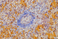
hVOD (also known as sinusoidal obstruction syndrome) may include fibrous concentric narrowing of zone 3 terminal hepatic venules, centrilobular hepatocyte necrosis, and sinusoidal congestion (see ).
Note: If hepatic biopsy is contraindicated, hepatic ultrasonography and Doppler ultrasonography may provide supportive evidence of hVOD.
Hepatic biopsy showing vascular obliteration, perivenular fibrosis, zone 3 fibrosis, and hepatocyte dropout from a girl who presented at age five months with hepatomegaly and ascites (Picro-Mallory stain 100x)
Establishing the Diagnosis
The diagnosis of VODI is established in a proband with the above clinical diagnostic criteria. Identification of biallelic pathogenic variants in SP110 on molecular genetic testing establishes the diagnosis if clinical features are inconclusive (see Table 1).
Molecular testing approaches can include single-gene testing, use of a multigene panel, and more comprehensive genomic testing.
Targeted analysis for the
c.642delC pathogenic variants can be performed first in individuals of Lebanese ancestry.
Sequence analysis of
SP110 may be performed in a tiered approach beginning with exons 2, 4, 5 and 8 (reference sequence: LRG_109). Sequencing of the entire
coding region of 19 exons and an alternatively spliced
exon 15 in the Sp110c isoform is performed if neither or only one
pathogenic variant is identified in exons 2, 4, 5 and 8.
A multigene panel that includes
SP110 and other genes of interest (see
Differential Diagnosis) may also be considered. Note: (1) The genes included in the panel and the diagnostic
sensitivity of the testing used for each
gene vary by laboratory and are likely to change over time. (2) Some multigene panels may include genes not associated with the condition discussed in this
GeneReview; thus, clinicians need to determine which multigene panel is most likely to identify the genetic cause of the condition while limiting identification of variants of
uncertain significance and pathogenic variants in genes that do not explain the underlying
phenotype. (3) In some laboratories, panel options may include a custom laboratory-designed panel and/or custom phenotype-focused
exome analysis that includes genes specified by the clinician. (4) Methods used in a panel may include
sequence analysis,
deletion/duplication analysis, and/or other non-sequencing-based tests.
For an introduction to multigene panels click
here. More detailed information for clinicians ordering genetic tests can be found
here.
More comprehensive genomic testing (when available) including
exome sequencing and
genome sequencing may be considered if single-
gene testing (and/or use of a
multigene panel that includes
SP110) fails to confirm a diagnosis in an individual with features of VODI. Such testing may provide or suggest a diagnosis not previously considered (e.g., mutation of a different gene or genes that results in a similar clinical presentation).
For an introduction to comprehensive
genomic testing click
here. More detailed information for clinicians ordering genomic testing can be found
here.
Table 1.
Molecular Genetic Testing Used in Hepatic Veno-Occlusive Disease with Immunodeficiency
View in own window
| Gene 1 | Method | Proportion of Probands with Pathogenic Variants 2 Detectable by Method |
|---|
|
SP110
| Sequence analysis 3 | 13/13 (100%) 4 |
| Gene-targeted deletion/duplication analysis 5 | Unknown 6 |
- 1.
- 2.
- 3.
- 4.
- 5.
Gene-targeted deletion/duplication analysis detects intragenic deletions or duplications. Methods used may include quantitative PCR, long-range PCR, multiplex ligation-dependent probe amplification (MLPA), and a gene-targeted microarray designed to detect single-exon deletions or duplications.
- 6.
Clinical Characteristics
Clinical Description
Hepatic veno-occlusive disease with immunodeficiency (VODI) is a primary immunodeficiency associated with terminal hepatic lobular vascular occlusion and hepatic lobule zone 3 fibrosis. All children in the cohort from Sydney, Australia presented prior to age six months, the majority with sequelae of the immunodeficiency either alone or concurrently with features of hepatic veno-occlusive disease (hVOD) (see Table 2).
Immunodeficiency is characterized by severe hypogammaglobulinemia, clinical evidence of T-cell immunodeficiency with normal numbers of circulating T and B cells, absent lymph node germinal centers, and absent tissue plasma cells [Roscioli et al 2006]. Bacterial and opportunistic infections including Pneumocystis jirovecii infection, mucocutaneous candidiasis, and enteroviral or cytomegalovirus infections occur.
Hepatic veno-occlusive disease (hVOD). Ninety percent of the children with VODI present ab initio either with hepatomegaly (83% with preceding infection) or hepatic failure (53% with preceding infection). Typically, an infant has been unwell for several weeks, has become tachypneic, is failing to thrive, is admitted with interstitial pneumonitis caused by Pneumocystis jirovecii, is found to be jaundiced with ascites, has hypogammaglobulinemia and thrombocytopenia, and has evidence of hVOD.
Neurologic. Overall, 30% of children with VODI had neurologic involvement; none of the individuals were reported to have veno-occlusive disease of the brain. Four individuals (sister of B II.1 and B II.2 [Table 3] and 3 unrelated children) had multiorgan failure associated with extensive cerebral necrosis on postmortem examination. A striking finding is the presence of cerebrospinal leukodystrophy in three individuals (20%) with VODI. Individual 5 had a leukodystrophy of unknown etiology and individual 6 developed this complication after a CMV-related gastroenteritis. In individual A II.1, the initial diagnosis of a cerebrovascular accident with a right-sided cerebral white matter lesion – presumed to be Toxoplasma or a porencephalic cyst – was revised as more consistent with cerebrospinal leukodystrophy. Individual B II.2 remained well on intravenous immunoglobulin (IVIG) and cotrimoxazole prophylaxis until age 18 years when she suddenly developed paraparesis and urinary retention associated with cerebral lesions. No infective cause or evidence of veno-occlusive disease was found on extensive investigation. There was partial response to high-dose IVIG and steroid therapy, suggesting an inflammatory etiology [MW Lin and M Wong, personal communication].
Other features. Infants and children with VODI do not have dysmorphic features. Thrombocytopenia is frequent at presentation but may improve with resolution of the hVOD. One child had the syndrome of inappropriate ADH secretion, presumably as a manifestation of his cerebral leukodystrophy.
Morbidity/mortality. VODI is associated with 100% mortality in the first year of life if unrecognized and untreated with IVIG or subcutaneous immunoglobulin (SCIG) replacement and Pneumocystis jirovecii prophylaxis, and a 90% mortality overall by the mid-teenage years [Roscioli et al 2006]. However, there have been only three deaths among eight recently ascertained affected individuals older than age one year, representing a markedly improved prognosis with early recognition and treatment [Cliffe et al 2012]. Should hVOD recovery occur, recurrence of hVOD appears to be prevented by continuation of IVIG and Pneumocystis
jirovecii prophylaxis. However, there may be an ongoing risk for neurologic inflammatory complications (B II.2). One child (Individual A II.1, Table 3) died following recurrence of hVOD after bone marrow transplantation at age six years. Table 2 summarizes the clinical and immunologic features of the 20 individuals from Sydney with the clinical diagnosis of VODI (including the 11 individuals who were able to be investigated by molecular analysis confirming the presence of SP110 pathogenic variants) and eight newly ascertained individuals [Cliffe et al 2012].
Table 2.
Clinical and Immunologic Features of Hepatic Veno-Occlusive Disease with Immunodeficiency
View in own window
| Clinical Features | Individuals from Sydney w/VODI 1 | Comments | Newly Ascertained Individuals w/VODI w/Novel Pathogenic Variants 2 |
|---|
| Presenting <6 months | 20/20 (100%) | | 7/8 |
| Hepatic failure at initial presentation | 4/20 (20%) | 1/12 post-HSCT
3/12 no obvious precipitant | 0/8 |
| Hepatomegaly at initial presentation | 9/20 (45%) | 3/6 P. jirovecii
2/6 hepatomegaly w/out SOS | 6/8
1/8 enterovirus & disseminated cytomegalovirus 3 |
| P. jirovecii infection | 12/20 (60%) | 7/12 proven
5/12 suspected | 1/8 suspected 3
1/8 proven 4 |
| Mucocutaneous candidiasis | 2/20 (10%) | | 1/8 |
| Other features | 1/20 (5%) | By age 19 years | 1/8 lung fibrosis 5 |
| Death | 19/20 (95%) | | 3/8 (38%) |
| Recovery from initial SOS | 4/20 (20%) | 1 completely well
1 chronic liver disease requiring hepatic transplantation
1 SOS post-HSCT
1 developmental disability, chronic aspiration | 4/8 |
| Neurologic abnormalities | 6/20 (35%) | 4/7 cerebral infarction
2/7 leukodystrophy | 1/8 leukodystrophy |
| Panhypogammaglobulinemia | 19/19 (100%) | 1/18 loss of normal immunoglobulins at age 4 mos | 5/5 tested
1/5 low normal levels of IgA & IgM after commencing IVIG |
| Normal number of lymphocytes | 10/11 (92%) | | 8/8 |
| Normal NK cells | 12/12 (100%) | | 3/3 3, 4, 5 |
| Decreased intracytoplasmic IFN-γ, IL-2, IL-4, IL-10 | 4/5 (80%) | Low levels at 4 hrs, normal/increased levels at 48 hrs | 1/1 3 |
| Decreased number of memory T & B cells | 3/4 (75%) | | 2/3 6 |
HSCT = hematopoietic stem cell transplantation
IVIG = intravenous immunoglobulin
SOS = sinusoidal obstruction syndrome
- 1.
- 2.
- 3.
- 4.
- 5.
- 6.
Memory T and B cells were present in affected individual 1 (see Table 3)
Table 3 outlines clinical features in individuals with identified homozygous SP110 pathogenic variants [Roscioli et al 2006, Cliffe et al 2012, Wang et al 2012, Ganaiem et al 2013].
Table 3.
Clinical Features of Individuals Homozygous for SP110 Pathogenic Variants
View in own window
| Affected Individual | SP110 (LRG_109) Pathogenic Variant | Presentation | Serum Igs | Memory T/B Cells | T Cell Cytokines | Clinical Findings | Deceased |
|---|
A II.1 1
Lebanese | c.642delC in exon 5 | Age 5 mos: immunodeficiency, thrombocytopenia, SOS | ↓ | N/A | N/A | Left hemiparesis 2, recurrent hVOD w/GVHD post-HSCT | Yes |
B II.1 1
Lebanese | Age 7 mos: immunodeficiency | ↓ | N/A | N/A | Chronic lung disease secondary to recurrent aspiration | Yes (age 19 yrs) |
B II.2 1
Lebanese | Age 6 mos: hepatosplenomegaly, ascites, SOS | ↓ | ↓ | ↓ | Well until 18 yrs (developed paraparesis, urinary retention, cerebral lesions) | No |
C II.1 1
Lebanese | Age 4 mos: hepatosplenomegaly, ascites, SOS, thrombocytopenia, mucocutaneous candidiasis | ↓ | ↓ | ↓ | Chronic liver disease, portal hypertension post-hepatic transplantation | Yes |
D II.1 1
Lebanese | Age 3 mos: hepatosplenomegaly, ascites, SOS | ↓ 3 | ↓ | ↓ | Hemophagocytic syndrome post-hepatic transplantation | Yes |
16 4
Lebanese | c.642delC in exon 5 | Age 3 mos: hepatosplenomegaly, ascites, SOS | ↓ | ↓ | ↓ | Pulmonary hemorrhage, multiorgan failure | Yes |
5 4
Lebanese | c.642delC in exon 5 | Age 3 mos, respiratory distress | ↓ | ↓ | N/A | SIADH, idiopathic cerebrospinal leukodystrophy | No |
6 4
Lebanese | c.642delC in exon 5 | Age 3 mos: chronic cough, diarrhea
Age 8 yrs: hepatosplenomegaly
Age ≥12 yrs: hVOD | ↓ | N/A | N/A | Idiopathic left frontal lobe calcified cyst, epilepsy, CMV colitis, post-diarrheal encephalomyelitis w/lower limb paralysis, cerebrospinal leukodystrophy, esophageal candidiasis, duodenal lymphocytic infiltrate | No |
7 4
Lebanese | c.642delC presumed 5 | Age 2 mos: chronic diarrhea, failure to thrive, middle ear & respiratory infections, hepatosplenomegaly, thrombocytopenia | N/A | N/A | N/A | Microcephaly, hepatic biopsy consistent w/SOS | Yes, 11 mos from diarrhea leading to septic shock |
8 4
Lebanese | c.642delC presumed 5 | Age 5 mos: upper respiratory illness
Age 8 mos: chronic diarrhea, hepatomegaly, thrombocytopenia | N/A | N/A | N/A | Hepatic biopsy consistent w/SOS | Yes, 3.5 yrs from diarrhea leading to septic shock |
9 4
Lebanese | c.642delC presumed 5 | Age 2 mos: ascites, hepatomegaly, anemia, thrombocytopenia | N/A | N/A | N/A | Hepatic biopsy consistent w/SOS | Yes, 2.5 mos from otitis, diarrhea, pneumonia |
E I.1 1
Lebanese | c.40delC in exon 2 | Age 3 mos: immunodeficiency, thrombocytopenia, hepatosplenomegaly w/out definite evidence of SOS | ↓ | N/A | N/A | Enteroviral & P. jirovecii infection | Yes |
4 4
Hispanic | c.78_79delinsAT (p.Ile27Leu) in exon 2 | Age 5 mos: hepatosplenomegaly, fever, respiratory distress, pancytopenia | ↓ | ↓ | ↓ | Stable & well | No |
1 4
Italian | c.319_325dup GGTGCTT in exon 4 | Age 11 mos: hepatosplenomegaly, disseminated CMV, rotavirus gastroenteritis, vulvar abscesses, SOS | ↓ initially | ↓ | N/A | Recovering from hVOD, well | No |
2 4
Italian | c.667+1dup exon 5 splice site | Age 3 mos: hepatosplenomegaly, failure to thrive, respiratory distress/lung fibrosis, diarrhea | ↓ | ↓ | N/A | Hepatic biopsy consistent w/sinusoidal dilatation, moderate central vein & perivenular subsinusoidal fibrosis; stable w/improvement | No |
3 4
Palestinian Arab | c.373del in exon 4 | Age 3 mos: diagnosis of VODI confirmed w/cascade testing prior to illness onset. No hepatomegaly or liver function abnormalities | N/A | N/A | N/A | Stable & well | No |
CMV = cytomegalovirus; GVHD = graft-versus-host disease; HSCT = hematopoietic stem cell transplantation; hVOD = hepatic veno-occlusive disease; SIADH = syndrome of inappropriate antidiuretic hormone secretion; SOS = sinusoidal obstruction syndrome
Note: Although families A, B, and C are not known to be related, they are believed to have a common ancestor. Individuals included in the initial homozygosity mapping analysis: A II.1, B II.1, B II.2, C II.1, 16 (‘G’ in initial analysis), and 5 (‘J’ in initial analysis).
Affected individual designations are those used in the articles cited.
- 1.
- 2.
Secondary to cerebral white matter abnormality
- 3.
IgA and IgM serum concentrations increased to lower limit of normal while on IVIG.
- 4.
- 5.
Genotype-Phenotype Correlations
No significant difference in the clinical manifestations of VODI is observed between individuals with different pathogenic variants.
The one child with an out-of-frame duplication in exon 4 (Individual 1, Table 3) presented at age 11 months (later than average) with disseminated CMV infection, which has not been noted in other children with VODI. In addition, the numbers of memory T and B cells were normal and intracellular cytokine production was normal – findings not observed in other children with VODI.
Penetrance
Penetrance for the combined B- and T-cell immunodeficiency has been 100% in individuals confirmed to have VODI caused by pathogenic variants in SP110. Likewise, hVOD has been described in all probands or their affected sibs.
Approximately 10% of children with VODI, ascertained at a young age because of an affected sib and treated early in the disease course with IVIG, may manifest immunodeficiency only at presentation.
Nomenclature
Hepatic veno-occlusive disease alone was known previously as Jamaican bush tea disease due to a dietary and geographic association. This term is now superseded by hepatic veno-occlusive disease (hVOD) or sinusoidal obstruction syndrome (SOS), terms less limiting given the occurrence of hVOD worldwide and it being secondary to other precipitants. The combination of hVOD and a combined immunodeficiency is termed VODI.
Prevalence
VODI was described originally in Australians of Lebanese origin by Mellis & Bale [1976]. Subsequently, the majority of children reported with VODI have been of Lebanese origin. The prevalence of VODI in the Lebanese population of Sydney, Australia has been calculated at one in 2,500 [Roscioli et al 2006]. Unpublished data from Australia and Lebanon suggest that VODI associated with the SP110 exon 5 pathogenic variant c.642delC is found not infrequently in families of Lebanese background, especially in association with consanguinity. This in turn suggests that the diagnosis has been missed in expatriate Lebanese communities.
The prevalence of VODI in children of non-Lebanese origin is unknown; however, the following reports suggest that the VODI phenotype is observed in other populations. Additional reports of VODI:
A
simplex case of VODI (i.e., a single occurrence in a family) with humoral and cellular immunodeficiency in the Spanish literature
Differential Diagnosis
Although sinusoidal obstruction syndrome in association with severe combined immunodeficiency (SCID) was described in an individual reported by Washington et al [1993], and in one postmortem HIV cohort reported by Buckley & Hutchins [1995], the lack of a recognized and replicated association of immunodeficiency with hepatic veno-occlusive disease (hVOD) in other classes of immunodeficiency suggests that hVOD may be a primary feature of VODI rather than secondary to an immunodeficiency per se.
The primary differential diagnosis for hVOD alone would be environmental alkaloid or sinusoidal cell toxicity. However, hVOD has also been reported in association with alcoholic cirrhosis [Kishi et al 1999], ataxia-telangiectasia [Srisirirojanakorn et al 1999], osteopetrosis [Corbacioglu et al 2006] (see CLCN7-Related Osteopetrosis), and hypereosinophilic syndrome (OMIM 607685). HIV should also be considered in the differential diagnosis for the immune phenotype.
Previous case-control studies using single-nucleotide polymorphisms (SNPs) have also reported associations between hVOD and SNPs in the carbamoyl phosphate synthetase 1 (CPS1) (see Urea Cycle Disorders Overview), factor V Leiden (FVL), HFE (see HFE-Associated Hereditary Hemochromatosis), and glutathione S-transferase (GSTM1 and GSTT1) genes. Relative risks of 8.6 for the homozygous HFE Cys282Tyr allele and 4.12 for the GSTM1 null allele have been reported [Srivastava et al 2004, Kallianpur 2005, Kallianpur et al 2005]. No independent replication of these findings has been performed.
There has been no report of SP110 pathogenic variants in individuals described to have hVOD without immunodeficiency.
Management
Evaluations Following Initial Diagnosis
To establish the extent of disease and needs in an individual diagnosed with hepatic veno-occlusive disease with immunodeficiency (VODI), the following evaluations are recommended:
Assessment of immune function including serum immunoglobulin levels, T- and B-cell numbers and percentages, and T-cell proliferative response to mitogens
More extensive immune testing for number of memory B and T cells and intracellular cytokine (IL2, IL4, IL6, and IFNγ) responses to stimulation, if available
Complete blood count
Assessment of hepatic function (including serum concentrations of aminotransferases, bilirubin, and albumin) and assessment for sequelae of portal hypertension (including anemia and thrombocytopenia)
Consultation with a clinical geneticist and/or genetic counselor
A clotting profile and a hepatic Doppler ultrasound examination should be undertaken prior to consideration of hepatic biopsy for a histologic diagnosis of hepatic veno-occlusive disease (hVOD). Evidence of impaired clotting and/or portal hypertension contraindicates hepatic biopsy.
Treatment of Manifestations
Hypogammaglobulinemia is treated via intravenous immunoglobulin (IVIG), which should commence at the diagnosis of VODI or in presymptomatic sibs confirmed to have homozygous SP110 pathogenic variants. An appropriate dose is 0.4 g/kg every four weeks adjusting the dose to maintain a trough IgG level greater than 6 g/L.
Pneumocystis jirovecii prophylaxis with cotrimoxazole pediatric suspension (5 mL = trimethoprim 40 mg and sulfamethoxazole 200 mg) should be ongoing in children with VODI who tolerate this medication. This may be administered as a single daily dose or as a single dose three days per week. The recommended dose is 5 mg trimethoprim per kg (0.625 mL/kg) or 150 mg/M2 (3.75 mL/M2).
Infections with specific agents should be treated with appropriate supportive care and antibacterials or antivirals.
Hepatic transplantation may be considered, but appears to have a high rate of complications in the VODI cohort studied to date (see Other).
Bone marrow transplantation.
Ganaiem et al [2013] reported that this may be an efficacious treatment modality with appropriate conditioning therapy (see Other).
Prevention of Primary Manifestations
Initiation of regular IVIG at the time of diagnosis to prevent infection related to severe hypogammaglobulinemia and cotrimoxazole prophylaxis to prevent Pneumocystis jirovecii infection is appropriate (see Treatment of Manifestations).
Prevention of Secondary Complications
Some evidence suggests that treatment of immunodeficiency early in VODI may reduce the risk for development or recurrence of hVOD.
Surveillance
The following are appropriate:
Regular surveillance of hepatic function, platelet count, and hemoglobin level in children with VODI as hepatic failure and portal hypertension may occur
Surveillance of serum and urine electrolytes as the syndrome of inappropriate anti-diuretic hormone (SIADH) may occur
Measurement of immunoglobulin concentrations prior to IVIG infusions
Broncho-alveolar lavage to diagnose Pneumocystis jirovecii infection; viral cultures or lung function studies as needed
Cerebrospinal imaging to diagnose leukodystrophy when clinically indicated
Agents/Circumstances to Avoid
Agents known to predispose to hVOD (e.g., cyclophosphamide and senecio alkaloids/bush teas) should be avoided.
Evaluation of Relatives at Risk
It is appropriate to evaluate sibs of a proband who are younger than age 12 months in order to identify as early as possible those who would benefit from initiation of treatment and preventive measures. The majority of children with VODI present before age six months; however, as one child presented at age 11 months, testing should be considered in sibs of a proband who are younger than age 12 months.
Evaluations include:
Molecular genetic testing if the pathogenic variants in the family are known;
Serum immunoglobulins, full blood count and liver function tests at birth and repeated at six months if the pathogenic variants in the family are not known.
Penetrance is complete (i.e., 100%) in the individuals with VODI described to date; thus, molecular genetic testing of healthy at-risk sibs of a proband who are older than age 12 months is not recommended.
See Genetic Counseling for issues related to testing of at-risk relatives for genetic counseling purposes.
Pregnancy Management
For an affected pregnant woman, ongoing intravenous immunoglobulin to prevent infection related to severe hypogammaglobulinemia and cotrimoxazole prophylaxis to prevent Pneumocystis jirovecii infection is appropriate during pregnancy. There is evidence that early treatment of a baby known to be homozygous for pathogenic SP110 pathogenic variants may result in improved long-term outcomes.
Therapies Under Investigation
Search ClinicalTrials.gov in the US and EU Clinical Trials Register in Europe for access to information on clinical studies for a wide range of diseases and conditions. Note: There may not be clinical trials for this disorder.
Other
Hepatic VOD (hVOD) has been reported in the Australian cohort with VODI following hematopoietic stem cell transplantation (HSCT); therefore, individuals with VODI are likely to have at least the population risk for hVOD following HSCT. The first successful HSCT was reported by Ganaiem et al [2013] suggesting that this may be an efficacious treatment modality with appropriate conditioning therapy. Ganaiem et al described a consanguineous Arab family with eight affected individuals; five individuals received HSCT, of which three were successful. All five individuals received matched sib or family member bone marrow (4 individuals) or peripheral stem cells (1 individual). All five individuals received conditioning with fludarabine and serotherapy and an alkylating agent (cyclophosphamide, busulfan, or treosulfan). The two individuals who died also received thiotepa, and following engraftment died with hVOD and multiorgan failure. One child has remained well for two years following uncomplicated HSCT in 2014. She was of consanguineous Lebanese background and presented at age four months with probable herpetic hepatitis which initially responded clinically to acyclovir, but then developed florid hVOD following a transfusion. She responded well to IVIG, prophylactic Bactrim™, and defibrotide. She was neurologically normal, but asymptomatic bilateral subdural hematomas were found on routine pre-transplant MRI despite a normal head ultrasound a week prior when coagulation studies and platelets had already normalized. At age six months she underwent an unrelated cord blood transplant with alemtuzumab, fludarabine, treosulfan and antithymocyte globulin conditioning. Defibrotide was prophylactically continued until two months post transplant. She remains neurologically normal and her MRI scan at age two years showed no anatomic abnormality [M Wong, personal communication].
In 2016, an Australian infant of consanguineous Lebanese background presented at age four months with Pneumocystis, CMV infection, and hVOD with hypogammaglobinemia. A homozygous SP110 pathogenic variant (c.642delC) was identified on molecular genetic testing. At age five months, after control of pneumonitis and hVOD, he received a haplo-identical paternal HSCT after depletion of α/β TCR and CD19+ cells. He received conditioning with fludarabine, treosulfan, and antithymocyte globulin. Defibrotide was given as hVOD prophylaxis. Two months post transplant he was well and had engrafted without GVHD or recurrence of hVOD but with moderate CMV hepatitis. Defibrotide was able to be discontinued at day 60. Both the affected individual and donor were CMV positive prior to transplant. Seventy days following HSCT he developed seizures. MRI showed extensive abnormalities throughout the brain and spinal cord, including areas of edema and ring-like enhancing lesions. CT showed widespread calcifications. CMV retinitis was apparent and both CSF and vitreous were CMV PCR positive [T Cole, J Smart, and T Soosay Raj, personal communication].
Inclusion of defibrotide prophylaxis in the transplant regimen may improve survival. Omission of a second alkylating agent in the conditioning regimen may reduce the risk for transplant-related hVOD.
Genetic Counseling
Genetic counseling is the process of providing individuals and families with
information on the nature, mode(s) of inheritance, and implications of genetic disorders to help them
make informed medical and personal decisions. The following section deals with genetic
risk assessment and the use of family history and genetic testing to clarify genetic
status for family members; it is not meant to address all personal, cultural, or
ethical issues that may arise or to substitute for consultation with a genetics
professional. —ED.
Mode of Inheritance
Hepatic veno-occlusive disease with immunodeficiency (VODI) is inherited in an autosomal recessive manner. Penetrance is complete.
Risk to Family Members
Parents of a proband
Sibs of a proband
Offspring of a proband. The offspring of an individual with hepatic veno-occlusive disease with immunodeficiency are obligate heterozygotes (carriers) for a pathogenic variant in SP110.
Other family members of a proband. Each sib of the proband's parents is at a 50% risk of being a carrier of an SP110 pathogenic variant.
Carrier Detection
Carrier testing for at-risk relatives requires prior identification of the SP110 pathogenic variants in the family.
Prenatal Testing and Preimplantation Genetic Testing
Once the SP110 pathogenic variants have been identified in an affected family member, prenatal testing for a pregnancy at increased risk and preimplantation genetic testing for VODI are possible.
Resources
GeneReviews staff has selected the following disease-specific and/or umbrella
support organizations and/or registries for the benefit of individuals with this disorder
and their families. GeneReviews is not responsible for the information provided by other
organizations. For information on selection criteria, click here.
International Patient Organization for Primary Immunodeficiencies (IPOPI)
United Kingdom
Phone: +44 01503 250 668
Fax: +44 01503 250 668
Email: info@ipopi.org
Jeffrey Modell Foundation/National Primary Immunodeficiency Resource Center
Email: info@jmfworld.org
European Society for Immunodeficiencies (ESID) Registry
Email: esid-registry@uniklinik-freiburg.de
Molecular Genetics
Information in the Molecular Genetics and OMIM tables may differ from that elsewhere in the GeneReview: tables may contain more recent information. —ED.
Table A.
Hepatic Veno-Occlusive Disease with Immunodeficiency: Genes and Databases
View in own window
Data are compiled from the following standard references: gene from
HGNC;
chromosome locus from
OMIM;
protein from UniProt.
For a description of databases (Locus Specific, HGMD, ClinVar) to which links are provided, click
here.
Gene structure.
SP110 encodes three major isoforms:
Sp110 isoform A,
NM_004509.3 (average mass 78.438 kd; transcript does not include
exon 17)
Isoform B,
NM_004510.3 (average mass 61.940 kd; transcript includes an alternate
exon 15 and terminates within exon 15)
Isoform C,
NM_080424.2 (average mass 81.211 kd; full-length transcript including
exon 17 and terminating at exon 19)
Pathogenic variants. See Table 4. The majority of these pathogenic variants cause a frameshift with consequent protein truncation. The c.642delC pathogenic variant has been identified in multiple probands of Lebanese ancestry [Roscioli et al 2006, Cliffe et al 2012].
Table 4.
SP110 Pathogenic Variants Discussed in This GeneReview
View in own window
Variants listed in the table have been provided by the authors. GeneReviews staff have not independently verified the classification of variants.
GeneReviews follows the standard naming conventions of the Human Genome Variation Society (varnomen.hgvs.org). See Quick Reference for an explanation of nomenclature.
- 1.
Variant designation that does not conform to current naming conventions
Normal gene product. Sp110 is expressed primarily in leukocytes and spleen; it is induced by interferon gamma and all-trans retinoic acid (ATRA). SP110 isoform B has been described as showing activity as a potent transcriptional co-repressor of retinoic acid receptor alpha (RARα) perhaps via competitive exclusion of activators at receptor [Watashi et al 2003].
The Sp110 nuclear body protein is a member of the Sp100/Sp140 promyelocytic leukemia nuclear body (PML NB) protein family. The protein has an Sp100 domain (amino acids 6-159), which is involved in dimerization with other Sp100 family proteins, a nuclear localization signal (amino acids 288-306) and a nuclear hormone interaction domain (LXXLL type), which may act as an ATRA response element. Other domains that are common features of modular proteins involved in chromatin-mediated gene transcription include a SAND domain (amino acids 452-532), a plant homeobox domain (amino acids 537-577), and a bromodomain (amino acids 606-674) [Bloch et al 2000].
The Sp110 nuclear body protein is associated with the PML NB, a nuclear macromolecular complex, which is deployed to areas of active host or viral DNA replication, transcription, and repair and has been reported to be involved in apoptosis, cell cycle control, and the immune response.
Abnormal gene product. EBV-transformed B cells from an individual with VODI and a homozygous inactivating SP110 variant have shown an absence of nuclear Sp100-specific immunolabeling in a setting of normal numbers of PML nuclear bodies. This finding is consistent with Sp110 protein having an important role in the immune response without being essential for PML nuclear body formation [Roscioli et al 2006].
Most pathogenic variants associated with VODI are loss-of-function variants. The c.78_79delinsAT (p.Ile27Leu) pathogenic variant occurs at the highly conserved p.Ile27 within the highly conserved Sp100 domain (see Normal gene product). The c.78_79delinsAT (p.Ile27Leu) pathogenic variant greatly reduces Sp110 stability [Cliffe et al 2012].
Pathophysiology. It is currently unknown whether the hVOD is a direct manifestation of SP110 sequence variants, related to altered apoptosis in the hepatic sinusoid, or secondary to infection; however, hVOD appears to develop after infections occur.
References
Literature Cited
Bloch DB, Nakajima A, Gulick T, Chiche JD, Orth D, de La Monte SM, Bloch KD. Sp110 localizes to the PML-Sp100 nuclear body and may function as a nuclear hormone receptor transcriptional coactivator.
Mol Cell Biol. 2000;20:6138–46. [
PMC free article: PMC86089] [
PubMed: 10913195]
Buckley JA, Hutchins GM. Association of hepatic veno-occlusive disease with the acquired immunodeficiency syndrome.
Mod Pathol. 1995;8:398–401. [
PubMed: 7567938]
Cliffe ST, Bloch DB, Suryani S, Kamsteeg EJ, Avery DT, Palendira U, Church JA, Wainstein BK, Trizzino A, Lefranc G, Akatcherian C, Megarbané A, Gilissen C, Moshous D, Reichenbach J, Misbah S, Salzer U, Abinun M, Ong PY, Stepensky P, Ruga E, Ziegler JB, Wong M, Tangye SG, Lindeman R, Buckley MF, Roscioli T. Clinical, molecular, and cellular immunologic findings in patients with SP110-associated veno-occlusive disease with immunodeficiency syndrome.
J Allergy Clin Immunol. 2012;130:735–742.e6. [
PubMed: 22621957]
Corbacioglu S, Honig M, Lahr G, Stohr S, Berry G, Friedrich W, Schulz AS. Stem cell transplantation in children with infantile osteopetrosis is associated with a high incidence of VOD, which could be prevented with defibrotide.
Bone Marrow Transplant. 2006;38:547–53. [
PubMed: 16953210]
Ganaiem H, Eisenstein EM, Tenenbaum A, Somech R, Simanovsky N, Roscioli T, Weintraub M, Stepensky P. The role of hematopoietic stem cell transplantation in SP110 associated veno-occlusive disease with immunodeficiency syndrome.
Pediatr Allergy Immunol. 2013;24:250–6. [
PubMed: 23448538]
Kallianpur AR. Genomic screening and complications of hematopoietic stem cell transplantation: has the time come?
Bone Marrow Transplant. 2005;35:1–16. [
PubMed: 15489868]
Kallianpur AR, Hall LD, Yadav M, Byrne DW, Speroff T, Dittus RS, Haines JL, Christman BW, Summar ML. The hemochromatosis C282Y allele: a risk factor for hepatic veno-occlusive disease after hematopoietic stem cell transplantation.
Bone Marrow Transplant. 2005;35:1155–64. [
PubMed: 15834437]
Kishi M, Maeyama S, Ogata S, Koike J, Uchikoshi T. Hepatic veno-occlusive lesions in severe alcoholic hepatitis and alcoholic liver cirrhosis: a comparative histopathological study in autopsy cases.
Alcohol Clin Exp Res. 1999;23:47S–51S. [
PubMed: 10235278]
Mellis C, Bale PM. Familial hepatic venoocclusive disease with probable immune deficiency.
J Pediatr. 1976;88:236–42. [
PubMed: 1249685]
Roscioli T, Cliffe ST, Bloch DB, Bell CG, Mullan G, Taylor PJ, Sarris M, Wang J, Donald JA, Kirk EP, Ziegler JB, Salzer U, McDonald GB, Wong M, Lindeman R, Buckley MF. Mutations in the gene encoding the PML nuclear body protein Sp110 are associated with immunodeficiency and hepatic veno-occlusive disease.
Nat Genet. 2006;38:620–2. [
PubMed: 16648851]
Ruga EM, Guariso G, Antiga LD, Guido M, Fassan M, Elia RD, Ziegler JB, Roscioli T, Cliffe ST, Buckley MF, Zancan L. Hepatic veno-occlusive disease with immunodeficiency syndrome: case report. Budapest, Hungary: 12th Meeting of the European Society for Immunodeficiencies. 2006.
Srisirirojanakorn N, Finegold MJ, Gopalakrishna GS, Klish WJ. Hepatic veno-occlusive disease in ataxia-telangiectasia.
J Pediatr. 1999;134:786–8. [
PubMed: 10356154]
Srivastava A, Poonkuzhali B, Shaji RV, George B, Mathews V, Chandy M, Krishnamoorthy R. Glutathione S-transferase M1 polymorphism: a risk factor for hepatic venoocclusive disease in bone marrow transplantation.
Blood. 2004;104:1574–7. [
PubMed: 15142875]
Wang T, Ong P, Roscioli T, Cliffe ST, Church JA. Hepatic veno-occlusive disease with immunodeficiency (VODI): first reported case in the U.S. and identification of a unique mutation in Sp110.
Clin Immunol. 2012;145:102–7. [
PubMed: 22982295]
Washington K, Gossage DL, Gottfried MR. Pathology of the liver in severe combined immunodeficiency and DiGeorge syndrome.
Pediatr Pathol. 1993;13:485–504. [
PubMed: 8372033]
Watashi K, Hijikata M, Tagawa A, Doi T, Marusawa H, Shimotohno K. Modulation of retinoid signaling by a cytoplasmic viral protein via sequestration of Sp110b, a potent transcriptional corepressor of retinoic acid receptor, from the nucleus.
Mol Cell Biol. 2003;23:7498–509. [
PMC free article: PMC207568] [
PubMed: 14559998]
Chapter Notes
Revision History
12 January 2017 (sw) Comprehensive update posted live
3 July 2013 (me) Comprehensive update posted live
15 September 2009 (me) Comprehensive update posted live
21 February 2007 (me) Review posted live
29 November 2006 (mb) Original submission