U.S. flag

An official website of the United States government

NCBI Bookshelf. A service of the National Library of Medicine, National Institutes of Health.

Adam MP, Feldman J, Mirzaa GM, et al., editors. GeneReviews® [Internet]. Seattle (WA): University of Washington, Seattle; 1993-2024.

Cover of GeneReviews®

GeneReviews® [Internet].

Show details

Primary Hyperoxaluria Type 1

, MD, , PhD, , DO, , BS, and , MD.

Author Information and Affiliations

Initial Posting: ; Last Revision: February 10, 2022.

Estimated reading time: 1 hour

Summary

Clinical characteristics.

Primary hyperoxaluria type 1 (PH1) is caused by a deficiency of the liver peroxisomal enzyme alanine:glyoxylate-aminotransferase (AGT), which catalyzes the conversion of glyoxylate to glycine. When AGT activity is absent, glyoxylate is converted to oxalate, which forms insoluble calcium oxalate crystals that accumulate in the kidney and other organs. Individuals with PH1 are at risk for recurrent nephrolithiasis (deposition of calcium oxalate in the renal pelvis / urinary tract), nephrocalcinosis (deposition of calcium oxalate in the renal parenchyma), or end-stage renal disease (ESRD). Age at onset of symptoms ranges from infancy to the sixth decade. Approximately 10% of affected individuals present in infancy or early childhood with nephrocalcinosis, with or without nephrolithiasis, and failure to thrive related to renal failure. The majority of individuals with PH1 present in childhood or early adolescence, usually with symptomatic nephrolithiasis and normal or reduced kidney function. The remainder of affected individuals present in adulthood with recurrent renal stones and a mild-to-moderate reduction in kidney function. The natural history of untreated PH1 is one of progressive decline in renal function as a result of calcium oxalate deposits in kidney tissue and complications of nephrolithiasis (e.g., obstruction and infection) with eventual progression to oxalosis (widespread tissue deposition of calcium oxalate) and death from ESRD and/or complications of oxalosis.

Diagnosis/testing.

The diagnosis of PH1 is established in a proband with hyperoxaluria or hyperoxalemia by identification biallelic pathogenic variants in AGXT on molecular genetic testing. Failure to identify at least one AGXT pathogenic variant should prompt examination for other types of primary hyperoxaluria and in occasional circumstances may require consideration of liver biopsy to assay the activity of the enzyme AGT. However, given the wide availability of genetic testing, liver biopsy to obtain tissue for enzymatic activity is now rarely employed.

Management.

Treatment of manifestations: The primary strategy in prevention and treatment of the renal and systemic complications of PH1 is reduction of hepatic overproduction of oxalate. Until recently, the two options were pharmacologic doses of pyridoxine (limited to those individuals responsive to pyridoxine determined by their AGXT genotype) or liver transplantation (most often combined with or performed sequentially with kidney transplantation in persons with ESRD). In 2020 the FDA and EMA approved lumasiran, an mRNAi therapeutic agent that reduces the amount of glyoxylate substrate available for metabolic conversion to oxalate by targeting the hepatic enzyme glycolate oxidase (an enzyme distinct from AGT that is in the same metabolic pathway). Since lumasiran targets glycolate oxidase, it is expected to be effective in all individuals with PH1, independent of AGXT genotype.

Strategies to minimize calcium oxalate crystal and stone formation by reducing urinary calcium oxalate supersaturation include: high oral fluid intake; alkalization of the urine using oral potassium citrate; and/or oral solutions that increase urinary pyrophosphate.

Surveillance: At regular intervals, obtain serum creatinine to estimate glomerular filtration rate (GFR), renal ultrasound examinations or other kidney imaging, urinalysis; and periodic fundoscopic eye examinations. Additionally:

  • In those with reduced GFR (<60 mL/min/1.73 m2): regular measurement of plasma oxalate and more frequent monitoring of kidney function.
  • In individuals with greatly reduced GFR (<30 mL/min/1.73 m2) or rapid deterioration in function: frequent assessment of serum creatinine and plasma oxalate. At regular intervals obtain x-ray examination of the long bones, electrocardiogram for conduction abnormalities, echocardiogram for evidence of oxalate cardiomyopathy, hemoglobin, thyroid function testing, and frequent clinical evaluation for additional complications of oxalosis.

Agents/circumstances to avoid: Dehydration from any cause can lead to irreversible kidney failure and should be strictly avoided. Individuals with PH1 should avoid intake of vitamin C that exceeds the recommended daily allowance, loop diuretics, high doses of nonsteroidal anti-inflammatory medications or other medications that can compromise kidney function; and large intake of foods high in oxalate (e.g., chocolate, rhubarb, starfruit).

Evaluation of relatives at risk: Early diagnosis of at-risk relatives enables early institution of treatment and preventive measures.

Genetic counseling.

PH1 is inherited in an autosomal recessive manner. At conception, each sib of a proband with PH1 has a 25% risk of being affected, a 50% risk of being an asymptomatic carrier, and a 25% risk of being unaffected and not a carrier. Carrier testing for at-risk relatives and prenatal testing for a pregnancy at increased risk are possible if both pathogenic variants have been identified in a family. Assay of AGT enzymatic activity prenatally is not generally offered because it requires a fetal liver biopsy.

Diagnosis

Suggestive Findings

Primary hyperoxaluria type 1 (PH1) should be suspected in a proband with any of the following clinical, radiographic, and/or laboratory features.

Clinical and radiographic features

  • Recurrent nephrolithiasis. Renal ultrasound examination often reveals multiple bilateral radiopaque calculi (computed tomography and kidney ureter bladder x-ray may demonstrate similar findings).
  • Nephrocalcinosis. In older children or adults, the strongest echoes are from the corticomedullary regions, whereas in infants the pattern is more likely to be that of diffuse nephrocalcinosis with few if any observable discrete stones.
  • A child with a first kidney stone [Cochat et al 2012]
  • A child younger than age 12 months with failure to thrive [Cochat & Rumsby 2013] and impaired kidney function
  • Reduced kidney function or end-stage renal disease at any age with a history of renal stones or nephrocalcinosis [Edvardsson et al 2013]
  • Stone composition of pure calcium oxalate monohydrate (whewellite)
  • Oxalate crystals identified in any biologic fluid or tissue [Cochat et al 2012]

Laboratory findings

  • Elevated urinary oxalate excretion persistently >0.7 mmol/1.73 m2/day or above the age-related reference range(s) (see Table 1)
  • Elevated urinary glycolic acid (glycolate) concentration; occurs in approximately 75% of individuals with PH1 (see Table 1).
  • Elevated plasma oxalate concentration. Individuals with PH1 and well-preserved GFR typically have mildly elevated values. Substantially elevated values are the rule when GFR is <30 mL/min/1.73 m2 (CKD stages 4 and 5). Plasma oxalate concentrations >50 µmol/L are very suggestive of PH1 [Cochat & Rumsby 2013, Perinpam et al 2017].

Table 1.

Normal Values for Urinary Oxalate, Glycolate, and L-Glycerate Excretion in 24-Hour Urine and Spot (Random) Urine Samples

ParameterAgeNormal Values 1
Urinary oxalate excretion 2 All ages In 24-hour
urine
samples
<0.50 mmol (<45 mg)/1.73 m2/day
Urinary glycolate excretion <0.50 mmol (<45 mg)/1.73 m2/day
Urinary L-glycerate
concentration
<5 μmol/L
Spot urinary oxalate-to-
creatinine molar ratio 3
0-6 mos 4In spot
urine
samples 2
<325-360 mmol/mol (<253-282 mg/g)
7-24 mos 4<132-174 mmol/mol (<103-136 mg/g)
2-5 yrs<98-101 mmol/mol (<76-79 mg/g)
5-14 yrs<70-82 mmol/mol (<55-64 mg/g)
>16 yrs<40 mmol/mol (<32 mg/g)
Spot urinary glycolate-to-
creatinine molar ratio
0-6 mos 4<363-425 mmol/mol (<241-282 mg/g)
7-24 mos 4<245-293 mmol/mol (<163-194 mg/g)
2-5 yrs<191-229 mmol/mol (<127-152 mg/g)
5-14 yrs<166-186 mmol/mol (<110-123 mg/g)
>16 yrs<99-125 mmol/mol (<66-83 mg/g)
Spot urinary L-glycerate-
to-creatinine molar ratio
0-6 mos 414-205 mmol/mol (<13-192 mg/g)
7-24 mos 414-205 mmol/mol (<13-192 mg/g)
2-5 yrs14-205 mmol/mol (<13-192 mg/g)
5-14 yrs23-138 mmol/mol (<22-129 mg/g)
>16 yrs<138 mmol/mol (<129 mg/g)

Adapted from Hoppe [2012]

1.

Values are laboratory and method dependent.

2.

Urinary excretion of creatinine on a per-kg basis differs between males and females and does not stabilize until ages 14 to 18 years [Remer et al 2002].

3.

To prevent alkaline conversion of ascorbate to oxalate in urine, the sample must be strongly acidified to stabilize ascorbate and minimize formation of calcium crystals [Marangella & Petrarulo 1995].

4.

In children younger than age 1.5-2.0 years, interpretation of random urine oxalate/creatinine ratios is challenging due to rapid age-related changes associated with normal maturation. Normal newborns and young infants/children can excrete ≥3-5x the amount of oxalate excreted by adults; this amount slowly decreases into the normal adult range in the older child [Leumann et al 1990, von Schnakenburg et al 1994, Marangella & Petrarulo 1995].

Establishing the Diagnosis

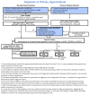
A guideline for diagnosis has been developed [Edvardsson et al 2013]; see Figure 1 for algorithm. The algorithm also assists the clinician in differentiating between PH types 1, 2, and 3.

Figure 1.

Figure 1.

Algorithm for diagnostic evaluation of primary hyperoxaluria From Edvardsson et al [2013]. Published with permission of SpringerNature.

The diagnosis of PH1 is established in a proband with hyperoxaluria or hyperoxalemia by ONE of the following:

Molecular genetic testing approaches can include single-gene testing, use of a multigene panel, and more comprehensive genomic testing:

  • Single-gene testing. Sequence analysis of AGXT is performed first and followed by gene-targeted deletion/duplication analysis if only one or no pathogenic variant is found.
    Sequence analysis includes testing for the common AGXT polymorphic variant, p.Pro11Leu. Although p.Pro11Leu, referred to as the "minor allele," does not cause PH1 by itself, it is known to exacerbate the deleterious effects of other pathogenic variants in cis configuration (see Molecular Genetics).
  • A multigene panel that includes AGXT and other genes of interest (e.g., GRHPR, HOGA1 [see Differential Diagnosis]) is now recommended. Note: (1) The genes included in the panel and the diagnostic sensitivity of the testing used for each gene vary by laboratory and are likely to change over time. (2) Some multigene panels may include genes not associated with the condition discussed in this GeneReview; thus, clinicians need to determine which multigene panel is most likely to identify the genetic cause of the condition at the most reasonable cost while limiting identification of variants of uncertain significance and pathogenic variants in genes that do not explain the underlying phenotype. (3) In some laboratories, panel options may include a custom laboratory-designed panel and/or custom phenotype-focused exome analysis that includes genes specified by the clinician. (4) Methods used in a panel may include sequence analysis, deletion/duplication analysis, and/or other non-sequencing-based tests.
    For an introduction to multigene panels click here. More detailed information for clinicians ordering genetic tests can be found here.
  • More comprehensive genomic testing (when available) including exome sequencing and genome sequencing may be considered. Such testing may provide or suggest a diagnosis not previously considered (e.g., mutation of a different gene or genes that results in a similar clinical presentation.
    For an introduction to comprehensive genomic testing click here. More detailed information for clinicians ordering genomic testing can be found here.

Table 2.

Molecular Genetic Testing Used in Primary Hyperoxaluria Type 1

Gene 1MethodProportion of Probands with Pathogenic Variants 2 Detectable by Method
AGXT Sequence analysis 3>97% 4
Gene-targeted deletion/duplication analysis 5<3% 6
Unknown 7NA
1.
2.

See Molecular Genetics for information on allelic variants detected in this gene.

3.

Sequence analysis detects variants that are benign, likely benign, of uncertain significance, likely pathogenic, or pathogenic. Variants may include small intragenic deletions/insertions and missense, nonsense, and splice site variants; typically, exon or whole-gene deletions/duplications are not detected. For issues to consider in interpretation of sequence analysis results, click here.

4.
5.

Gene-targeted deletion/duplication analysis detects intragenic deletions or duplications. Methods used may include a range of techniques such as quantitative PCR, long-range PCR, multiplex ligation-dependent probe amplification (MLPA), and a gene-targeted microarray designed to detect single-exon deletions or duplications.

6.

Out of more than 170 known AGXT pathogenic variants, six large deletions have been reported [Nogueira et al 2000, Coulter-Mackie et al 2001, Coulter-Mackie et al 2005, Monico et al 2007, Williams et al 2009, Tammachote et al 2012].

7.

The phenotype in PH2 and PH3 can appear similar to PH1 (without detailed biochemical analysis), 11.3% of families with a PH-like phenotype were not found to have pathogenic variants in AGXT, GRHPR, or HOGA1 [Hopp et al 2015]. It is not known what percentage of these individuals have PH1.

Liver biopsy. If urinary glycerate, 4-hydroxyoxoglutarate (HOG), and dihydroxyglutarate (DHG) are normal, and no AGXT, GRHPR, or HOGA1 pathogenic variants are identified, liver biopsy can be performed to assay AGT, glyoxylate reductase (GR), and 4-hydroxy-2-oxoglutarate aldolase (HOGA) enzyme activity. Normal GR and HOGA enzyme activity will rule out PH types 2 and 3 as the cause of the hyperoxaluria (see Primary Hyperoxaluria Type 2 and Primary Hyperoxaluria Type 3). Individuals with clinical features suggestive of PH who have AGT enzyme deficiency on liver biopsy and do not have identified AGXT pathogenic variants have a presumed diagnosis of PH1. Individuals who do not have identified pathogenic variants in AGXT, GRHPR, or HOGA1 accounted for 11.3% of individuals with hyperoxaluria tested in one recent series [Hopp et al 2015].

Clinical Characteristics

Clinical Description

In primary hyperoxaluria type 1 (PH1), high concentrations of oxalate in the urine combine with calcium resulting in calcium oxalate crystal formation. Crystal deposition leads to nephrolithiasis in the urinary tract, renal tubular damage, and nephrocalcinosis when the crystals deposit in the renal parenchyma. Progressive kidney damage and ultimately renal failure ensue. As declining glomerular filtration rate (GFR) limits excretion of the excess oxalate, plasma levels rise rapidly. Supersaturation of calcium oxalate in plasma then results in calcium oxalate deposition in many organs and tissues (oxalosis) with systemic manifestations.

The clinical presentation of PH1 is variable. Age at onset of symptoms ranges from early infancy to the sixth decade (median age: 4-6 years) [Cochat & Rumsby 2013, Mandrile et al 2014, Hopp et al 2015]. The majority of individuals present with symptoms related to nephrolithiasis. Some present with a severe, very early-onset form of PH1 in the first year of life characterized by failure to thrive and end-stage renal disease (ESRD). At the other end of the spectrum of clinical severity seen in PH1, some individuals remain free of symptoms or minimally symptomatic into the sixth decade of life [Cochat & Rumsby 2013, Hopp et al 2015].

Renal manifestations. The renal manifestations of PH1 can be quite variable, with individuals generally falling into one of five groups:

  • Early nephrocalcinosis and renal failure in infancy or early childhood (accounts for ~10% of individuals with PH1 in Europe and North America)
  • Recurrent nephrolithiasis and progressive renal failure with diagnosis in childhood, adolescence, or early to mid-adulthood (accounts for majority of affected individuals)
  • Diagnosis after ESRD that is first recognized due to oxalate deposits on renal biopsy, recurrent oxalate nephropathy in a renal allograft following renal transplantation or due to systemic oxalosis developing during chronic dialysis. (~10% of affected individuals)
  • Late-onset form with occasional stones diagnosed in adulthood (<10%)
  • Diagnosis in a presymptomatic individual because of a positive family history (<10%)

In individuals with severe, early-onset (infantile) disease, the presenting signs and symptoms include nephrocalcinosis with or without nephrolithiasis and failure to thrive related to renal failure. ESRD can appear as early as age four to six months. In most series in Europe and the US severe infantile oxalosis with kidney failure accounts for 10% or less of individuals with PH1 [Harambat et al 2010, Mandrile et al 2014, Hopp et al 2015]. However, the proportion appears higher in some areas of the world [Cochat et al 2006]. One limitation of current understanding of the natural history of PH1 is that a large majority of individuals and nearly all large reported series are from Europe and North America.

Presenting symptoms from early childhood through adolescence are most often related to kidney stone disease including hematuria, dysuria, pain, urinary tract infection, or stone passage [Hoppe et al 2009]. Nephrocalcinosis is common [Tang et al 2014]. Initial symptoms occur in most individuals before age ten years, and in 85%-90% by age 20 years [Hoppe et al 2009, van der Hoeven et al 2012, Mandrile et al 2014]. Median age of onset of symptoms was 3.9 to 5.2 years in several case series [Mandrile et al 2014, Hopp et al 2015].

The remainder of affected individuals present in adulthood with recurrent renal stones [van der Hoeven et al 2012]. Many adults report a history of unrecognized symptoms and missed or delayed diagnosis. A mild-to-moderate reduction in kidney function is common, though the rate of decline is highly variable. Some individuals, mainly adults, may present with acute renal failure secondary to bilateral renal obstruction caused by calcium oxalate stones, or due to other illnesses that compromise fluid intake and thus urine volume. Acute renal failure has also been reported following stone removal procedures [Carrasco et al 2015]. Up to 20%-50% of individuals with adult-onset PH1 were diagnosed when they presented in the late stages of chronic kidney disease (CKD) or ESRD [Harambat et al 2012, van der Hoeven et al 2012, Zhao et al 2016]. A higher prevalence of ESRD without a prior history of renal stones was observed in a national study of individuals with PH1 from the Netherlands [van Woerden et al 2003]. The proportion of individuals with ESRD at the time of diagnosis is higher in adults than in children [van der Hoeven et al 2012, Mandrile et al 2014]. The median age of individuals with PH1 who reach ESRD prior to diagnosis is 25 years (range: early childhood to late middle age) [Zhao et al 2016]. In approximately 10% of individuals the diagnosis of PH1 is made only following recurrence in a renal transplant when calcium oxalate crystals are found on allograft biopsy performed for early nonfunction of the transplanted kidney or a subsequent increase in serum creatinine [Cochat & Rumsby 2013].

Progression to renal failure has been shown to correlate with urine oxalate excretion rate and with nephrocalcinosis [Tang et al 2014, Zhao et al 2016]. AGXT pathogenic variant p.Gly170Arg is associated with a milder phenotype, with lower urine oxalate excretion, responsiveness to pyridoxine treatment, and later onset of ESRD [Monico et al 2005a, Harambat et al 2010, Mandrile et al 2014, Hopp et al 2015]. Later onset of ESRD has also been reported with the mistargeting pathogenic variant p.Phe152Ile [Mandrile et al 2014]; other mistargeting pathogenic variants do not appear to delay the onset of ESRD [Hopp et al 2015]. The natural history of untreated PH1 is one of progressive decline in renal function as a result of calcium oxalate deposits in kidney tissue and complications of nephrolithiasis such as obstruction and infection. A recent study found renal survival in individuals with PH1 to be 76%, 43%, and 12% at ages 20, 40, and 60 years, respectively [Hopp et al 2015]. Another study noted renal survival of 59%, 41%, and 10% at ages 20, 30, and 50 years, respectively [Harambat et al 2010]. In the absence of treatment there is eventual progression to oxalosis and death from ESRD and/or complications of oxalosis [Hoppe et al 2009, Harambat et al 2010, van der Hoeven et al 2012, Cochat & Groothoff 2013].

Among 54 children and adolescents with PH1 who developed ESRD from 2000 through 2009, five-year survival was 83% following initiation of renal replacement therapy. Results showed improvement compared with renal replacement therapy prior to 2000, particularly in children younger than age two years. However, outcomes were less favorable in children with PH1 who required renal replacement compared to children with other renal diseases [Harambat et al 2012]. In a series of six individuals with PH1 with infantile oxalosis, excellent outcomes were reported following combined kidney/liver transplantation at a mean age of 14.8 months following a mean of 11.8 months of dialysis [Millan et al 2003].

Oxalosis. When the GFR is <30 mL/min/1.73 m2, the daily production of oxalate outstrips renal oxalate clearance, resulting in an accelerated rise in plasma oxalate concentration associated with declining GFR, and further loss in residual renal function. Supersaturation in plasma leads to deposition of calcium oxalate crystals in multiple body tissues (oxalosis), including the kidneys, retina, myocardium, blood vessels, bone, bone marrow, and subcutaneous tissue [Hoppe et al 2009, Cochat et al 2012, Cochat & Rumsby 2013]. Peripheral nerves, synovia, and other tissues may also be involved [Hoppe et al 2009]. Clinical manifestations may include visual disturbance, cardiac conduction disturbances such as heart block, and cardiomyopathy. Vascular involvement can lead to ischemia, most often manifest as non-healing cutaneous ulcers. Though unusual, cerebral infarcts have been reported from calcium oxalate in cerebral vessels [Rao et al 2014]. Refractory hypotension can be seen in advanced oxalosis. Bone is the largest repository for excess oxalate. Oxalate osteodystrophy causes bone pain and pathologic fractures. Involvement of bone marrow can result in anemia refractory to erythropoietin-stimulating agents (ESA). Dental pain due to oxalate deposition and root resorption have been described [Mitsimponas et al 2012]. Peripheral neuropathy can be caused by crystal deposition within nerves [Berini et al 2015]. Progressive oxalosis, observed over time in most individuals with PH on dialysis, eventually leads to death. Oxalosis is slowly reversible following successful liver and kidney transplantation, but mobilization of the tissue deposits poses risk for a transplanted kidney.

Bone mineral density measurements, as opposed to the gold standard of bone biopsy, allow for noninvasive assessment of oxalate burden [Behnke et al 2001], though interpretation of bone density is complicated by multiple bone changes related to ESRD.

Pathophysiology

The stone type most prevalent in individuals with PH1 is calcium oxalate monohydrate or whewellite [Daudon et al 2008]. These authors noted that stones from individuals with PH1 differ in morphologic characteristics from idiopathic calcium oxalate stone formers: they appear white or pale yellow on the surface and form a loose, unorganized section whereas idiopathic oxalate stone formers appear as a dark-brown surface with a well-organized radiating inner structure. These differences in morphology suggest a different mechanism of stone formation from idiopathic oxalate stones.

Genotype-Phenotype Correlations

Pathogenic variants that result in mistargeting of alanine:glyoxylate-aminotransferase (AGT) enzyme activity (see Molecular Genetics, Abnormal gene product) are the most likely to be associated with B6 (pyridoxine) responsiveness [Monico et al 2005a].

  • Homozygotes for either c.508G>A (p.Gly170Arg) or c.454T>A (p.Phe152Ile) show B6 responsiveness and benefit from early treatment (see Management).
  • Response to B6 therapy is relative to the number of copies of the p.Gly170Arg pathogenic variant present [Monico et al 2005a, Monico et al 2005b]. Compound heterozygotes for these pathogenic variants also demonstrate a reduction in urine oxalate following B6 treatment, though less than observed in homozygotes.
  • Results of in vitro studies suggest that additional pathogenic variants may respond to B6, reinforcing the recommendation of testing all individuals for B6 responsiveness [Fargue et al 2013b].

The presence of the p.Gly170Arg pathogenic variant is associated with lower urine oxalate excretion rates even in the absence of treatment and could even predict disease severity and/or rate of progression [Monico et al 2005a, Hoyer-Kuhn et al 2014, Hopp et al 2015]. A large retrospective study of individuals with PH1 suggested that the p.Gly170Arg pathogenic variant is associated with longer preservation of renal function with conservative treatment compared to other pathogenic variants [Harambat et al 2010].

Although it may be possible to establish a relationship between the AGXT genotype and AGT enzyme activity in vitro, in most cases it is difficult to correlate enzyme activity with clinical severity [Danpure 2001, Pirulli et al 2003, Danpure & Rumsby 2004, van Woerden et al 2004].

The clinical course of PH1 in affected sibs is usually similar; however, families in which affected relatives with identical pathogenic variants had different disease manifestations have been described [Frishberg et al 2005, Beck & Hoppe 2006, Lorenzo et al 2006, Mandrile et al 2008, Alfadhel et al 2012, Hopp et al 2015]. Possible causes of this intrafamilial variation include differences in activity level of other enzymes important in oxalate synthesis, modifier genes, the quantity of oxalate precursors in the diet, renal oxalate handling, absorption of dietary oxalate, hydration status, infections, and urinary crystallization factors [Danpure 2001].

Other than potential delay in diagnosis due to the rarity of PH1, the reason for a poorer outcome in infants with the same pathogenic variants as older individuals is not clear. Possibilities include:

  • The low glomerular filtration rate (both absolute and corrected) in children younger than age six to 12 months, which predisposes to earlier oxalate deposition [Quigley 2012];
  • High oxalate excretion in infants (see Table 1);
  • Reduced number of nephrons present at birth, which may be associated with low birth weight as well as environmental and genetic factors [Luyckx & Brenner 2005, Puddu et al 2009, Luyckx & Brenner 2010].

Nomenclature

With the identification of oxalate in human urine in the 1800s, hyperoxaluria fell into a category referred to as "oxalate diathesis" – a catch-all term for a wide variety of ailments.

PH1 can be considered a peroxisomal disorder, as it results from deficiency of a single peroxisomal enzyme. PH1, however, is distinct from disorders of peroxisomal biogenesis (see Zellweger Spectrum Disorder and Rhizomelic Chondrodysplasia Punctata Type 1) in that a normal number of relatively normal-looking peroxisomes are observed on liver biopsy.

Prevalence

PH1 is estimated to account for 1%-2% of children with ESRD [Harambat et al 2012].

When considering the following statistics, it is important to remember that PH1 remains underdiagnosed because of the wide variability in its clinical presentation and age of onset.

Clinical estimates of prevalence of PH1, primarily from European studies, range from one to three in 1,000,000 [van Woerden et al 2003, Cochat & Rumsby 2013]. PH1 is estimated to occur in 1:120,000 live births in Europe [Cochat & Rumsby 2013]. Since phenotypic heterogeneity ranges from severe disease in infancy to adults with recurrent stones, and since advanced disease is present in 20%-50% at the time of diagnosis, underdiagnosis likely occurs. Population analysis based on AGXT pathogenic variants identified in the National Heart, Lung, and Blood Institute Exome Sequencing Project suggest a PH1 prevalence of 1:149,000 in European Americans and 1:157,000 in African Americans [Hopp et al 2015].

A higher frequency of PH1 has been reported in Tunisia and Kuwait [Kamoun & Lakhoua 1996, Al-Eisa et al 2004, Cochat et al 2006], in Arabs and Druze families of Israel [Rinat et al 1999], and in Iran [Madani et al 2001] as a result of the high rate of consanguinity in these populations. No common founder variant has been identified in these populations [Rinat et al 1999, Coulter-Mackie 2005, Frishberg et al 2005].

Differential Diagnosis

Primary hyperoxaluria type 2 (PH2) is caused by pathogenic variants in GRHPR, resulting in deficiency of the cytosolic enzyme glyoxylate reductase/hydroxypyruvate reductase (GRHPR), which catalyzes the reduction of glyoxylate and hydroxypyruvate to D-glycerate. While found in many tissues, GRHPR is mainly found in hepatocytes with some expression in mitochondria. In PH2, glyoxylate removal is impaired, resulting in the metabolism of glyoxylate by lactate dehydrogenase to oxalate and L-glycerate.

The diagnosis of PH2 can be established by sequence analysis of GHPRH or assay of GRHPR enzymatic activity in liver. Inheritance is autosomal recessive.

PH2 is rarer than PH1. It is associated with better preservation of kidney function than PH1 [Hopp et al 2015]. However, many individuals with PH2 still experience renal failure [Garrelfs et al 2019].

From a small cohort of individuals with PH1 and PH2 from one center, PH1 as a group appears to differ from PH2 in the following respects:

  • PH2 is considered a less aggressive disease than PH1, even when onset is early.
  • Individuals with PH1 have statistically higher urine oxalate excretions and more stone-forming activity and thus require more frequent stone removal.
  • Individuals with PH1 have statistically lower urine osmolalities and lower urine concentration of calcium, citrate, and magnesium. (For a single individual with hyperoxaluria, the differences observed cannot reliably distinguish PH1 from PH2.)
  • In individuals with PH1, urinary glycolate (as well as oxalate) is often elevated, although some individuals with PH1 have normal urine glycolate.
  • In individuals with PH2, urinary L-glycerate and oxalate are generally elevated; a single individual with PH2 and normal urine glycerate has been reported [Rumsby et al 2001].

Primary hyperoxaluria type 3 (PH3) is caused by a defect in the hepatocyte-specific mitochondrial enzyme 4-hydroxy-2-oxoglutarate aldolase (HOGA), which appears to lead to excess metabolism of hydroxyproline with the forward reaction product (HOG) itself being converted to pyruvate or glyoxalate or perhaps actually inhibiting mitochondrial GRHPR (effectively causing "secondary" PH2). Pathogenic variants in HOGA1 are causative. Diagnosis is by exclusion of PH1 and PH2 with confirmation by DNA sequence analysis of HOGA1. Inheritance is autosomal recessive.

Enteric hyperoxaluria. Diseases affecting the small bowel that result in fat malabsorption including celiac disease, Crohn disease, chronic pancreatitis, and short bowel syndrome can be associated with hyperoxaluria. The precipitation of enteric calcium by non-absorbed free fatty acids leads to loss of the normal inhibition in oxalate reabsorption from the gut, increasing plasma oxalate concentration by increasing paracellular and transcellular transport. Delivery of excess fatty acids and bile salts to the colon also injures the mucosa and increases oxalate absorption [Milliner 2005, Hoppe et al 2009]. By contrast, individuals with PH1 show low-to-normal levels of oxalate absorption [Sikora et al 2008]. Gastric bypass procedures used in the treatment of obesity have been associated with increased oxalate absorption, high levels of hyperoxaluria, increased risk of kidney stone formation, and ESRD caused by oxalate nephropathy [Asplin & Coe 2007, Kleinman 2007, Duffey et al 2008, Lieske et al 2008]. Urinary risk factors for stones such as hyperoxaluria occur more commonly in individuals with Roux-en-Y gastric bypass than gastric banding [Semins et al 2010, Kumar et al 2011, Tasca 2011].

Dietary hyperoxaluria. Excess intake of foods high in oxalate including chocolate, cocoa, leafy greens (especially rhubarb and spinach), black tea, nuts, peanut butter, or starfruit [Holmes & Kennedy 2000, Monk & Bushinsky 2000] may lead to elevated plasma concentration of oxalate and hence increased urinary concentration of oxalate. It was previously thought that dietary oxalate accounted for little of the oxalate in the urine (<10%), but Holmes et al [2001b] showed that between 24% and 53% of urinary oxalate is attributable to oxalate from the diet [Holmes & Assimos 2004]. Therapy consists of dietary oxalate restriction and use of calcium carbonate or calcium citrate at meal times to bind dietary oxalate [Penniston & Nakada 2009].

Idiopathic calcium oxalate urolithiasis is associated with "mild metabolic hyperoxaluria." Features that often differentiate this condition from PH1:

  • Lower urinary oxalate excretion
  • Less severe stone disease
  • The hyperoxaluria rarely leads to ESRD.
  • Tendency to hypercalciuria (as opposed to hypocalciuria in PH1)
  • Day-to-day variability in the levels of urinary oxalate excretion (in contrast to PH1, in which levels are persistently elevated in the urine [Milliner 2005])

Dent disease. The clinical features of Dent disease may overlap those of PH1. Both are associated with nephrocalcinosis and urolithiasis in childhood and progress to renal failure (Table 3).

Table 3.

Clinical and Diagnostic Features of Dent Disease and Primary Hyperoxaluria Type 1

Clinical and Diagnostic FeaturesDent DiseasePH1
Nephrocalcinosis3+2+
Urolithiasis3+4+
Osteodystrophy1+1+ 1
Renal failure2+2+
Hypercalciuria1+
Hyperoxaluria3+
Low-molecular-weight proteinuria1+— 2
Gene CLCN5 AGXT
InheritanceX linkedAutosomal recessive
1.

After renal failure established

2.

May be observed following renal damage but not an early or characteristic finding

Other hereditary causes of kidney stones. A number of common and rare forms of nephrocalcinosis or urolithiasis may be associated with kidney disease [Monico & Milliner 2011, Edvardsson et al 2013].

  • Another condition that may present with findings of nephrolcalcinosis is familial hypomagnesemia with hypercalciuria and nephrocalcinosis (FHHNC) (OMIM 248250).
  • While not associated with nephrocalcinosis, children with adenine phosphoribosyltransferase (APRT) deficiency (OMIM 614723) can develop crystal nephropathy and chronic kidney disease, as do children with PH1. Hyperoxaluria is not a feature of APRT deficiency, though 2,8-dihydroxyadenine (DHA) crystals in kidney tissue can resemble those of calcium oxalate.

Other. Acute renal failure secondary to oxalate deposition in the kidneys has occurred in persons taking large doses ("megadoses") of ascorbic acid (vitamin C) [Petrarulo et al 1998, Mashour et al 2000] as well as in a number of persons who were "juicing" (extraction of juice from vegetables or fruit) with high oxalate-containing foods [Getting et al 2013].

Ingestion of ethylene glycol, an oxalate precursor, can lead to excess production and increased concentrations of oxalate in both the plasma and urine [Milliner 2005].

Hyperoxaluria in association with total parenteral nutrition has been described in premature infants [Sikora et al 2003] and adults [Buchman et al 1995].

Hyperoxaluria has been documented in Zellweger spectrum disorder despite the apparent cytoplasmic stability of alanine:glyoxylate aminotransferase [van Woerden et al 2006]. The presence of hyperoxaluria was statistically correlated with the degree of neurologic involvement.

Management

Evaluations Following Initial Diagnosis

The following initial evaluations are recommended for individuals with primary hyperoxaluria type 1 (PH1) in part based on the guidelines and suggestions of the European hyperoxaluria consortium (OxalEurope) [Cochat et al 2012] (see Table 3), and Hoppe [2012].

  • For those with preserved renal function (i.e., glomerular filtration rate [GFR] >60 mL/min/1.73 m2):
    • Renal ultrasound examination and fundoscopic eye examination to identify the presence/extent of oxalate deposition
    • Baseline urinalysis and measurement of oxalate and creatinine, preferably in a 24-hour collection
  • For those with a GFR <60 mL/min/1.73 m2, in addition to the above evaluations: measurement of plasma oxalate
  • For those with a GFR <30 mL/min/1.73 m2 or in whom there is a rapid deterioration in function, in addition to the above evaluations:
    • Bone x-rays to evaluate for radiodense metaphyseal bands and diffuse demineralization or evidence of pathologic fractures
    • Hemoglobin to evaluate for anemia associated with either renal dysfunction or marrow deposition of oxalate. Bone marrow examination may be needed.
    • Electrocardiogram to evaluate for an associated atrioventricular block
    • Echocardiogram for evidence of oxalate cardiomyopathy which may show changes in refractile characteristics of myocardium and/or reduced ejection fraction
    • Ultrasound and/or CT scan of the heart and viscera for calcifications
    • History and physical examination to assess the risk of arterial insufficiency or ischemia based on vessel wall deposition
    • Thyroid function testing

Consultation with a clinical geneticist and/or genetic counselor is recommended.

Treatment of Manifestations

In PH1, hepatic overproduction of oxalate is the underlying cause of hyperoxaluria, kidney stones, nephrocalcinosis, progressive chronic kidney disease (CKD) and end-stage renal disease (ESRD), and oxalosis (deposition of calcium oxalate crystals in multiple body tissues). Despite efforts to mitigate oxalate-induced kidney damage and stone formation with conservative therapies including hyperhydration and crystallization inhibitors, most individuals with PH1 ultimately progress to ESRD. Thus, the primary strategy in prevention and treatment of disease manifestations is to reduce hepatic oxalate production.

Until recently, options to reduce hepatic overproduction of oxalate were limited to pharmacologic doses of pyridoxine or liver transplantation (most often combined with or performed sequentially with kidney transplantation in persons with ESRD). Pyridoxine is most effective only in certain genotypes of PH1, however, and liver transplantation can be associated with significant morbidity. Early experience with lumasiran, an RNAi therapeutic agent approved in late 2020 by the FDA and EMA, suggests that it significantly reduces hepatic oxalate production and urinary oxalate excretion in individuals with PH1 [Scott & Keam 2021]. However, additional data are needed to confirm long-term safety and efficacy of lumasiran and to determine if treatment with lumasiran can reverse the renal effects of hepatic overproduction of oxalate.

Because disease manifestations, including CKD or even ESRD, are often present by the time of diagnosis, several approaches to treatment are necessary.

Reduction of Oxalate Biosynthesis

Pyridoxine (vitamin B6, i.e., pyridoxal phosphate) can reduce oxalate production in some individuals with PH1 by enhancing activity of the pyridoxal phosphate-dependent enzyme alanine:glyoxylate aminotransferase (AGT) [Cochat & Rumsby 2013]. Approximately 30%-50% of individuals with PH1 are pyridoxine responsive, defined as greater than 30% reduction in urine oxalate concentration or normalization of urinary oxalate excretion after a minimum of three months of maximal therapy [Cochat et al 2012]. In order for deficient AGT to respond to pyridoxine, some degree of enzyme activity must be preserved, as occurs with certain pathogenic AGXT variants [Fargue et al 2016]. See Genotype-Phenotype Correlations for AGXT pathogenic variants associated with pyridoxine responsiveness.

While most pyridoxine-responsive individuals show maximum benefit at a dose of 5-8 mg/kg/day [Monico et al 2005a, Hoyer-Kuhn et al 2014], some have required higher doses.

  • A starting pyridoxine dose of 5 mg/kg/day is recommended. Response should be assessed by comparing the 24-hour urine oxalate excretion rate before treatment and after three to six months of treatment. Note that pyridoxine should be continued during the follow-up urine collection.
  • Stepwise increases in pyridoxine dose to a maximum of 10-20 mg/kg/day with assessments of response at each step is appropriate to determine effectiveness. If the individual is responsive, this approach also facilitates identification of lowest pyridoxine dose at which the maximum response occurs.
  • Several authors have noted little additional benefit at pyridoxine doses >10 mg/kg/day [Bobrowski & Langman 2008, Hoppe et al 2009].
  • Pyridoxine response can be difficult to determine in individuals with advanced CKD or ESRD since urinary oxalate excretion rates may be influenced by the low GFR. In individuals with GFR <30 mL/min/1.73 m2, changes in plasma oxalate concentration may be valuable in assessing response.

Pyridoxine has an excellent safety profile in individuals with PH1, even after decades of use. In contrast, peripheral neuropathy (including paresthesias) has been reported in individuals who do not have PH1 who were receiving very large doses of pyridoxine (typically, adults receiving 1-2 g/day) [Toussaint 1998]. Of note, paresthesias reported in one individual with PH1 on a dose of pyridoxine of 2.1 mg/kg/day resolved following its discontinuation.

Pyridoxine-responsive individuals should continue this therapy indefinitely or until orthotopic liver transplantation (as a result of which normal functioning AGT has replaced the deficient AGT). Additionally, the recent availability of the mRNAi therapeutic lumasiran calls for comparative examination of pyridoxine and lumasiran regarding efficacy, durability of effect, long-term tolerability and safety, and use under special circumstances such as pregnancy.

Lumasiran (Oxlumo®), an mRNAi therapeutic agent approved for use in the care of individuals with PH1 by the FDA and EMA in 2020, reduces the amount of glyoxylate substrate available for metabolic conversion to oxalate by targeted reduction of hepatic glycolate oxidase [Scott & Keam 2021]. Since lumasiran targets an enzyme that is in the same metabolic pathway as AGT but distinct from AGT, lumasiran is expected to be effective in all individuals with PH1, independent of specific AGXT pathogenic variants (a limiting factor in pyridoxine responsiveness).

Since the currently approved lumasiran dose is administered subcutaneously on a quarterly basis (see Table 4 for loading and maintenance dose schedule), it has the advantage of less medication burden than other agents used historically to manage PH1. Except for mild and transient injection-site reactions observed in early clinical trials, lumasiran appears to be well tolerated; no serious adverse effects have been identified to date.

Table 4.

Schedule for Weight-Based Subcutaneous Administration of Lumasiran

WeightDose Schedule
First 3 mos of treatment
(loading dosage)
Subsequent mos of treatment
(maintenance dosage)
20 kg3 mg/kg 1x/mo3 mg/kg every 3 mos
10 to <20 kg6 mg/kg 1x/mo6 mg/kg every 3 mos
<10 kg6 mg/kg 1x/mo3 mg/kg 1x/mo

In double-blind, placebo-controlled trials of 39 individuals with PH1 (lumasiran treated n=26, placebo n=13), lumasiran was highly effective in reducing urine and plasma oxalate [Garrelfs et al 2021]. After six months of treatment, 84% of individuals treated with lumasiran had 24-hour urinary oxalate levels ≤1.5 times the upper limit of the normal range, as compared with none of the individuals who received placebo (P<0.001). Half (52%) of lumasiran-treated individuals had 24-hour urinary oxalate levels within normal range [Garrelfs et al 2021].

Although early clinical trial experience demonstrated promising reductions in plasma and urinary oxalate with lumasiran treatment [Garrelfs et al 2021, Sas et al 2021], longer term study is needed to confirm benefit for clinical outcome measures such as reduction in symptomatic kidney stone events and stabilization of kidney function. Initial studies were performed in individuals with PH1 with preserved kidney function (eGFR >30 mL/min/1.73 m2); Thus, current experience using lumasiran in individuals with advanced CKD, on dialysis, or following kidney transplantation alone is limited.

Reduction of Urinary Calcium Oxalate Supersaturation

The general therapies for nephrolithiasis benefit all individuals with PH1. Early diagnosis and initiation of conservative therapy are critical in preserving adequate renal function for as long as possible [Fargue et al 2009].

  • Drinking large volumes of fluid (2-3 L/m2/24 hours) at regular intervals over the entire day/night prevents calcium oxalate supersaturation. Small children may require a gastrostomy or nasogastric tube for feeds and fluid supplementation. Extreme care should be taken during any illness that could lead to hypovolemia or decreased oral fluid intake; individuals should be advised to seek early medical attention with initiation of intravenous fluids if needed to maintain urine volume.
  • Alkalinization of urine (pH 6.2-6.8) to inhibit calcium oxalate crystallization using oral potassium citrate at a dose of 0.1-0.15 mg/kg or 0.3-0.5 mmol/kg/day in 3-4 divided doses so long as the GFR is preserved. When the GFR is reduced or concerns about potassium levels ensue, alkalinization can be achieved with sodium citrate.
  • Moderate doses of pyrophosphate-containing solutions may also inhibit crystal formation, and may be dosed as 20-30 mg/kg/day of phosphate in divided doses.
  • Since the excess oxalate in PH1 is derived from endogenous metabolism and is not related to dietary oxalate intake, dietary restriction of oxalate is of little benefit. The principle of careful food choices (avoid high oxalate food and drink) is appropriate without strict limits.
  • Some have advocated use of thiazide medications in PH1. However, urinary calcium is usually in the low normal range or below in individuals with PH1. There is also potential for intravascular volume depletion with thiazide medication. The benefits of thiazide diuretics have not been systematically studied in PH. For these reasons, the authors suggest that they be used on an individual basis only, when specific clinical circumstances warrant.

Treatment of Kidney Stones

Individuals with PH1 must be counseled regarding the different success rates of the following surgical modalities:

  • Shockwave lithotripsy (SWL) is a viable first option, but has a lower success rate and higher rate of subsequent endoscopic surgery. Calcium oxalate monohydrate stones are among the hardest stones, and are thus more likely to be resistant to SWL [Williams et al 2003]. Stones in the lower pole of the kidney or parenchymal stones did not respond as well to SWL as stones in other locations [Al-Abadi & Hulton 2013]. There is good evidence that any given stone should not be treated with more than two SWL sessions, particularly if no change is observed with treatment [Pace et al 2000].
  • Ureteroscopy holds a very good record for success of stone clearance with minimal complication rates and may be supplanting SWL as first-line therapy at many centers.
  • Percutaneous nephrolithotomy should be considered as first-line therapy for larger, bulky stone burdens (>15 mm).

Dialysis

The issues related to the removal of oxalate and current modalities of dialysis are complex. Interested readers are directed to papers outlining the specific details of extracorporeal removal of oxalate [Cochat et al 2012, Plumb et al 2013, Tang et al 2014]. Oxalate removal is much more effective with hemodialysis than peritoneal dialysis. For that reason, peritoneal dialysis should not be relied on as the primary dialysis modality in any individual with PH1.

In brief, despite the small size of the oxalate molecule (90 daltons), the rate of oxalate production in persons with PH1-related ESRD (frequently 2-7 mmol/1.73 m2/day) vastly surpasses the ability to remove it via conventional dialysis. The sequestration of oxalate in tissue compartments outside the vascular space makes it difficult to effectively remove it from the body. Current guidelines suggest that the intent of dialysis is to reduce and maintain the plasma oxalate level below 30-45 µmol/L (the calcium/oxalate supersaturation threshold at which tissue deposition occurs) as much of the time between dialysis sessions as possible.

While more aggressive strategies using high-flux dialyzers, daily hemodialysis, combined hemodialysis and peritoneal dialysis, hemodiafiltration, and even charcoal perfusion have all been reported [Plumb et al 2013, Tang et al 2014], the end result is that while plasma removal rates of oxalate can be achieved in the range of greater than 60%, the total body store of oxalate rebounds to a level of 80% of the predialysis levels within 24 hours of the last hemodialysis session.

A case report of one individual treated with aggressive hemodialysis (8-10 hrs, 7 nights/week) does suggest that this level of intense dialysis allows for maintenance of the predialysis oxalate levels at or just below the tissue saturation point (30-45 µmol/L) [Plumb et al 2013]. In most individuals with PH1, four or more dialysis sessions per week are needed to maintain predialysis plasma oxalate concentrations in a range to minimize risk of oxalosis [Tang et al 2014]. Since oxalate production varies significantly among individuals with PH1, it is important that dialysis prescriptions are individualized [Tang et al 2014]. In addition, continuous dialytic therapies such as continuous veno-venous hemodialysis can maintain plasma oxalate below 20 μmol/L for prolonged periods and may be useful in acute situations such as following transplantation in an individual with extensive oxalosis whose kidney allograft is functioning poorly.

Though dialysis is most often used as a bridge to transplantation, it may also be needed as an adjunct therapy to decrease oxalate burden in the presence of delayed or poor renal function after liver/kidney transplantation, in older individuals if liver/kidney transplantation is not deemed an option, and in countries with no access to organ transplantation.

Organ Transplantation

As dialysis cannot prevent systemic oxalosis in an individual with a GFR <25-30 mL/min/1.73 m2, organ transplantation is the preferred option for treatment. The reader is encouraged to review the best practice recommendations on organ transplant of non-pyridoxine-sensitive individuals with PH1 [Cochat et al 2012]. These guidelines take into consideration the individual's age, residual GFR, and evidence of systemic oxalate deposition in extrarenal organs.

The percentage of survivors after liver-kidney transplant surpasses the percentage of survivors after isolated kidney transplant: adult five-year survival percentage for individuals after kidney vs dual transplant are 45% vs 67% [Bergstralh et al 2010]; those for children are 14% vs 76% [Harambat et al 2012].

Isolated liver transplant may be considered in an individual with significant residual renal function (e.g., GFR >60 mL/min/1.73 m2), with the presumption that decline in renal function will be arrested and only a single organ transplant required. However, the concerns regarding the risks of liver transplant, the uncertain and often slow rate of decline in renal function, and emerging novel treatments for PH1 are such that most experts defer the decision to proceed until dual transplantation is required.

Most of the published literature includes reports of transplants using organs from deceased donors; however, living/living related donation of a split liver graft (if the recipient is small enough) and/or living donor kidney or liver allografts are viable alternatives in some situations. It is important to note that the appropriateness of using a parent or sib who is heterozygous for an AGXT pathogenic variant as a donor remains unclear. Heterozygotes can have reduced AGT enzyme activity in the liver, though they typically have normal urine oxalate excretion and remain free of stones or oxalate-related CKD.

It is believed that simultaneous liver/kidney transplant is the most logical for any adult or child with CKD Category 4 or below, given the need for renal function to excrete the body burden of oxalate. However, if the wait for a suitable liver graft will significantly delay transplantation, simultaneous liver/kidney transplant may not be the best option for individuals with CKD Category 5 or on dialysis, as they often face severe oxalate burden that can overwhelm a new renal graft and lead to graft failure caused by oxalate stones/deposition. The choice in such individuals, most often small children/infants where anatomy may preclude a simultaneous approach, is to proceed with sequential (i.e., liver followed by kidney) transplantation.

It is also important to note that in all forms of transplantation, mobilization of systemic oxalate places the renal allograft at risk until tissue oxalate stores are completely cleared. For this reason, both liver and kidney transplant recipients must be monitored closely following transplantation with maintenance of high urine volume and crystal inhibitor medication, and even dialyzed following surgery if delayed or otherwise compromised kidney allograft function results in high plasma oxalate levels. In the post-kidney- and/or liver-transplant period, daily hemodialysis should be maintained until the renal clearance of oxalate maintains plasma oxalate well below 30 μmol/L, in order to minimize risk of oxalate nephropathy. It is important to note that complete clearance of tissue oxalate stores by a well-functioning renal allograft requires several months to more than five years following successful liver transplantation [Monico & Milliner 2001, Tang et al 2014] and is confirmed by sustained normalization of urine oxalate excretion. Pyridoxine supplementation can be discontinued at the time of liver transplantation, since normal AGT activity will have been restored.

Surveillance

Individuals with PH type 1 require lifelong surveillance, the frequency of which is related to kidney function [Hoppe et al 2009, Cochat et al 2012, Cochat & Rumsby 2013].

  • Individuals with preserved renal function (i.e., measured or estimated GFR [eGFR] >60 mL/min/1.73 m2) require the following to evaluate/ensure treatment efficacy:
    • Regular monitoring of kidney function. Serum creatinine and/or cystatin C for determination of eGFR should be performed at least annually and more frequently in children and adolescents and in individuals with changing kidney function or clinically active stone disease.
    • Regular renal ultrasound examinations to monitor and manage stone forming activity
    • Urinalysis and measurements of urine oxalate excretion, urine volume, and calcium oxalate saturation (spot and 24-hour collections)
    • Periodic fundoscopic eye examination to identify the extent of any oxalate deposition
  • When GFR is known to be reduced to <60 mL/min/1.73 m2 plasma oxalate concentration should be measured and monitored regularly (at least annually or more often as indicated by the clinical course) in addition to the above evaluations. More frequent monitoring of kidney function is indicated.
  • Individuals with a GFR <30 mL/min/1.73 m2 or a rapid deterioration in function should have the above evaluations as well as the following testing (performed prior to initiation of dialysis if possible, and repeated as needed):
    • Frequent assessment of serum creatinine and plasma oxalate, typically at least monthly or more often depending on clinical circumstances
    • X-ray examination of the long bones to evaluate for radiodense metaphyseal bands and diffuse demineralization. Bone marrow examination may also be required.
    • Electrocardiogram to evaluate for an associated atrioventricular block or other oxalate related conduction abnormalities
    • Echocardiogram for evidence of oxalate cardiomyopathy
    • Hemoglobin to evaluate for anemia associated with either renal dysfunction or marrow deposition of oxalate
    • History and physical examination to assess the risk of arterial insufficiency or ischemia based on vessel wall deposition
    • Thyroid function testing
    • Regular dental examinations/care
    • Ophthalmologic follow up
    • Frequent clinical evaluation to monitor for additional complications of oxalosis (see Clinical Description, Oxalosis) – noting that at GFR <30 mL/min/1.73 m2 the ongoing deposition of tissue oxalate (oxalosis) will predispose all individuals to multiorgan involvement, and this will worsen/accelerate as the person enters ESRD and initiates dialysis.

Note: Investigations should occur more often in newly diagnosed symptomatic individuals or in children younger than age two to three years.

Agents/Circumstances to Avoid

Avoid the following:

  • Intravascular volume depletion. The importance of maintaining dilute urine cannot be overemphasized.
  • Intake of vitamin C that exceeds the recommended daily allowance
  • Loop diuretics to maintain dilute urine, as they can lead to hypercalciuria and increase the production of calcium oxalate stones
  • High doses of nonsteroidal anti-inflammatory medications or any pharmacologic agent that can compromise kidney function
  • Large intake of foods high in oxalate (e.g., chocolate, rhubarb, starfruit)

Evaluation of Relatives at Risk

It is appropriate to evaluate all at-risk family members, whether or not they are symptomatic, in order to identify those individuals who would benefit from early treatment, monitoring, and preventive intervention [Cochat et al 2012]. If the AGXT pathogenic variants in the family are known, the genetic status of at-risk family members can be clarified by molecular genetic testing. The benefits of early initiation of conservative measures cannot be ignored [Chand & Kaskel 2009, Fargue et al 2009, Martín et al 2011].

Asymptomatic individuals found to have biallelic AGXT pathogenic variants are at risk for stones and kidney damage and should be managed according to the same guidelines as those who present with symptoms.

See Genetic Counseling for issues related to testing of at-risk relatives for genetic counseling purposes.

Pregnancy Management

Pregnancy does not appear to be an important risk factor for the development of ESRD in the majority of women with PH1 [Norby & Milliner 2004]; however, women in whom renal function deteriorated during the pregnancy and remained abnormal post delivery have been reported [Cimino et al 2005]. Maintenance of adequate fluid intake throughout pregnancy can be challenging, especially during the first trimester, and in some with hyperemesis gravidarum intravenous fluid administration may be required. Continuation of pyridoxine (for pyridoxine-sensitive individuals) and careful monitoring of kidney function and stones throughout pregnancy and the early postpartum period are important to good outcomes.

Women with PH1 have had successful pregnancies following liver/kidney transplantation. In one woman liver function was apparently preserved, but renal graft function declined transiently after the birth of her first child and permanently after the birth of her second child [Pruvot et al 1997].

Of particular note, women with PH1 warrant close monitoring during pregnancy and the postpartum period by both an obstetrician and nephrologist. Lumasiran, recently approved for use in individuals with PH1, has not been studied during human pregnancy. Although preclinical animal studies were reassuring, pending more clinical experience the authors would advise women taking lumasiran to discontinue it prior to becoming pregnant and to use alternative (traditional PH1) treatments throughout the pregnancy. To date, no information is available regarding lumasiran transmission in breast milk; thus, risk to breast-feeding infants cannot be ruled out.

Generally, the offspring of women with PH1 have done well [Norby & Milliner 2004].

Therapies Under Investigation

Several novel therapies are under investigation.

Oxalate-Degrading Bacteria

Oral administration of bacteria such as Oxalobacter formigenes (O formigenes) or lactic acid bacteria to degrade oxalate and reduce the amount of oxalate available for intestinal absorption [Azcarate-Peril et al 2006, Ivanovski & Drüeke 2013] is being investigated. O formigenes shows the most promise as a potential therapy for the hyperoxalurias. Although it is a normal component of the intestinal flora, many individuals do not maintain colonization. O formigenes is also thought to stimulate secretion of endogenous oxalate into the intestine for its own metabolic use [Hatch et al 2006, Hatch & Freel 2008, Arvans et al 2017]. A human strain of O formigenes (HC-1) has been shown to promote oxalate secretion into the intestine of a mouse model of primary hyperoxaluria [Hatch & Freel 2013]. Administration of the HC-1 strain reduced the amount of oxalate excreted via the kidney. Despite these findings in animal studies, and promising early findings in pilot studies in individuals with PH [Hoppe et al 2006], three double-blind, placebo-controlled trials of O formigenes administered orally to individuals with PH have failed to show benefit [Hoppe et al 2011, Hoppe et al 2017, Milliner et al 2018].

Gene Modification

A variety of strategies are currently being explored.

Salido et al [2011] have demonstrated successful replacement of AGT enzyme activity in the livers of a knockout mouse model of PH1 utilizing a somatic gene transfer via two adeno-associated viral vectors. However, applicability of these approaches to treatment of individuals with PH1 will be challenging. Limitations include transduction efficiency (due to the need to modify a significant proportion of liver cells), loss of target expression over time, and potential adverse effects of viral vectors. An alternative strategy is coupling of functional AGT to a PEG-PGA polymer which will target the enzyme to the peroxisomes of hepatic cells [Roncador et al 2017]. This approach is currently in early stages of exploration.

Pluripotent cells induced from leukocytes or fibroblasts from individuals with PH1 could potentially be genetically modified to contain normal AGXT. If the genetically modified cells were then differentiated in vitro into hepatocytes, they could theoretically be used for autotransplantation to repopulate the liver of the cell donor with AGT-competent cells. Zapata-Linares and colleagues recently described an early step in this process in which they were able to generate pluripotent stem cells from an individual with PH1 with the p.Ile244Thr pathogenic variant [Zapata-Linares et al 2016]. While autotransplantation of cells could bypass the immune barrier of hepatocyte transplantation, considerable work remains to establish this as viable treatment.

Small Molecule Therapies

Chemical chaperones facilitate folding of newly translated proteins offering protection from cellular quality-control degradative processes. Stabilization of AGT with a missense pathogenic variant may permit the protein to achieve a folded state with some degree of enzymatic activity [Danpure 2005a, Danpure 2005b]. In PH1 specifically, this type of effect has been demonstrated in vitro for both mistargeting variants and AGT variants that induce aggregation/accelerated degradation [Coulter-Mackie & Lian 2008, Hopper et al 2008]. Chemical chaperones may have general stabilizing functions or they may be designed to target proteins with specific pathogenic variants. Missense pathogenic variants that do not cause loss of protein stability are not suitable candidates for this pharmacogenetic approach; nor are insertions, deletions, nonsense variants, or splice junction changes, which usually do not produce a protein product.

Pyridoxine, commonly used in treatment of PH1, has been shown in vitro to act as a chemical chaperone increasing AGT expression and correcting peroxisomal targeting in cells with three common pathogenic variants p.Gly170Arg, p.Phe152Ile, and p.Ile244Thr [Fargue et al 2013a, Fargue et al 2013b, Cellini et al 2014].

Dequalinium chloride has been shown in vitro to rescue AGT mistargeting defects caused by the pathogenic variant p.Gly170Arg [Miyata et al 2014] by blocking import into mitochondria. No clinical trials of this agent have been reported. High throughput methods are now being used to screen for other compounds with AGT chaperone effects.

Pyridoxamine, a drug explored as therapy for human diabetic nephropathy, has been shown in a CHO cell model for PH1 to be more effective than pyridoxine in rescuing folding-defective variants of human AGT causing PH1 [Oppici et al 2015]. In addition, pyridoxamine has been hypothesized to trap metabolic precursors of oxalate, glycoaldehyde, and glyoxylate [Chetyrkin et al 2005]. Animal studies have shown 50% reduction of urinary oxalate excretion in an ethylene glycol model of hyperoxaluria [Chetyrkin et al 2005, Scheinman et al 2005]. A proposed clinical trial in hyperoxaluric stone formers and individuals with PH was withdrawn prior to enrollment (ClinicalTrials.gov).

Manipulation of the Metabolic Pathway

Substrate reduction targets precursors in the metabolic pathway, reducing them and thus preventing their eventual metabolism to oxalate. In addition to lumasiran, a second mRNAi therapeutic (nedosiran) that targets hepatic lactate dehydrogenase A to reduce conversion of glyoxylate to oxalate has shown promise in preclinical studies in PH1 and is now in late-stage clinical trials.

Hydroxyproline is found in the diet as well as derived from normal bone turnover. Intravenous infusion of hydroxyproline labeled with a stable isotope demonstrated that its metabolism contributes 17% of the oxalate in urine in individuals with PH1, while it contributes 46% and 33% in individuals with PH2 and PH3, respectively. Thus, hydroxyproline reduction would appear more valuable in forms of PH other than PH1 [Fargue et al 2018].

L-2-oxothiazolindine-4-carboxylate is converted in tissues to cysteine, forms an adduct with glyoxylate, and has been shown to reduce urine oxalate in healthy subjects. Though its administration to two individuals with PH1 led to a slight reduction in plasma oxalate, there was no change in urine oxalate in these individuals [Holmes et al 2001a].

An Agxt knockout mouse model has been developed to explore the effects of substrate depletion and to clarify the various adjustments in the metabolic pathway that result from absence of AGT [Hernández-Fernaud & Salido 2010, Knight et al 2012]. A model system developed in CHO cells uses stable transfection with all combinations of recombinant genes that encode glycolate oxidase, glyoxylate reductase, and AGT, allowing investigation of the interaction of these enzymes and the effects of deficiencies of one or more enzymes [Behnam et al 2006].

Hepatocyte Transplantation

Repopulation of the liver of an individual with PH1 with normal or genetically corrected hepatocytes is less invasive than liver transplantation. However, host cells must be ablated as they would continue to produce oxalate and the donor hepatocytes would then require a growth advantage to achieve repopulation. This approach has been explored in a mouse model of PH1 [Guha et al 2005, Jiang et al 2008]. Koul et al [2005] transfected AGXT (genetically engineered for selective peroxisomal delivery) into cultured human hepatocytes by amplifying the cDNA and using liposomal transfection techniques. They demonstrated high efficiency of transfection and appropriate intracellular localization to peroxisomes [Koul et al 2005].

Search ClinicalTrials.gov in the US and EU Clinical Trials Register in Europe for access to information on clinical studies for a wide range of diseases and conditions.

Other

Auxiliary liver transplant. Oxalate is an end product of metabolism that cannot be degraded by humans. Accordingly, elimination of all or a substantial portion of the native liver that is overproducing oxalate – in addition to replacement of functional AGT enzyme – is required [Trotter & Milliner 2014]. Simply adding normal liver cells cannot reduce the oxalate overproduction of the AGT-deficient liver that remains. Though it is possible for some degree of hyperoxaluria caused by remaining native liver to be adequately tolerated, it increases long-term risk to the renal allograft and is further complicated in most circumstances by the hyperoxaluria that follows liver transplantation for months or years due to gradual mobilization of oxalate from tissue stores. For these reasons, the authors cannot recommend auxiliary liver transplantation in individuals with PH1.

Genetic Counseling

Genetic counseling is the process of providing individuals and families with information on the nature, mode(s) of inheritance, and implications of genetic disorders to help them make informed medical and personal decisions. The following section deals with genetic risk assessment and the use of family history and genetic testing to clarify genetic status for family members; it is not meant to address all personal, cultural, or ethical issues that may arise or to substitute for consultation with a genetics professional. —ED.

Mode of Inheritance

Primary hyperoxaluria type 1 (PH1) is inherited in an autosomal recessive manner.

Risk to Family Members

Parents of a proband

  • The parents of a child affected with PH1 are presumed to be obligate heterozygotes (i.e., carriers of one AGXT pathogenic variant).
  • Molecular genetic testing to confirm the carrier status of both parents is appropriate.
  • Heterozygotes are asymptomatic and are not at risk of developing the disorder.

Sibs of a proband

  • At conception, each sib of a proband with PH1 has a 25% chance of being affected, a 50% chance of being an asymptomatic carrier, and a 25% chance of being unaffected and not a carrier.
    In a very rare exception to this generalization, one individual with PH1 caused by maternal chromosome 2 telomeric isodisomy has been reported. The affected individual was homozygous for the common p.Lys12GlnfsTer156 pathogenic variant. The mother was heterozygous for the variant; the variant was absent in the father [Chevalier-Porst et al 2005]. This situation would alter the recurrence risk.
  • Heterozygotes (carriers) are asymptomatic and are not at risk of developing the disorder.

Offspring of a proband

Other family members. Each sib of the proband's parents is at a 50% risk of being a carrier.

Carrier Detection

Carrier testing for at-risk relatives is most informative if the AGXT pathogenic variants have been identified in an affected family member. If the affected family member is not available for testing, molecular genetic testing of AGXT, GRHPR, and HOGA1 can be considered.

Related Genetic Counseling Issues

See Management, Evaluation of Relatives at Risk for information on evaluating at-risk relatives for the purpose of early diagnosis and treatment.

Family planning

  • The optimal time for determination of genetic risk, clarification of carrier status, and discussion of the availability of prenatal/preimplantation genetic testing is before pregnancy.
  • It is appropriate to offer genetic counseling (including discussion of potential risks to offspring and reproductive options) to young adults who are affected, are carriers, or are at risk of being carriers.

DNA banking. Because it is likely that testing methodology and our understanding of genes, pathogenic mechanisms, and diseases will improve in the future, consideration should be given to banking DNA from probands in whom a molecular diagnosis has not been confirmed (i.e., the causative pathogenic mechanism is unknown).

Prenatal Testing and Preimplantation Genetic Testing

Molecular genetic testing. Once the AGXT pathogenic variants have been identified in an affected family member, prenatal testing for a pregnancy at increased risk and preimplantation genetic testing are possible.

Biochemical testing

  • Biochemical testing has been supplanted by molecular genetic testing.
  • Assay of alanine:glyoxylate aminotransferase (AGT) enzymatic activity prenatally is not generally offered because the enzyme is not expressed in amniocytes or chorionic villi and, thus, the assay of enzyme activity requires a fetal liver biopsy. AGT is not detectable in fetal liver until after 14 weeks' gestation [Danpure et al 1989].

Differences in perspective may exist among medical professionals and within families regarding the use of prenatal testing, particularly if the testing is being considered for the purpose of pregnancy termination rather than early diagnosis. While most centers would consider use of prenatal testing to be a personal decision, discussion of these issues may be helpful. Side effects of renal and/or liver transplantation and scarcity of suitable organs for transplantation may be a consideration for parents who already have one affected child.

Resources

GeneReviews staff has selected the following disease-specific and/or umbrella support organizations and/or registries for the benefit of individuals with this disorder and their families. GeneReviews is not responsible for the information provided by other organizations. For information on selection criteria, click here.

Molecular Genetics

Information in the Molecular Genetics and OMIM tables may differ from that elsewhere in the GeneReview: tables may contain more recent information. —ED.

Table A.

Primary Hyperoxaluria Type 1: Genes and Databases

GeneChromosome LocusProteinLocus-Specific DatabasesHGMDClinVar
AGXT 2q37​.3 Alanine-glyoxylate transaminase AGXT database AGXT AGXT

Data are compiled from the following standard references: gene from HGNC; chromosome locus from OMIM; protein from UniProt. For a description of databases (Locus Specific, HGMD, ClinVar) to which links are provided, click here.

Table B.

OMIM Entries for Primary Hyperoxaluria Type 1 (View All in OMIM)

259900HYPEROXALURIA, PRIMARY, TYPE I; HP1
604285ALANINE-GLYOXYLATE AMINOTRANSFERASE; AGXT

Molecular Pathogenesis

When alanine:glyoxylate aminotransferase (AGT) enzymatic activity is deficient, the substrate glyoxylate accumulates and is converted to oxalate by glycolate oxidase in peroxisomes or in the cytosol by lactate dehydrogenase [Holmes & Assimos 1998, Danpure 2001]. Oxalate forms insoluble calcium oxalate salts that the body cannot readily eliminate. In the most common pathogenic allele c.508G>A (p.Gly170Arg), the AGT enzyme is mistargeted to the mitochondria rather than to the peroxisomes, where the substrate is localized. The mistargeted AGT enzyme retains substantial enzymatic activity but has no contact with its substrate, and thus the functional consequences are the same as for other pathogenic variants that result in no enzymatic activity. Mistargeting and high residual activity are seen in heterozygotes and homozygotes for the p.Gly170Arg variant [Danpure 1998, Danpure 2001]. (See "Major" and "minor" AGXT alleles.)

Gene structure. AGXT (NM_000030.2) spans approximately 10 kb and comprises 11 exons. For a detailed summary of gene and protein information, see Table A, Gene.

"Major" and "minor" AGXT alleles. Two common normal alleles of AGXT are known: the most frequent is termed the "major allele" (80% frequency in individuals of European origin) and the less frequent the "minor allele" (20% frequency in individuals of European origin, 2% in Japanese, 3% in South African blacks) [Danpure et al 1994b, Coulter-Mackie et al 2003].

The major allele is the haplotype defined by the transcript variant NM_000030.2, while the minor allele haplotype has two single amino acid substitutions, p.Pro11Leu and p.Ile340Met, among other genomic changes in strong disequilibrium [reviewed by Pey et al 2013].

In the minor allele, the only normal allelic variant of functional significance is p.Pro11Leu, which alters the amino acid sequence and creates a cryptic N-terminal mitochondrial targeting sequence [Purdue et al 1991, Fargue et al 2013a]. The mitochondrial targeting sequence of the minor allele is functionally ineffective due to protein conformation; only about 5% of AGT encoded by the minor allele is found in the mitochondria (see Pey et al [2013] and references therein). However, certain pathogenic variants on the minor allele disrupt AGT folding, thereby unmasking the mitochondrial targeting signal, resulting in efficient mislocalization of AGT. Therefore, when in cis configuration the minor allele acts synergistically with some pathogenic variants.

Other AGXT allelic haplotypes have been reported [Danpure et al 1994a, Tarn et al 1997, Coulter-Mackie et al 2003]. These normal variants may be useful intragenic markers for determination of phase of pathogenic variants.

Pathogenic variants. More than 190 AGXT pathogenic variants have been documented [Williams et al 2009, Hopp et al 2015]. Missense variants make up approximately 67% of PH1-causing variants [Hopp et al 2015].

There are four common pathogenic variants and a few with ethnic associations. The other pathogenic variants have been detected just once or in small number of families.

The four common pathogenic variants p.Gly170Arg, p.Phe152Ile, and p.Ile244Thr (which occur on the minor allele haplotype) and c.33dupC (p.Lys12GlnfsTer156) (on the major allele haplotype) together account for more than 65% of PH1-causing alleles.

  • An AGXT minor allele haplotype may exacerbate at least one copy of the AGXT minor allele with one of the following common pathogenic variants in cis configuration:
    • p.Gly170Arg, the most common pathogenic variant, accounts for approximately one third of PH1-causing alleles. When in cis configuration with the p.Pro11Leu variant of the minor allele, p.Gly170Arg slows the rate of dimerization of AGT monomers, exposing the cryptic mitochondrial targeting signal resulting in efficient import of monomers to the mitochrondrion, rather than to the peroxisome [Lumb et al 1999, Lumb & Danpure 2000]. Denaturation studies support a destabilizing effect of p.Gly170Arg [Cellini et al 2010a]. Analysis of the crystal structure of AGT with p.Gly170Arg indicates significant local structural changes that may be associated with decreased protein stability [Djordjevic et al 2010].
      In individuals with the p.Gly170Arg variant, the therapeutic response to pyridoxine is likely attributable at least in part to enhancement of the dimerization process by increased pyridoxal phosphate (PLP) [Cellini et al 2011].
    • p.Phe152Ile. When in cis configuration with the p.Pro11Leu variant of the minor allele haplotype, p.Phe152Ile is also associated with mitochondrial mistargeting. In the absence of saturating PLP, p.Phe152Ile is thought to monomerize and be susceptible to mistargeting [Cellini et al 2009, Cellini et al 2011, Fargue et al 2013b].
      This is consistent with the positive response to pyridoxine in affected individuals with the p.Phe152Ile variant.
    • p.Ile244Thr appears to be the result of a founder effect within the Canary Islands population [Santana et al 2003]. AGT with the p.Ile244Thr pathogenic variant on the minor allele apparently has an altered conformation [Santana et al 2003]. This variant is also apparently associated with mistargeting [Fargue et al 2013a].
  • On the AGXT major allele haplotype:

Most missense variants have not had specific biochemical phenotypes associated with them other than degradation and loss of enzymatic activity [Coulter-Mackie & Lian 2006]. The pathogenic mechanism of a few of the rarer missense variants is known:

In addition to the missense variants, splicing and nonsense variants and several small insertions and deletions are known (see databases in Table A; and Coulter-Mackie & Rumsby [2004], Williams et al [2009]).

Large documented deletions that are typically detected by gene-targeted deletion/duplication analysis include:

Table 5.

AGXT Variants Discussed in This GeneReview

Variant ClassificationDNA Nucleotide ChangePredicted Protein ChangeReference Sequences
Defines "minor AGXT allele" c.32C>Tp.Pro11Leu 1 NM_000030​.2
NP_000021​.1
c.1020A>Gp.Ile340Met 1
Pathogenic c.33dupCp.Lys12GlnfsTer156
c.121G>Ap.Gly41Arg
c.245G>Ap.Gly82Glu
c.454T>Ap.Phe152Ile
c.466G>Ap.Gly156Arg
c.508G>Ap.Gly170Arg
c.560C>Tp.Ser187Phe
c.613T>Cp.Ser205Pro
c.697C>Tp.Arg233Cys
c.731T>Cp.Ile244Thr
c.738G>Ap.Trp246Ter

Variants listed in the table have been provided by the authors. GeneReviews staff have not independently verified the classification of variants.

GeneReviews follows the standard naming conventions of the Human Genome Variation Society (varnomen​.hgvs.org). See Quick Reference for an explanation of nomenclature.

1.

Two of the variants that define the haplotype of the minor AGXT allele

Normal gene product. The mRNA (NM_000030.2) encodes a 392-amino-acid protein with a molecular mass of 43 kd. In humans, AGT is synthesized mainly in the liver and is normally located exclusively in the peroxisomes [Danpure 2001]. The enzyme is translated in the cytosol and transported into the peroxisomes. A C-terminal peroxisomal targeting signal is recognized by the peroxisomal receptor, Pex5p, allowing translocation into the peroxisome [Fodor et al 2012]. AGT is a key enzyme in the detoxification of glyoxylate, converting glyoxylate to glycine [Holmes & Assimos 1998, Danpure 2001]. In humans, glyoxylate is produced in the peroxisomes. PLP is an essential cofactor for AGT activity. The PLP site in AGT lies in a highly conserved amino acid sequence and is critical in the catalytic activity of the enzyme. The crystal structure of the normal AGT protein has been determined [Zhang et al 2003], allowing a delineation of the active site and the dimerization interface.

Note that AGXT encodes AGT (EC 2.6.1.44), whose activity is largely confined to peroxisomes in the liver. This protein also shows serine:pyruvate aminotransferase activity (SPT; EC 2.6.1.51) (OMIM 604285). AGT and SPT are two separate enzymatic activities on the same protein coded by AGXT. AGT is the major activity; when it is deficient, PH1 results.

Abnormal gene product. Approximately 50% of all individuals with PH1 show no AGT enzymatic activity and produce no immunologically detectable AGT protein.

Pathogenic variants resulting in nonsense codons, frameshifts, partial gene deletion, or splice junction variants are usually predicted to result in little or no functional protein.

Approximately 30% of affected individuals display a high level of residual AGT activity. Most of these individuals exhibit the mistargeting defect in which an otherwise functional AGT enzyme is synthesized in adequate amounts but is mislocalized to mitochondria instead of peroxisomes, where it is normally found and where the substrate glyoxylate remains. These individuals have classic PH1 despite the residual AGT enzymatic activity.

Pathogenic variants, apart from mistargeting ones, that cause true partial enzymatic activity appear to be rare but may be associated with late-onset or mild disease.

With many genetic diseases, it is now clear that a common consequence of pathogenic missense variants is protein misfolding and subsequent elimination by intracellular quality-control processes [Waters 2001]. This biologic instability of protein carrying a missense change has been documented in p.Ser205Pro [Nishiyama et al 1993] and with a variety of other pathogenic missense variants in AGT [Coulter-Mackie & Lian 2006, Coulter-Mackie & Lian 2008, Hopper et al 2008]. Biochemical studies of a broad range of individual pathogenic variants has revealed a diversity of effects both structural and functional, such as altered PLP or substrate binding, thermostability changes, altered interactions with peroxisomal targeting components, and misfolding with subsequent aggregation or degradation [Cellini et al 2007, Cellini et al 2012, Fodor et al 2012, Oppici et al 2012, Mesa-Torres et al 2013, Pey et al 2013]. The findings may provide clues to potential therapeutic strategies as well as clues to the response to PLP. For instance, p.Gly82Glu has been demonstrated to have a reduced affinity for the pyridoxal phosphate cofactor [Cellini et al 2007].

It has been reported recently that the protein encoded by four pathogenic variants that occur on the minor allele (p.Gly170Arg, p.Ile244Arg, p.Phe152Ile, and p.Gly41Arg) undergo mistargeting [Fargue et al 2013a]. It is speculated that this is a common feature of variants occurring on the minor allele. Variant AGT proteins with p.Gly170Arg, p.Ile244Thr, p.Ile244Arg, or p.Phe162Ile are able to dimerize and are catalytically active, although functionally ineffective if located in the mitochondria. The variant p.Gly41Arg tends to aggregate and is inactive.

The effect of a given pathogenic missense variant may be exacerbated if it occurs on the AGT minor allele. In vitro studies have shown increased stability and enzymatic activity for some pathogenic variants when expressed on a major allele haplotype compared to a minor allele [Williams & Rumsby 2007, Coulter-Mackie & Lian 2008, Williams et al 2009]. It has been speculated that some missense variants found on the minor allele in association with PH1 may not cause disease if they occurred on the major allele. However, some missense variants (e.g., p.Gly41Arg) found on both major and minor alleles cause disease in both instances.

The determination of a crystal structure for AGT [Zhang et al 2003] has permitted the rationalization of the functional consequences of selected missense pathogenic protein variants: p.Gly170Arg (mitochondrial mistargeting), p.Gly82Glu (prevention of cofactor binding), p.Gly41Arg (protein aggregation) [Danpure 2004, Danpure & Rumsby 2004, Danpure 2006], p.Gly47Arg (affects dimerization), and p.Ser81Leu (no effect on dimerization) [Robbiano et al 2010]. See Pathogenic variants for additional descriptions of abnormal proteins.

Chapter Notes

Author History

Ben H Chew, MD, MSc, FRCSC; University of British Columbia (2009-2017)
Andrea G Cogal (2017-present)
Marion B Coulter-Mackie, PhD; BC Children's Hospital and BC Women's Hospital & Health Centre (2002-2017)
Peter C Harris, PhD (2017-present)
R Morrison Hurley, MD, MSc, FRCPC; BC Children's Hospital (2002-2014)
Dirk Lange, PhD; University of British Columbia (2009-2017)
John C Lieske, MD (2017-present)
Dawn S Milliner, MD (2017-present)
David J Sas, DO (2022-present)
Colin T White, MD; BC Children's Hospital and BC Women's Hospital & Health Centre (2002-2017)

Revision History

  • 10 February 2022 (aa) Revision: lumasiran approved for treatment of PH1 (See Management.)
  • 30 November 2017 (sw) Comprehensive update posted live
  • 17 July 2014 (me) Comprehensive update posted live
  • 17 November 2011 (me) Comprehensive update posted live
  • 11 August 2009 (me) Comprehensive update posted live
  • 21 December 2006 (me) Comprehensive update posted live
  • 25 June 2004 (me) Comprehensive update posted live
  • 19 June 2002 (me) Review posted live
  • 4 January 2002 (rmh) Original submission

References

Literature Cited

  • Al-Abadi E, Hulton SA. Extracorporal shock wave lithotripsy in the management of stones in children with oxalosis--still the first choice? Pediatr Nephrol. 2013;28:1085–9. [PubMed: 23397522]
  • Al-Eisa AA, Samhan M, Naseef M. End-stage renal disease in Kuwaiti children: an 8-year experience. Transplant Proc. 2004;36:1788–91. [PubMed: 15350478]
  • Alfadhel M, Alhasan KA, Alotaibi M, Al Fakeeh K. Extreme intrafamilial variability of Saudi brothers with primary hyperoxaluria type 1. Ther Clin Risk Manag. 2012;8:373–6. [PMC free article: PMC3431957] [PubMed: 22956877]
  • Arvans D, Jung YC, Antonopoulos D, Koval J, Granja I, Bashir M, Karrar E, Roy-Chowdhury J, Musch M, Asplin J, Chang E, Hassan H. Oxalobacter formigenes-derived bioactive factors stimulate oxalate transport by intestinal epithelial cells. J Am Soc Nephrol. 2017;28:876–87. [PMC free article: PMC5328155] [PubMed: 27738124]
  • Asplin JR, Coe FL. Hyperoxaluria in kidney stone formers treated with modern bariatric surgery. J Urol. 2007;177:565–9. [PubMed: 17222634]
  • Azcarate-Peril MA, Bruno-Barcena JM, Hassan HM, Klaenhammer TR. Transcriptional and functional analysis of oxalyl-coenzyme A (CoA) decarboxylase and formyl-CoA transferase genes from Lactobacillus acidophilus. Appl Environ Microbiol. 2006;72:1891–9. [PMC free article: PMC1393175] [PubMed: 16517636]
  • Beck BB, Hoppe B. Is there a genotype-phenotype correlation in primary hyperoxaluria type1? Kidney Int. 2006;70:984–6. [PubMed: 16957746]
  • Behnam JT, Williams EL, Brink S, Rumsby G, Danpure CJ. Reconstruction of human hepatocyte glyoxylate metabolic pathways in stably transformed Chinese hamster ovary cells. Biochem J. 2006;394:409–16. [PMC free article: PMC1408671] [PubMed: 16309382]
  • Behnke B, Kemper MJ, Kruse HP, Müller-Wiefel DE. Bone mineral density in children with primary hyperoxaluria type I. Nephrol Dial Transplant. 2001;16:2236–9. [PubMed: 11682674]
  • Bergstralh EJ, Monico CG, Lieske JC, Herges RM, Langman CB, Hoppe B, Milliner DS. IPHR Investigators. Transplantation outcomes in primary hyperoxaluria. Am J Transplant. 2010;10:2493–501. [PMC free article: PMC2965313] [PubMed: 20849551]
  • Berini SE, Tracy JA, Engelstad JK, Lorenz EC, Milliner DS, Dyck PJ. Progressive polyradiculoneuropathy due to intraneural oxalate deposition in type 1 primary hyperoxaluria. Muscle Nerve. 2015;51:449–54. [PMC free article: PMC4577279] [PubMed: 25363903]
  • Bobrowski AE, Langman CB. The primary hyperoxalurias. Semin Nephrol. 2008;28:152–62. [PubMed: 18359396]
  • Buchman AL, Moukarzel AA, Ament ME. Excessive urinary oxalate excretion occurs in long-term TPN patients both with and without ileostomies. J Am Coll Nutr. 1995;14:24–8. [PubMed: 7706606]
  • Carrasco A Jr, Granberg CF, Gettman MT, Milliner DS, Krambeck AE. Surgical management of stone disease in patients with primary hyperoxaluria. Urology. 2015;85:522–6. [PMC free article: PMC4559267] [PubMed: 25733260]
  • Cellini B, Bertoldi M, Montioli R, Paiardini A, Voltattorni C. Human wild-type alanine:glyoxylate aminotransferase and its naturally occurring G82E variant: functional properties and physiological implications. Biochem J. 2007;408:39–50. [PMC free article: PMC2049084] [PubMed: 17696873]
  • Cellini B, Lorezetto A, Montioli R, Oppici E, Voltattorni CB. Human liver peroxisomal alanine:glyoxylate aminotransferase: Different stability under chemical stress of the major allele, the minor allele, and its pathogenic G170R variant. Biochimie. 2010a;92:1801–11. [PubMed: 20713123]
  • Cellini B, Montioli R, Oppici E, Astegno A, Voltattorni CB. The chaperone role of the pyridoxal 5'-phosphate and its implications for rare diseases involving B6-dependent enzymes. Clin Biochem. 2014;47:158–65. [PubMed: 24355692]
  • Cellini B, Montioli R, Paiardini A, Lorenzetto A, Maset F, Bellini T, Oppici E, Voltattorni CB. Molecular defects of the glycine 41 variants of alanine:glyoxylate aminotransferase associated with primary hyperoxaluria type 1. Proc Nat Acad Sci. 2010b;107:2896–901. [PMC free article: PMC2840350] [PubMed: 20133649]
  • Cellini B, Montioli R, Paiardini A, Lorenzetto A, Voltatorni CB. Molecular insight into the synergism between the minor allele of human liver peroxisomal alanine:glyoxylate aminotransferase and the F152I mutation. J Biol Chem. 2009;284:8349–58. [PMC free article: PMC2659193] [PubMed: 19155213]
  • Cellini B, Montioli R, Voltattorni CB. Human liver peroxisomal alanine:glyoxylate aminotransferase: characterization of the two allelic forms and their pathogenic variants. Biochim Biophys Acta. 2011;1814:1577–84. [PubMed: 21176891]
  • Cellini B, Oppici E, Paiardini A, Montioli R. Molecular insights into primary hyperoxaluria type 1 pathogenesis. Front Biosci (Landmark Ed). 2012;17:621–34. [PubMed: 22201765]
  • Chand AQ, Kaskel FJ. Timely diagnosis of primary hyperoxaluria type 1. Nat Rev Nephrol. 2009;5:670–1. [PubMed: 19935811]
  • Chetyrkin SV, Kim D, Belmont JM, Scheinman JI, Hudson BG, Voziyan PA. Pyridoxamine lowers kidney crystals in experimental hyperoxaluria: a potential therapy for primary hyperoxaluria. Kidney Int. 2005;67:53–60. [PubMed: 15610227]
  • Chevalier-Porst F, Rolland MO, Cochat P, Bozon D. Maternal isodisomy of the telomeric end of chromosome 2 is responsible for a case of primary hyperoxaluria type 1. Am J Med Genet A. 2005;132A:80–3. [PubMed: 15580638]
  • Cimino S, Ranno S, Garozzo M, Reggio M, Battaglia G, Maugeri O, Ventimiglia B. Minerva Urol Nefrol. 2005;57:341–3. [Primary hyperoxaluria and pregnancy] [PubMed: 16247357]
  • Cochat P, Groothoff J. Primary hyperoxaluria type 1: practical and ethical issues. Pediatr Nephrol. 2013;28:2273. [PubMed: 23494551]
  • Cochat P, Liutkus A, Fargue S, Basmaison O, Ranchin B, Rolland MO. Primary hyperoxaluria type 1: still challenging! Pediatr Nephrol. 2006;21:1075–81. [PubMed: 16810517]
  • Cochat P, Hulton SA, Acquaviva C, Danpure CJ, Daudon M, De Marchi M, Fargue S, Groothoff J, Harambat J, Hoppe B, Jamieson NV, Kemper MJ, Mandrile G, Marangella M, Picca S, Rumsby G, Salido E, Straub M, van Woerden CS. Primary hyerpoxaluria type 1: indications for screening and guidance for diagnosis and treatment. Nephrol Dial Transplant. 2012;27:1729–36. [PubMed: 22547750]
  • Cochat P, Rumsby G. Primary hyperoxaluria. New Engl J Med. 2013;369:649–58. [PubMed: 23944302]
  • Coulter-Mackie MB. Preliminary evidence for ethnic differences in primary hyperoxaluria type 1 genotype. Am J Nephrol. 2005;25:264–8. [PubMed: 15961945]
  • Coulter-Mackie MB, Applegarth D, Toone JR, Henderson H. The major allele of the alanine:glyoxylate aminotransferase gene: seven novel mutations causing primary hyperoxaluria type 1. Mol Genet Metab. 2004;82:64–8. [PubMed: 15110324]
  • Coulter-Mackie MB, Lian Q. Partial trypsin digestion as an indicator of mis-folding of mutant alanine:glyoxylate aminotransferase and chaperone effects of specific ligands. Study of a spectrum of missense mutants. Mol Genet Metab. 2008;94:368–74. [PubMed: 18448374]
  • Coulter-Mackie MB, Lian Q, Applegarth D, Toone J. The major allele of the alanine:glyoxylate aminotransferase gene: nine novel mutations and polymorphisms associated with primary hyperoxaluria type 1. Mol Genet Metab. 2005;86:172–8. [PubMed: 15963748]
  • Coulter-Mackie MB, Lian Q, Applegarth D, Toone J, Waters PJ, Vallance H. Mutation-based diagnostic testing for primary hyperoxaluria type 1: survey of results. Clin Biochem. 2008;41:598–602. [PubMed: 18282470]
  • Coulter-Mackie MB, Lian Q. Consequences of missense mutations for dimerization and turnover of alanine:glyoxylate aminotransferase: Study of a spectrum of mutations. Mol Genet Metab. 2006;89:349–59. [PubMed: 16971151]
  • Coulter-Mackie MB, Rumsby G. Genetic heterogeneity in primary hyperoxaluria type 1: impact on diagnosis. Mol Genet Metab. 2004;83:38–46. [PubMed: 15464418]
  • Coulter-Mackie MB, Rumsby G, Applegarth DA, Toone JR. Three novel deletions in the alanine:glyoxylate aminotransferase gene of three patients with type 1 hyperoxaluria. Mol Genet Metab. 2001;74:314–21. [PubMed: 11708860]
  • Coulter-Mackie MB, Tung A, Henderson HE, Toone JR, Applegarth DA. The AGT gene in Africa: a distinctive minor allele haplotype, a polymorphism (V326I), and a novel PH1 mutation (A112D) in Black Africans. Mol Genet Metab. 2003;78:44–50. [PubMed: 12559847]
  • Danpure CJ. Molecular aetiology of primary hyperoxaluria type 1. Nephron Exp Nephrol. 2004;98:e39–e44. [PubMed: 15499210]
  • Danpure CJ. Primary hyperoxaluria type 1: AGT mistargeting highlights the fundamental differences between the peroxisomal and mitochondrial protein import pathways. Biochim Biophys Acta. 2006;1763:1776–84. [PubMed: 17027096]
  • Danpure CJ. The molecular basis of alanine: glyoxylate aminotransferase mistargeting: the most common single cause of primary hyperoxaluria type 1. J Nephrol. 1998;11:8–12. [PubMed: 9604801]
  • Danpure CJ. Primary hyperoxaluria. In: Valle D, Beaudet AL, Vogelstein B, Kinzler KW, Antonarakis SE, Ballabio A, Gibson K, Mitchell G, eds. The Metabolic and Molecular Bases of Inherited Disease. Chap 133. 8 ed. New York, NY: McGraw-Hill. 2001:3323-67.
  • Danpure CJ. Primary hyperoxaluria: from gene defects to designer drugs? Nephrol Dial Transplant. 2005a;20:1525–9. [PubMed: 15956068]
  • Danpure CJ. Molecular etiology of primary hyperoxaluria type 1: new directions for treatment. Am J Nephrol. 2005b;25:303–10. [PubMed: 15961951]
  • Danpure CJ, Birdsey GM, Rumsby G, Lumb MJ, Purdue PE, Allsop J. Molecular characterization and clinical use of a polymorphic tandem repeat in an intron of the human alanine:glyoxylate aminotransferase gene. Hum Genet. 1994a;94:55–64. [PubMed: 8034295]
  • Danpure CJ, Jennings PR, Fryer P, Purdue PE, Allsop J. Primary hyperoxaluria type 1: genotypic and phenotypic heterogeneity. J Inherit Metab Dis. 1994b;17:487–99. [PubMed: 7967498]
  • Danpure CJ, Jennings PR, Penketh RJ, Wise PJ, Cooper PJ, Rodeck CH. Fetal liver alanine: glyoxylate aminotransferase and the prenatal diagnosis of primary hyperoxaluria type 1. Prenat Diagn. 1989;9:271–81. [PubMed: 2717533]
  • Danpure CJ, Purdue PE, Fryer P, Griffiths S, Allsop J, Lumb MJ, Guttridge KM, Jennings PR, Scheinman JI, Mauer SM, Davidson NO. Enzymological and mutational analysis of a complex primary hyperoxaluria type 1 phenotype involving alanine:glyoxylate aminotransferase peroxisome-to-mitochondrion mistargeting and intraperoxisomal aggregation. Am J Hum Genet. 1993;53:417–32. [PMC free article: PMC1682352] [PubMed: 8101040]
  • Danpure CJ, Rumsby G. Molecular aetiology of primary hyperoxaluria and its implications for clinical management. Expert Rev Mol Med. 2004;6:1–16. [PubMed: 14987413]
  • Daudon M, Jungers P, Bazin D. Peculiar morphology of stones in primary hyperoxaluria. N Engl J Med. 2008;359:100–2. [PubMed: 18596285]
  • Djordjevic S, Zhang X, Bartlam M, Ye S, Rao Z, Danpure CJ. Structural implications of a G170R mutation of alanine:glyoxylate aminotransferase that is associated with peroxisome-to-mitochondrion mistargeting. Acta Crystallogr Sect F Struct Biol Cryst Commun. 2010;66:233–6. [PMC free article: PMC2833026] [PubMed: 20208150]
  • Duffey BG, Pedro RN, Makhlouf A, Kriedberg C, Stessman M, Hinck B, Ikramuddin S, Kellogg T, Slusarek B, Monga M. Roux-en-Y gastric bypass is associated with early increased risk factors for development of calcium oxalate nephrolithiasis. J Am Coll Surg. 2008;206:1145–53. [PubMed: 18501812]
  • Edvardsson VO, Goldfarb DS, Lieske JC, Beara-Lasic L, Auglani F, Milliner DS, Palsson R. Hereditary causes of kidney stones and chronic kidney disease. Pediatr Nephrol. 2013;28:1923–42. [PMC free article: PMC4138059] [PubMed: 23334384]
  • Fargue S, Harambat J, Gagnadoux M-F, Tsimaratos M, Janssen F, Llanas B, Bertheleme J-P, Boudailliez B, Champion G, Guyot G, Macher M-Q, Nivet H, Ranchin B, Salomon R, Taque S, Rolland M-O, Cochat P. Effect of conservative treatment on the renal outcome of children with primary hyperoxaluria type 1. Kidney Int. 2009;76:767–73. [PubMed: 19571789]
  • Fargue S, Lewin J, Rumsby G, Danpure CJ. Four of the most common mutations in primary hyperoxaluria type 1 unmask the cryptic mitochondrial targeting sequence of alanine:glyoxylate aminotransferase encoded by the polymorphic minor allele. J Biol Chem. 2013a;288:2475–84. [PMC free article: PMC3554916] [PubMed: 23229545]
  • Fargue S, Milliner DS, Knight J, Olson JB, Lowther WT, Holmes RP. Hydroxyproline metabolism and oxalate synthesis in primary hyperoxaluria. J Am Soc Nephrol. 2018;29:1615–23. [PMC free article: PMC6054332] [PubMed: 29588429]
  • Fargue S, Knight J, Holmes RP, Rumsby G, Danpure CJ. Effects of alanine:glyoxylate aminotransferase variants and pyridoxine sensitivity on oxalate metabolism in a cell-based cytotoxicity assay. Biochim Biophys Acta. 2016;1862:1055–62. [PMC free article: PMC5110007] [PubMed: 26854734]
  • Fargue S, Rumsby G, Danpure CJ. Multiple mechanisms of action of pyridoxine in primary hyperoxaluria type 1. Biochim Biophys Acta. 2013b;1832:1776–83. [PubMed: 23597595]
  • Frishberg Y, Rinat C, Shalata A, Khatib I, Feinstein S, Becker-Cohen R, Weismann I, Wanders RJ, Rumsby G, Roels F, Mandel H. Intra-familial clinical heterogeneity: absence of genotype-phenotype correlation in primary hyperoxaluria type 1 in Israel. Am J Nephrol. 2005;25:269–75. [PubMed: 15961946]
  • Fodor K, Wolf J, Erdmann R, Schliebs W, Wilmanns M. Molecular requirements for peroxisomal targeting of alanine-glyoxylate aminotransferase as an essential determinant in primary hyperoxaluria type 1. PLoS Biol. 2012;10:e1001309. [PMC free article: PMC3328432] [PubMed: 22529745]
  • Garrelfs SF, Frishberg Y, Hulton SA, Koren MJ, O'Riordan WD, Cochat P, Deschênes G, Shasha-Lavsky H, Saland JM, Van't Hoff WG, Fuster DG, Magen D, Moochhala SH, Schalk G, Simkova E, Groothoff JW, Sas DJ, Meliambro KA, Lu J, Sweetser MT, Garg PP, Vaishnaw AK, Gansner JM, McGregor TL, Lieske JC, et al. Lumasiran, an RNAi therapeutic for primary hyperoxaluria type 1. N Engl J Med. 2021;384:1216–26. [PubMed: 33789010]
  • Garrelfs SF, Rumsby G, Peters-Sengers H, Erger F, Groothoff JW, Beck BB, Oosterveld MJS, Pelle A, Neuhaus T, Adams B, Cochat P, Salido E, Lipkin GW, Hoppe B, Hulton SA, et al. Patients with primary hyperoxaluria type 2 have significant morbidity and require careful follow-up. Kidney Int. 2019;96:1389–99. [PubMed: 31685312]
  • Getting JE, Gregoire JR, Phul A, Kasten MJ. Oxalate nephropathy due to ‘juicing': case report and review. Am J Med. 2013;126:768–72. [PubMed: 23830537]
  • Guha C, Yamanouchi K, Jiang J, Wang X, Roy Chowdhury N, Santana A, Shapiro LJ, Salido E, Roy-Chowdhury J. Feasibility of hepatocyte transplantation-based therapies for primary hyperoxalurias. Am J Nephrol. 2005;25:161–70. [PubMed: 15849463]
  • Harambat J, Fargue S, Acquaviva C, Gagnadoux MF, Janssen F, Liutkus A, Mourani C, Macher MA, Abramowicz D, Legendre C, Durrbach A, Tsimaratos M, Nivet H, Girardin E, Schott AM, Rolland MO, Cochat P. Genotype-phenotype correlation in primary hyperoxaluria type 1: the p.Gly170Arg AGXT mutation is associated with a better outcome. Kidney Int. 2010;77:443–9. [PubMed: 20016466]
  • Harambat J, van Stralen KJ, Espinosa L, Groothoff JQ, Hulton SA, Cerkausiene R, Schaefer F, Verrina E, Jager KJ, Cochat P. Characteristics and outcomes of children with primary oxalosis requiring renal replacement therapy. Clin J Am Soc Nephrol. 2012;7:458–65. [PMC free article: PMC3302673] [PubMed: 22223608]
  • Hatch M, Cornelius J, Sidhu H, Peck A, Freel RW. Oxalobacter sp. reduces urinary oxalate excretion by promoting enteric oxalate secretion. Kidney Int. 2006;69:691–8. [PubMed: 16518326]
  • Hatch M., Freel RW. A human strain of Oxalobacter (HC-1) promotes enteric oxalate secretion in the small intestine of mice and reduces urinary oxalate excretion. Urolithiasis. 2013;41:379–84. [PMC free article: PMC3815490] [PubMed: 23959075]
  • Hatch M, Freel RW. The roles and mechanisms of intestinal oxalate transport in oxalate homeostasis. Semin Nephrol. 2008;28:143–51. [PMC free article: PMC2430047] [PubMed: 18359395]
  • Hernández-Fernaud JR, Salido E. Differential expression of liver and kidney proteins in a mouse model for primary hyperoxaluria type 1. FEBS J. 2010;277:4766–74. [PubMed: 20977670]
  • Holmes RP, Assimos DG. Glyoxylate synthesis, and its modulation and influence on oxalate synthesis. J Urol. 1998;160:1617–24. [PubMed: 9783918]
  • Holmes RP, Assimos DG, Wilson DM, Milliner DS. (L)-2-oxothiazolidine-4-carboxylate in the treatment of primary hyperoxaluria type 1. BJU Int. 2001a;88:858–62. [PubMed: 11851603]
  • Holmes RP, Assimos DG. The impact of dietary oxalate on kidney stone formation. Urol Res. 2004;32:311–6. [PubMed: 15221245]
  • Holmes RP, Kennedy M. Estimation of the oxalate content of foods and daily oxalate intake. Kidney Int. 2000;57:1662–7. [PubMed: 10760101]
  • Holmes RP, Goodman HO, Assimos DG. Contribution of dietary oxalate to urinary oxalate excretion. Kidney Int. 2001b;59:270–6. [PubMed: 11135080]
  • Hopp K, Cogal AG, Bergstralh EJ, Seide BM, Olson JB, Meek AM, Lieske JC, Milliner DS, Harris PC., Rare Kidney Stone Consortium. Phenotype-genotype correlations and estimated carrier frequencies of primary hyperoxaluria. J Am Soc Nephrol. 2015;26:2559–70. [PMC free article: PMC4587693] [PubMed: 25644115]
  • Hoppe B. An update on primary hyperoxaluria. Nat Rev Nephrol. 2012;8:467–75. [PubMed: 22688746]
  • Hoppe B, Beck B, Gatter N, von Unruh G, Tischer A, Hesse A, Laube N, Kaul P, Sidhu H. Oxalobacter formingenes: a potential tool for the treatment of primary hyperoxaluria type 1. Kidney Int. 2006;70:1305–11. [PubMed: 16850020]
  • Hoppe B, Beck B, Milliner DS. The primary hyperoxalurias. Kidney Int. 2009;75:1264–71. [PMC free article: PMC4577278] [PubMed: 19225556]
  • Hoppe B, Danpure CJ, Rumsby G, Fryer P, Jennings PR, Blau N, Schubiger G, Neuhaus T, Leumann E. A vertical (pseudodominant) pattern of inheritance in the autosomal recessive disease primary hyperoxaluria type 1: lack of relationship between genotype, enzymic phenotype, and disease severity. Am J Kidney Dis. 1997;29:36–44. [PubMed: 9002528]
  • Hoppe B, Groothoff JW, Hulton SA, Cochat P, Niaudet P, Kemper MJ, Deschenes G, Unwin R, Milliner D. Efficacy and safety of Oxalobacter formigenes to reduce urinary oxalate in primary hyperoxaluria. Nephrol Dial Transplant. 2011;26:3609–15. [PubMed: 21460356]
  • Hoppe B, Niaudet P, Salomon R, Harambat J, Hulton SA, Van’t Hoff W, Moochhal SH, Deschênes G, Lindner E, Sjögren A, Cochat P. A randomised Phase I/II trial to evaluate the efficacy and safety of orally administered Oxalobacter formigenes to treat primary hyperoxaluria. Pediatr Nephrol. 2017;32:781–90. [PubMed: 27924398]
  • Hopper ED, Pittman A, Fitzgerald M, Tucker C. In vivo and in vitro examination of stability of primary hyperoxaluria-associated human alanine:glyoxylate aminotransferase. J Biol Chem. 2008;283:30493–502. [PMC free article: PMC2576547] [PubMed: 18782763]
  • Hoyer-Kuhn H, Kohbrok S, Volland R, Franklin J, Beck BB, Hoppe B. Vitamin B6 in primary hyperoxaluria 1: first prospective trial after 40 years of practice. Clin J Am Soc Nephrol. 2014;9:468–77. [PMC free article: PMC3944765] [PubMed: 24385516]
  • Ivanovski O, Drüeke TB. A new era in the treatment of calcium oxalate stones? Kidney Int. 2013;83:998–1000. [PubMed: 23728004]
  • Jiang J, Salido EC, Guha C, Wang X, Moitra R, Liu L, Roy-Chowdhury J, Roy-Chowdhury N. Correction of hyperoxaluria by liver repopulation with hepatocytes in a mouse model of primary hyperoxaluria type1. Transplantation. 2008;85:1253–60. [PubMed: 18475180]
  • Kamoun A, Lakhoua R. End-stage renal disease of the Tunisian child: epidemiology, etiologies, and outcome. Pediatr Nephrol. 1996;10:479–82. [PubMed: 8865247]
  • Kleinman JG. Bariatric surgery, hyperoxaluria, and nephrolithiasis: a plea for close postoperative management of risk factors. Kidney Int. 2007;72:8–10. [PubMed: 17597787]
  • Knight J, Holmes RP, Cramer SD, Takayama T, Salido E. Hydroxyproline metabolism in mouse models of primary hyperoxaluria. Am J Physiol Renal Physiol. 2012;302:F688–93. [PMC free article: PMC3311317] [PubMed: 22189945]
  • Koul S, Johnson T, Pramanik S, Koul H. Cellular transfection to deliver alanine-glyoxylate aminotransferase to hepatocytes: a rational gene therapy for primary hyperoxaluria-1 (PH-1). Am J Nephrol. 2005;25:176–82. [PMC free article: PMC1242120] [PubMed: 15849465]
  • Kumar R, Lieske JC, Collazo-Clavell ML, Sarr MG, Olson ER, Vrtiska TJ, Bergstralh EJ, Li X. Fat malabsorption and increased intestinal oxalate absorption are common after Roux-en-Y gastric bypass surgery. Surgery. 2011;149:654–61. [PMC free article: PMC3078956] [PubMed: 21295813]
  • Leumann EP, Dietl A, Matasovic A. Urinary oxalate and glycolate excretion in healthy infants and children. Pediatr Nephrol. 1990;4:493–7. [PubMed: 2242313]
  • Lieske JC, Kumar R, Collazo-Clavell ML. Nephrolithiasis after bariatric surgery for obesity. Semin Nephrol. 2008;28:163–73. [PMC free article: PMC2831413] [PubMed: 18359397]
  • Lorenzo V, Alvarez A, Torres A, Torregrosa V, Hernandez D, Salido E. Presentation and role of transplantation in adult patients with type 1 primary hyper¬oxaluria and the I244T AGXT mutation: single-centre experience. Kidney Int. 2006;70:1115–9. [PubMed: 16912707]
  • Lumb MJ, Danpure CJ. Functional synergism between the most common polymorphism in human alanine: glyoxylate aminotransferase and four of the most common disease-causing mutations. J Biol Chem. 2000;275:36415–22. [PubMed: 10960483]
  • Lumb MJ, Drake AF, Danpure CJ. Effect of N-terminal alpha-helix formation on the dimerization and intracellular targeting of alanine:glyoxylate aminotransferase. J Biol Chem. 1999;274:20587–96. [PubMed: 10400689]
  • Luyckx VA, Brenner BM. Low birth weight, nephron number, and kidney disease. Kidney Int Suppl. 2005;97:S68–77. [PubMed: 16014104]
  • Luyckx VA, Brenner BM. The Clinical Importance of Nephron Mass. J Am Soc Nephrol. 2010;21:898–910. [PubMed: 20150537]
  • Madani K, Otoukesh H, Rastegar A, Van Why S. Chronic renal failure in Iranian children. Pediatr Nephrol. 2001;16:140–4. [PubMed: 11261681]
  • Mandrile G, Robbiano A, Giachino D, Sebastriano R, Dondi E, Fenoglio R, Stratta P, Caruso M, Petrarulo M, Marangella M, De Marchi M. Primary hyperoxaluira: report of an Italian family with clear sex conditioned penetrance. Urol Res. 2008;36:309–12. [PubMed: 18985333]
  • Mandrile G, van Woerden CS, Berchialla P, Beck BB, Acquaviva Bourdain C, Hulton SA, Rumsby G. OxalEurope Consortium. Data from a large European study indicate that the outcome of primary hyperoxaluria type 1 correlates with the AGXT mutation type. Kidney Int. 2014;86:1197–204. [PubMed: 24988064]
  • Marangella M, Petrarulo M. Oxalate measurements in biological fluids. In: Khan SR, ed. Calcium Oxalate in Biological Systems. New York, NY: CRC Press; 1995:239-63.
  • Martín M, Martín Reyes G, Torres de Rueda A, Toledo Rojas R, Jironda C, García I, García de la Oliva T, Pérez Vaca ML, Hernández D. Delayed diagnosis of primary hyperoxaluria type in a young patient with advanced chronic renal failure. Nefrologia. 2011;31:227–9. [PubMed: 21461022]
  • Mashour S, Turner JF Jr, Merrell R. Acute renal failure, oxalosis, and vitamin C supplementation: a case report and review of the literature. Chest. 2000;118:561–3. [PubMed: 10936161]
  • Mesa-Torres N, Fabelo-Rosa I, Riverol D, Yunta C, Albert A, Salido E, Pey AL. The role of protein denaturation energetics and molecular chaperones in the aggregation and mistargeting of mutants causing primary hyperoxaluria type I. PLoS One. 2013;8:e71963. [PMC free article: PMC3796444] [PubMed: 24205397]
  • Millan MT, Berquist WE, So SK, Sarwal MM, Wayman KI, Cox KL, Filler G, Salvatierra O Jr, Esquivel CO. One hundred percent patient and kidney allograft survival with simultaneous liver and kidney transplantation in infants with primary hyperoxaluria: a single-center experience. Transplantation. 2003;76:1458–63. [PubMed: 14657686]
  • Milliner DS. Stones, bones, and heredity. Acta Paediatr Suppl. 2006;95:27–30. [PubMed: 16801162]
  • Milliner DS. The primary hyperoxalurias: an algorithm for diagnosis. Am J Nephrol. 2005;25:154–60. [PubMed: 15855742]
  • Milliner D, Hoppe B, Groothoff J. A randomised Phase II/III study to evaluate the efficacy and safety of orally administered Oxalobacter formigenes to treat primary hyperoxaluria. Urolithiasis. 2018;46:313–23. [PMC free article: PMC6061479] [PubMed: 28718073]
  • Mitsimponas KT, Wehrhan T, Falk S, Wehrhan R, Neukam FW, Schlegel KA. Oral findings associated with primary hyperoxaluria type 1. J Craniomaxillofac Surg. 2012;40:e301–6. [PubMed: 22417769]
  • Miyata N, Steffen J, Johnson ME, Fargue S, Danpure CJ, Koehler CM. Pharmacologic rescue of an enzyme-trafficking defect in primary hyperoxaluria 1. Proc Natl Acad Sci U S A. 2014;111:14406–11. [PMC free article: PMC4210028] [PubMed: 25237136]
  • Monico CG, Milliner DS. Combined liver-kidney and kidney-alone transplantation in primary hyperoxaluria. Liver Transpl. 2001;7:954–63. [PubMed: 11699031]
  • Monico CG, Milliner DS. Genetic determinants of urolithiasis. Nat Rev Nephrol. 2011;8:151–62. [PMC free article: PMC3901084] [PubMed: 22183508]
  • Monico CG, Olson JB, Milliner DS. Implications of genotype and enzyme phenotype in pyridoxine response of patients with type I primary hyperoxaluria. Am J Nephrol. 2005a;25:183–8. [PubMed: 15849466]
  • Monico CG, Rossetti S, Olson JB, Milliner DS. Pyridoxine effect in type I primary hyperoxaluria is associated with the most common mutant allele. Kidney Int. 2005b;67:1704–9. [PubMed: 15840016]
  • Monico CG, Rossetti S, Schwanz HA, Olson J, Lundquist P, Dawson D, Harris P, Milliner DS. Comprehensive mutation screening in 55 probands with type 1 primary hyperoxaluria shows feasibiity of a gene-based diagnosis. J Am Soc Nephrol. 2007;18:1905–14. [PubMed: 17460142]
  • Monk RD, Bushinsky DA. Nephrolithiasis and nephrocalcinosis. In: Johnson RJ, Feehally J, eds. Comprehensive Clinical Nephrology. London: Mosby; 2000:12731-44.
  • Nishiyama K, Funai T, Yokota S, Ichiyama A. ATP-dependent degradation of a mutant serine: pyruvate/alanine:glyoxylate aminotransferase in a primary hyperoxaluria type 1 case. J Cell Biol. 1993;123:1237–48. [PMC free article: PMC2119878] [PubMed: 8245128]
  • Nogueira PK, Vuong TS, Bouton O, Maillard A, Marchand M, Rolland MO, Cochat P, Bozon D. Partial deletion of the AGXT gene (EX1_EX7del): a new genotype in hyperoxaluria type 1. Hum Mutat. 2000;15:384–5. [PubMed: 10737993]
  • Norby SM, Milliner DS. Outcomes and complications of pregnancy in women with primary hyperoxaluria. Am J Kidney Dis. 2004;43:277–85. [PubMed: 14750093]
  • Oppici E, Fargue S, Reid ES, Mills PB, Clayton PT, Danpure CJ, Cellini B. Pyridoxamine and pyridoxal are more effective than pyridoxine in rescuing folding-defective variants of human alanine: glyoxylate aminotransferase causing primary hyperoxaluria type I. Hum Mol Genet. 2015;24:5500–11. [PubMed: 26199318]
  • Oppici E, Montioli R, Lorenzetto A, Bianconi S, Borri Voltattorni C, Cellini B. Biochemical analyses are instrumental in identifying the impact of mutations on holo and/or apo-forms and on the region(s) of alanine:glyoxylate aminotransferase variants associated with primary hyperoxaluria type I. Mol Genet Metab. 2012;105:132–40. [PMC free article: PMC3271384] [PubMed: 22018727]
  • Pace KT, Weir MJ, Tariq N, Honey RJ. Low success rate of repeat shock wave lithotripsy for ureteral stones after failed initial treatment. J Urol. 2000;164:1905–7. [PubMed: 11061877]
  • Pey AL, Albert A, Salido E. Protein homeostasis defects of alanine-glyoxylate aminotransferase: new therapeutic strategies in primary hyperoxaluria type I. Biomed Res Int. 2013;2013:687658. [PMC free article: PMC3730394] [PubMed: 23956997]
  • Penniston KL, Nakada SY. Effects of dietary changes on urinary oxalate excretion and calcium oxalate supersaturation in patients with hyperoxaluric stone formation. Urology. 2009;73:484–9. [PubMed: 19118878]
  • Perinpam M, Enders FT, Mara KC, Vaughan LE, Mehta RA, Voskoboev N, Milliner DS, Lieske JC. Plasma oxalate in relation to eGFR in patients with primary hyperoxaluria, enteric hyperoxaluria and urinary stone disease. Clin Biochem. 2017;50:1014–9. [PMC free article: PMC5705406] [PubMed: 28764885]
  • Petrarulo M, Vitale C, Facchini P, Marangella M. Biochemical approach to diagnosis and differentiation of primary hyperoxalurias: an update. J Nephrol. 1998;11:23–8. [PubMed: 9604805]
  • Pirulli D, Marangella M, Amoroso A. Primary hyperoxaluria: genotype-phenotype correlation. J Nephrol. 2003;16:297–309. [PubMed: 12768081]
  • Plumb TJ, Swee ML, Fillaus JA. Nocturnal home hemodialysis for a patient with type 1 hyperoxaluria. Am J Kidney Dis. 2013;62:1155–9. [PubMed: 23830800]
  • Pruvot FR, Noel C, Declerck N, Valat-Rigot AS, Roumilhac D, Hazzan M, Puech F, Lelievre G. Consecutive successful pregnancies in a combined liver and kidney transplant recipient with type 1 primary hyperoxaluria. Transplantation. 1997;63:615–6. [PubMed: 9047162]
  • Puddu M, Fanos V, Podda F, Zaffanello M. The kidney from prenatal to adult life: perinatal programming and reduction of number of nephrons during development. Am J Nephrol. 2009;30:162–70. [PubMed: 19339773]
  • Purdue PE, Allsop J, Isaya G, Rosenberg LE, Danpure CJ. Mistargeting of peroxisomal L-alanine:glyoxylate aminotransferase to mitochondria in primary hyperoxaluria patients depends upon activation of a cryptic mitochondrial targeting sequence by a point mutation. Proc Natl Acad Sci U S A. 1991;88:10900–4. [PMC free article: PMC53039] [PubMed: 1961759]
  • Quigley R. Developmental changes in renal function. Curr Opin Pediatr. 2012;24:184–90. [PubMed: 22426155]
  • Rao NM, Yallapragada A, Winden KD, Saver J, Liebeskind DS. Stroke in primary hyperoxaluria type I. J Neuroimaging. 2014;24:411–3. [PMC free article: PMC4156598] [PubMed: 23551880]
  • Remer T, Neubert A, Maser-Gluth C. Anthropometry-based reference values for 24-h urinary creatinine excretion during growth and their use in endocrine and nutritional research. Am J Clin Nutr. 2002;75:561–9. [PubMed: 11864864]
  • Rinat C, Wanders RJ, Drukker A, Halle D, Frishberg Y. Primary hyperoxaluria type I: a model for multiple mutations in a monogenic disease within a distinct ethnic group. J Am Soc Nephrol. 1999;10:2352–8. [PubMed: 10541294]
  • Robbiano A, Frecer V, Miertus J, Zadro C, Ulivi S, Bevilacqua E, Mandrile G, De Marchi M, Miertus S, Amoroso A. Modeling the effect of 3 missense AGXT mutations on the dimerization of the AGT enzyme in primary hyperoxaluria type 1. J Nephrol. 2010;23:667–76. [PubMed: 20564000]
  • Roncador A, Oppici E, Talelli M, Pariente AN, Donini M, Dusi S, Voltattorni CB, Vicent MJ, Cellini B. Use of polymer conjugates for the intraperoxisomal delivery of engineered human alanine: glyoxylate aminotransferase as a protein therapy for primary hyperoxaluria type I. Nanomedicine. 2017;13:897–907. [PubMed: 27993722]
  • Rumsby G, Sharma A, Cregeen DP, Solomon LR. Primary hyperoxaluria type 2 without L-glycericaciduria: is the disease under-diagnosed? Nephrol Dial Transplant. 2001;16:1697–9. [PubMed: 11477177]
  • Salido E, Rodriguez-Pena M, Santana A, Beattie SG, Petry H, Torres A. Phenotypic correction of a mouse model for primary hyperoxaluria with adeno-associated virus gene transfer. Mol Ther. 2011;19:870–5. [PMC free article: PMC3098628] [PubMed: 21119625]
  • Santana A, Salido E, Torres A, Shapiro LJ. Primary hyperoxaluria type 1 in the Canary Islands: a conformational disease due to I244T mutation in the P11L-containing alanine:glyoxylate aminotransferase. Proc Natl Acad Sci U S A. 2003;100:7277–82. [PMC free article: PMC165866] [PubMed: 12777626]
  • Sas DJ, Magen D, Hayes W, Shasha-Lavsky H, Michael M, Schulte I, Sellier-Leclerc AL, Lu J, Seddighzadeh A, Habtemariam B, McGregor TL, Fujita KP, Frishberg Y, Workgroup ILLUMINATE-B. Phase 3 trial of lumasiran for primary hyperoxaluria type 1: A new RNAi therapeutic in infants and young children. Genet Med. 2021. Epub ahead of print. [PubMed: 34906487]
  • Scheinman JI, Voziyan PA, Belmont JM, Chetyrkin SV, Kim D, Hudson BG. Pyridoxamine lowers oxalate excretion and kidney crystals in experimental hyperoxaluria: a potential therapy for primary hyperoxaluria. Urol Res. 2005;33:368–71. [PubMed: 16292584]
  • Scott LJ, Keam SJ. Lumasiran: first approval. Drugs. 2021;81:277–82. [PubMed: 33405070]
  • Semins MJ, Asplin JR, Steele K, Assimos DG, Lingeman JE, Donahue S, Magnuson T, Schweitzer M, Matlaga BR. The effect of restrictive bariatric surgery on urinary stone risk factors. Urology. 2010;76:826–9. [PubMed: 20381135]
  • Sikora P, Roth B, Kribs A, Michalk D, Hesse A, Hoppe B. Hypocitraturia is one of the major risk factors for nephrocalcinosis in very low birth weight (VLBW) infants. Kidney Int. 2003;63:2194–9. [PubMed: 12753307]
  • Sikora P, Unruh G, Beck B, Feldkotter M, Zajaczkowska M, Hesse A, Hoppe B. [13C2]oxalate absorption in children with idiopathic calcium oxalate urolithiasis or primary hyperoxaluria. Kidney Int. 2008;73:1181–6. [PubMed: 18337715]
  • Tammachote R, Kingsuwannapong N, Tongkobpetch S, Srichomthong C, Yeetong P, Kingwatanakul P, Monico CG, Suphapeetiporn K, Shotelersuk V. Primary hyperoxaluria type 1 and brachydactyly mental retardation syndrome caused by a novel mutation in AGXT and a terminal deletion of chromosome 2. Am J Med Genet A. 2012;158A:2124–30. [PubMed: 22821680]
  • Tang X, Voskoboev NV, Wannarka SL, Olson JB, Milliner DS, Lieske JC. Oxalate quantification in hemodialysate to assess dialysis adequacy for primary hyperoxaluria. Am J Nephrol. 2014;39:376–82. [PMC free article: PMC4485608] [PubMed: 24776840]
  • Tarn AC, von Schnakenburg C, Rumsby G. Primary hyperoxaluria type 1: diagnostic relevance of mutations and polymorphisms in the alanine:glyoxylate aminotransferase gene (AGXT). J Inherit Metab Dis. 1997;20:689–96. [PubMed: 9323564]
  • Tasca A. Metabolic syndrome and bariatric surgery in stone disease etiology. Curr Opin Urol. 2011;21:129–33. [PubMed: 21191301]
  • Toussaint C. Pyridoxine-responsive PH1: treatment. J Nephrol. 1998;11 Suppl 1:49–50. [PubMed: 9604811]
  • Trotter JF, Milliner D. Auxiliary liver transplant is an ineffective treatment of primary hyperoxaluria. Am J Transplant. 2014;14:241. [PubMed: 24330139]
  • van der Hoeven SM, van Woerden CS, Groothoff JW. Primary hyperoxaluria type 1, a too often missed diagnosis and potentially treatable cause of end-stage renal disease in adults: results of the Dutch cohort. Nephrol Dial Transplant. 2012;27:3855–62. [PubMed: 22844106]
  • van Woerden CS, Groothoff JW, Wanders RJ, Davin JC, Wijburg FA. Primary hyperoxaluria type 1 in The Netherlands: prevalence and outcome. Nephrol Dial Transplant. 2003;18:273–9. [PubMed: 12543880]
  • van Woerden CS, Groothoff JW, Wijburg FA, Annink C, Wanders RJ, Waterham HR. Clinical implications of mutation analysis in primary hyperoxaluria type 1. Kidney Int. 2004;66:746–52. [PubMed: 15253729]
  • van Woerden CS, Groothoff JW, Wijburg FA, Duran M, Wanders RJA, Barth PG, Poll-The BT. High incidence of hyperoxaluria in generalized peroxisomal disorders. Mol Genet Metab. 2006;88:346–50. [PubMed: 16621644]
  • von Schnakenburg C, Byrd DJ, Latta K, Reusz GS, Graf D, Brodehl J. Determination of oxalate excretion in spot urines of healthy children by ion chromatography. Eur J Clin Chem Clin Biochem. 1994;32:27–9. [PubMed: 8167190]
  • Waters PJ. Degradation of mutant proteins, underlying "loss of function" phenotypes, plays a major role in genetic disease. Curr Issues Mol Biol. 2001;3:57–65. [PubMed: 11488412]
  • Williams EL, Acquaviva A, Chevalier F, Coulter-Mackie B, Monico C, Giachino D, Robbiano A, Salido E, Waterham H, Rumsby G. Primary hyperoxaluria type 1: Update and additional mutation analysis of the AGXT gene. Hum Mut. 2009;30:910–7. [PubMed: 19479957]
  • Williams E, Rumsby G. Selected exonic sequencing of the AGXT gene provides a genetic diagnosis in 50% of patients with primary hyperoxaluria type 1. Clin Chem. 2007;53:1216–21. [PubMed: 17495019]
  • Williams JC Jr, Saw KC, Paterson RF, Hatt EK, McAteer JA, Lingeman JE. Variability of renal stone fragility in shock wave lithotripsy. Urology. 2003;61:1092–6. [PubMed: 12809867]
  • Zapata-Linares N, Rodriguez S, Salido E, Abizanda G, Iglesias E, Prosper F, Gonzalez-Aseguinolaza G, Rodriguez-Madoz JR. Generation and characterization of human iPSC lines derived from a primary hyperoxaluria type I patient with p.I244T mutation. Stem Cell Res. 2016;16:116–9. [PubMed: 27345794]
  • Zhang X, Roe SM, Hou Y, Bartlam M, Rao Z, Pearl LH, Danpure CJ. Crystal structure of alanine:glyoxylate aminotransferase and the relationship between genotype and enzymatic phenotype in primary hyperoxaluria type 1. J Mol Biol. 2003;331:643–52. [PubMed: 12899834]
  • Zhao F, Bergstralh EJ, Mehta RA, Vaughan LE, Olson JB, Seide BM, Meek AM, Cogal AG, Lieske JC, Milliner DS., Investigators of Rare Kidney Stone Consortium. Predictors of incident ESRD among patients with primary hyperoxaluria presenting prior to kidney failure. Clin J Am Soc Nephrol. 2016;11:119–26. [PMC free article: PMC4702224] [PubMed: 26656319]
Copyright © 1993-2024, University of Washington, Seattle. GeneReviews is a registered trademark of the University of Washington, Seattle. All rights reserved.

GeneReviews® chapters are owned by the University of Washington. Permission is hereby granted to reproduce, distribute, and translate copies of content materials for noncommercial research purposes only, provided that (i) credit for source (http://www.genereviews.org/) and copyright (© 1993-2024 University of Washington) are included with each copy; (ii) a link to the original material is provided whenever the material is published elsewhere on the Web; and (iii) reproducers, distributors, and/or translators comply with the GeneReviews® Copyright Notice and Usage Disclaimer. No further modifications are allowed. For clarity, excerpts of GeneReviews chapters for use in lab reports and clinic notes are a permitted use.

For more information, see the GeneReviews® Copyright Notice and Usage Disclaimer.

For questions regarding permissions or whether a specified use is allowed, contact: ude.wu@tssamda.

Bookshelf ID: NBK1283PMID: 20301460

Views

Tests in GTR by Gene

Related information

  • MedGen
    Related information in MedGen
  • OMIM
    Related OMIM records
  • PMC
    PubMed Central citations
  • PubMed
    Links to PubMed
  • Gene
    Locus Links

Similar articles in PubMed

See reviews...See all...

Recent Activity

Your browsing activity is empty.

Activity recording is turned off.

Turn recording back on

See more...