U.S. flag

An official website of the United States government

NCBI Bookshelf. A service of the National Library of Medicine, National Institutes of Health.

Adam MP, Feldman J, Mirzaa GM, et al., editors. GeneReviews® [Internet]. Seattle (WA): University of Washington, Seattle; 1993-2024.

Cover of GeneReviews®

GeneReviews® [Internet].

Show details

Pendred Syndrome / Nonsyndromic Enlarged Vestibular Aqueduct

Synonyms: PDS/NSEVA, PDS/DFNB4

, MD, , MD, PhD, and , MS, LGC.

Author Information and Affiliations

Initial Posting: ; Last Revision: June 18, 2020.

Estimated reading time: 28 minutes

Summary

Clinical characteristics.

Pendred syndrome / nonsyndromic enlarged vestibular aqueduct (PDS/NSEVA) comprises a phenotypic spectrum of sensorineural hearing loss (SNHL) that is usually congenital and often severe to profound (although mild-to-moderate progressive hearing impairment also occurs), vestibular dysfunction, and temporal bone abnormalities (bilateral enlarged vestibular aqueduct with or without cochlear hypoplasia). PDS also includes development of euthyroid goiter in late childhood to early adulthood whereas NSEVA does not.

Diagnosis/testing.

In at least 50% of probands with Pendred syndrome and/or NSEVA, the molecular diagnosis is established by identification of biallelic pathogenic variants in SLC26A4 or double heterozygosity for one pathogenic variant in SLC26A4 and one pathogenic variant in either FOXI1 or KCNJ10.

The clinical diagnosis of Pendred syndrome is established in a proband with SNHL, characteristic temporal bone abnormalities identified on thin-cut CT, and euthyroid goiter. In comparison, the clinical diagnosis of nonsyndromic enlarged vestibular aqueduct (NSEVA) is established in a proband with SNHL and the temporal bone finding of enlargement of the vestibular aqueducts. It is important to note that in PDS, the temporal bone abnormality can include both EVA and cochlear hypoplasia, an anomaly in which the cochlea has only 1.5 turns instead of the expected 2.75 turns. In NSEVA, the temporal bone abnormality is restricted to EVA, defined as a vestibular aqueduct that exceeds 1.5 mm in width at its midpoint. This distinction is relevant because thyroid enlargement is variably present, depending on methods used to assess thyroid size and nutritional iodine intake. Some studies have suggested that a goiter is present in only 50% of affected individuals.

Management.

Treatment of manifestations: Hearing habituation, hearing aids, and educational programs designed for the hearing impaired; consideration of cochlear implantation in individuals with severe-to-profound deafness; standard treatment of abnormal thyroid function.

Surveillance: Repeat audiometry every three to six months initially if hearing loss is progressive, then semiannually or annually. Baseline ultrasound examination of the thyroid with periodic physical examination and/or ultrasonography to monitor volumetric changes; thyroid function tests every two to three years.

Agents/circumstances to avoid: Some evidence suggests that dramatic increases in intracranial pressure can be associated with a sudden drop in hearing. For this reason, advisability of weightlifting and/or contact sports should be discussed with a physician/health care provider prior to participation.

Genetic counseling.

PDS/NSEVA is inherited in an autosomal recessive manner. At conception, each sib of an affected individual has a 25% chance of being affected, a 50% chance of being an asymptomatic carrier, and a 25% chance of being unaffected and not a carrier. When the family-specific pathogenic variants are known, carrier testing for at-risk family members, prenatal testing for pregnancies at increased risk, and preimplantation genetic testing are possible.

Diagnosis

Suggestive Findings

The diagnosis of Pendred syndrome / nonsyndromic enlarged vestibular aqueduct (PDS/NSEVA) spectrum is suggested by the following clinical, temporal bone imaging, and endocrine findings.

Clinical Findings

Sensorineural hearing impairment is usually congenital (or prelingual), non-progressive, and severe to profound as measured by auditory brain stem response (ABR) testing or pure tone audiometry. For evaluation of hearing loss, see Genetic Hearing Loss Overview.

Temporal Bone Imaging Findings

The identification and interpretation of temporal bone defects require both the appropriate test (i.e., thin-cut CT as a routine CT of the temporal bones typically will not suffice) and detailed familiarity with cochlear anatomy:

  • Mondini malformation or dysplasia (bilateral enlarged vestibular aqueduct [EVA] with cochlear hypoplasia) is detected on thin-cut CT of the temporal bones. The cochlea is hypoplastic and has 1.5 cochlear turns instead of the expected 2.75 turns, and the vestibular aqueduct is enlarged, with a midpoint width exceeding 1.5 mm. The presence of both cochlear hypoplasia and EVA is known as a Mondini malformation or dysplasia.
  • While the temporal bones are abnormal radiologically in all persons with PDS [Goldfeld et al 2005], a range of findings can be present. In a study of individuals homozygous for the same SLC26A4 pathogenic variant, high-resolution CT revealed that 100% had deficiency of the modiolus (i.e., the bony polyhedral structure centered on the cochlea was not apparent on a mid-modiolar section); 80% had EVA (i.e., width in the middle portion of the descending limb of the vestibular aqueduct >1.5 mm); and 75% had absence of the upper turn of the cochlea (i.e., the interscalar septum was not seen between the upper and middle turns) [Goldfeld et al 2005] (Figure 1).
  • Note: A radiologic diagnosis of EVA with or without cochlear hypoplasia does not equate to a clinical diagnosis of Pendred syndrome as there are other causes of these types of temporal bone malformations without associated thyroid abnormality (see Differential Diagnosis).
  • Nonsyndromic enlarged vestibular aqueduct (NSEVA) is detected on thin-cut CT of the temporal bones. The vestibular aqueduct is enlarged when its midpoint width exceeds 1.5 mm.
Figure 1. . Computed tomography in a proband with PDS shows absence of the upper turn of the cochlea and deficiency of the modiolus (white arrow).

Figure 1.

Computed tomography in a proband with PDS shows absence of the upper turn of the cochlea and deficiency of the modiolus (white arrow). EVA is also present (black arrow). Inset shows a normal right cochlea and no enlargement of the vestibular aqueduct, (more...)

Endocrine Findings

  • Euthyroid goiter, the typical thyroid defect of Pendred syndrome resulting from an organification defect of iodide, can be detected by volumetric studies to assess thyroid size; however, the ability to document thyroid enlargement depends on the method used to assess thyroid size. In addition, nutritional iodine intake may prevent thyroid enlargement. Some studies suggest that a goiter develops in only 50% of individuals with PDS [Reardon et al 1999, Wémeau & Kopp 2017]. If the thyroid is enlarged, thyroid hormone levels can be checked.
  • Goiter generally becomes apparent after age ten years [Suzuki et al 2007, Reardon et al 1999] and continues to increase 2.6-fold with each decade [Madeo et al 2009]. The thyroid status of these individuals should be monitored throughout their lifetime by physical examination and ultrasonography [Madeo et al 2009]. (See Management.)

Note: In the past, an iodine perchlorate discharge test was used to diagnose an organification defect of iodide. Click here (pdf) for details of the perchlorate discharge test.

Establishing the Diagnosis

The clinical diagnosis of PDS is established in a proband with SNHL, characteristic temporal bone abnormalities identified on thin-cut CT and euthyroid goiter; the clinical diagnosis of NSEVA is established in a proband with SNHL and the temporal bone finding of enlargement of the vestibular aqueducts (see Suggestive Findings).

The molecular diagnosis of PDS/NSEVA is established by identification of biallelic pathogenic (or likely pathogenic) variants in SLC26A4 or double heterozygosity for one pathogenic (or likely pathogenic) variant in SLC26A4 and one pathogenic (or likely pathogenic) variant in either FOXI1 or KCNJ10 (Table 1).

Note: (1) Per ACMG/AMP variant interpretation guidelines, the terms "pathogenic variant" and "likely pathogenic variant" are synonymous in a clinical setting, meaning that both are considered diagnostic and can be used for clinical decision making [Richards et al 2015]. Reference to "pathogenic variants" in this section is understood to include any likely pathogenic variants.

The outcome of testing varies by ethnicity and phenotype.

Ethnicity:

Phenotype:

  • The number of pathogenic variants in people of northern European descent is strongly correlated with the auditory and thyroid phenotypes: those with PDS are more likely than those with NSEVA to have biallelic pathogenic variants [Azaiez et al 2007].
  • The degree of hearing loss in persons with NSEVA is greater if two (as opposed to 1 or 0) SCL26A4 pathogenic variants are identified [King et al 2010, Rose et al 2017].

An explanation for these molecular findings has been described by Chattaraj et al [2017], who identified a haplotype "Caucasian EVA" (CEVA) – comprising 12 variants upstream of SLC26A4 – which is frequently found in persons with NSEVA in trans with a coding or splice site variant.

Approach to molecular genetic testing. For all persons with hearing loss, the use of a multigene panel for hearing loss and deafness maximizes the diagnostic rate while minimizing the diagnostic expense.

Hearing loss and deafness multigene panels typically include SLC26A4, FOXI1, KCNJ10, and other genes of interest (see Differential Diagnosis). Note: (1) The genes included in panels of this type and the diagnostic sensitivity of the testing vary by laboratory and over time. (2) Some multigene panels may include genes not associated with the condition discussed in this GeneReview; thus, clinicians need to determine which multigene panel provides the best opportunity to identify the genetic cause of the condition while limiting identification of variants of uncertain significance and pathogenic variants in genes that do not explain the underlying phenotype. (3) Methods used in a panel may include sequence analysis, deletion/duplication analysis, and/or other non-sequencing-based tests.

For an introduction to multigene panels click here. More detailed information for clinicians ordering genetic tests can be found here.

Note: Comprehensive genome sequencing (i.e., exome sequencing and genome sequencing) is currently not justified as a primary screen for genetic causes of deafness [Sloan-Heggen et al 2016].

Table 1.

Molecular Genetic Testing Used in Pendred Syndrome (PDS) and Nonsyndromic Enlarged Vestibular Aqueduct (NSEVA)

Gene 1, 2Proportion of PDS and NSEVA Attributed to Pathogenic Variants in GeneProportion of Pathogenic Variants 3 Detectable by Method
PDSNSEVASequence analysis 4Gene-targeted deletion/duplication analysis 5
FOXI1 None described<1% 62/2 6Unknown
KCNJ10 None described<1% 72/2 7Unknown
SLC26A4 ~90% 850%-90% 8~90%~10% 9
UnknownUnknown~50%NA
1.

Genes are listed alphabetically.

2.
3.

See Molecular Genetics for information on variants detected in this gene.

4.

Sequence analysis detects variants that are benign, likely benign, of uncertain significance, likely pathogenic, or pathogenic. Variants may include small intragenic deletions/insertions and missense, nonsense, and splice site variants; typically, exon or whole-gene deletions/duplications are not detected. For issues to consider in interpretation of sequence analysis results, click here.

5.

Gene-targeted deletion/duplication analysis detects intragenic deletions or duplications. Methods used may include a range of techniques such as quantitative PCR, long-range PCR, multiplex ligation-dependent probe amplification (MLPA), and a gene-targeted microarray designed to detect single-exon deletions or duplications.

6.

In two families, persons with NSEVA had a heterozygous pathogenic variant in both SLC26A4 and FOXI1 [Yang et al 2007].

7.

In two families, persons with NSEVA had a heterozygous pathogenic variant in both KCNJ10 and SLC26A4 [Yang et al 2009].

8.

The proportion of PDS and NSEVA attributable to SLC26A4 varies by ascertainment, inheritance, and ethnicity. In affected individuals ascertained for inner ear malformations (specifically enlarged vestibular aqueduct with or without cochlear hypoplasia), the proportion of cases attributable to SLC26A4 is ~40%-50% in the European-American population and higher in multiplex families and Asian populations [Campbell et al 2001, Tsukamoto et al 2003, Berrettini et al 2005, Huang et al 2011, Chattaraj et al 2017, Rose et al 2017].

9.

Single-exon and multiexon SLC26A4 deletions have been reported [Pera et al 2008].

Clinical Characteristics

Clinical Description

Pendred syndrome / nonsyndromic enlarged vestibular aqueduct (PDS/NSEVA) comprises a phenotypic spectrum of sensorineural hearing loss (SNHL), vestibular dysfunction, and temporal bone abnormalities. PDS also includes development of euthyroid goiter in late childhood to early adulthood whereas NSEVA does not.

Pendred Syndrome (PDS)

Variability in hearing loss and thyroid disease is considerable, even within the same family [Tsukamoto et al 2003, Napiontek et al 2004].

Hearing impairment. The degree of hearing impairment and its presentation vary. Classically, the hearing loss is bilateral, severe to profound, and congenital (or prelingual). However, hearing loss may be later in onset and progressive. The progression can be rapid in early childhood [Stinckens et al 2001] and may be associated with head injury, infection, or delayed secondary hydrops [Luxon et al 2003]. Vertigo can precede or accompany fluctuations in hearing [Sugiura et al 2005a, Sugiura et al 2005b]. The often-observed low-frequency air-bone gap in combination with normal tympanometry may represent a "third window" effect caused by the dilated vestibular aqueduct [Merchant et al 2007].

Vestibular dysfunction. Objective evidence of vestibular dysfunction can be demonstrated in 66% of individuals with PDS and ranges from mild unilateral canal paresis to gross bilateral absence of function. Vestibular dysfunction should be suspected in infants with normal motor development who episodically experience difficulty walking.

Temporal bone abnormalities. The temporal bones are abnormal radiologically in all persons with PDS [Goldfeld et al 2005]; however, universal agreement as to the type of abnormality is lacking. (See Suggestive Findings.)

Affected sibs may be discordant for temporal bone anomalies [Goldfeld et al 2005].

Goiter. Approximately 75% of individuals with PDS have evidence of goiter on clinical examination. Goiter is incompletely penetrant and develops in late childhood or early puberty in approximately 40% of individuals; in the remainder, it develops in early adult life.

Marked intrafamilial variability exists [Reardon et al 1999, Madeo et al 2009], making the distinction between NSEVA and PDS difficult during childhood.

While many individuals with PDS are started on thyroxine, only approximately 10% have abnormal thyroid function as defined by a serum TSH level >5 mU/L.

Abnormal thyroid function studies in the absence of a goiter have not been reported.

Nonsyndromic Enlarged Vestibular Aqueduct (NSEVA)

NSEVA is characterized by sensorineural hearing impairment in the absence of other obvious abnormalities (i.e., nonsyndromic hearing loss), although CT or MRI of the temporal bones reveals enlarged vestibular aqueduct (EVA). Thyroid defects are not seen.

Hearing impairment. The degree of hearing impairment and its presentation vary. Many persons with NSEVA are born with normal hearing and progressively become hearing impaired during childhood. The majority of persons with NSEVA (~80%) report fluctuations in hearing [Rose et al 2017]. Although several reports have described a correlation between the size of the EVA and the degree of hearing loss, a strict correlation has not been established [Berrettini et al 2005].

Vestibular dysfunction. Persons with EVA may deny vestibular disturbances, although vestibular deficits can be demonstrated by caloric testing. When EVA is unilateral, there is no strict correlation between the side of the vestibular deficit and the side of the vestibular enlargement [Berrettini et al 2005].

Temporal bone abnormalities. EVA is the most common imaging finding in persons with sensorineural hearing loss dating from infancy or childhood. EVA can be bilateral or unilateral.

Genotype-Phenotype Correlations

An understanding of the relationship between genotype and phenotype in the PDS/NSEVA spectrum is helpful in patient care.

The phenotypes PDS and NSEVA are distinguishable based on the presence of thyroid dysfunction in PDS. The thyroid phenotype is dependent on the degree of residual iodide transport function in pendrin, the protein encoded by SLC26A4 [Pryor et al 2005, Pera et al 2008].

The correlation between variant type (missense vs nonsense) and development of thyroid enlargement is not robust and individuals who have biallelic pathogenic/likely pathogenic variants in SLC26A4 are at increased risk of developing thyroid-related manifestations regardless of variant type [Pryor et al 2005, Ladsous et al 2014, Suzuki et al 2007]. (See Management.)

Pathogenic variants can occur anywhere in the 780-amino-acid protein. If a novel missense pathogenic variant is identified, it can be very difficult to predict the phenotype (i.e., hearing loss, whether moderate, severe, or profound; thyroid enlargement) in the absence of additional in vitro functional testing.

Nomenclature

Pendred syndrome (PDS) and nonsyndromic enlarged vestibular aqueduct (NSEVA) should be considered part of a disease continuum [Reardon et al 1999, Azaiez et al 2007].

PDS is also referred to as autosomal recessive sensorineural hearing impairment, enlarged vestibular aqueduct, and goiter.

NSEVA is also referred to as:

  • Nonsyndromic enlarged vestibular aqueduct hearing loss;
  • Autosomal recessive nonsyndromic deafness 4 (DFNB4);
  • DFNB4 nonsyndromic hearing impairment and EVA.

EVA is also referred to as dilated vestibular aqueduct (DVA).

Prevalence

When PDS/NSEVA are considered part of the same disease spectrum, prevalence rates are very high as pathogenic variants in SLC26A4 are the third most frequent cause of hearing loss (Figure 2).

Figure 2. . In an unbiased screen of 2434 persons who underwent comprehensive genetic testing for hearing loss, Pendred syndrome / nonsyndromic enlarged vestibular aqueduct (PDS/NSEVA) caused by biallelic pathogenic variants in SLC26A4 was the third most common diagnosis of 79 different genetic diagnoses, comprising 6% of the total [Sloan-Heggen et al 2016; Smith et al, unpublished data].

Figure 2.

In an unbiased screen of 2434 persons who underwent comprehensive genetic testing for hearing loss, Pendred syndrome / nonsyndromic enlarged vestibular aqueduct (PDS/NSEVA) caused by biallelic pathogenic variants in SLC26A4 was the third most (more...)

Differential Diagnosis

Congenital inherited hearing impairment. Congenital (or prelingual) inherited hearing impairment affects approximately one in 1,000 newborns; 30% of these infants have additional anomalies, making the diagnosis of a syndromic form of hearing impairment possible. (See Genetic Hearing Loss Overview.)

Although enlarged vestibular aqueduct (EVA) with or without cochlear hypoplasia are seen in virtually all individuals with Pendred syndrome (PDS), neither EVA nor cochlear hypoplasia is specific for PDS. Other causes of these types of temporal bone malformations include congenital cytomegalovirus and branchiootorenal syndrome, in which there is no associated thyroid abnormality.

Congenital hypothyroidism with sensorineural hearing loss. Sporadic and endemic congenital hypothyroidism associated with sensorineural hearing impairment is clinically similar to PDS but genetically distinct.

Resistance to thyroid hormone. Although the syndrome of resistance to thyroid hormone (RTH) is typically inherited in an autosomal dominant manner, one exceptional consanguineous kindred in which RTH was inherited in an autosomal recessive manner has been described. Two of six children had severe sensorineural hearing impairment and goiter and a large deletion (detected by karyotyping) on chromosome 3 that included the thyroid hormone receptor β gene (THRB; OMIM 190160).

Autoimmune thyroid diseases. Autoimmune thyroid diseases, including Graves' disease, Hashimoto thyroiditis, and primary idiopathic myxedema, are caused by multiple genetic and environmental factors. Candidate genes involved in this group of diseases include genes that regulate immune response and/or thyroid physiology.

See OMIM Autosomal Recessive Deafness Phenotypic Series to view genes associated with this phenotype.

Management

Evaluations Following Initial Diagnosis

To establish the extent of involvement in an individual with molecularly confirmed Pendred syndrome / nonsyndromic enlarged vestibular aqueduct (PDS/NSEVA) or clinically confirmed Pendred syndrome, the following evaluations are recommended if they have not already been completed:

  • Assessment of auditory acuity (ABR emission testing, pure tone audiometry)
  • Thyroid ultrasonography to measure the size of the thyroid and thyroid function tests (T3, T4, and TSH)
  • Consultation with an endocrinologist as needed
  • Consultation with a clinical geneticist and/or genetic counselor

Treatment of Manifestations

The following are appropriate:

  • Hearing habilitation (hearing aids as early as possible)
  • Consideration of cochlear implantation in individuals with severe to profound deafness
  • Educational programs designed for individuals with hearing impairment
  • Medical and/or surgical treatment of thyromegaly and/or abnormal thyroid function (requires consultation with an endocrinologist)

Surveillance

Surveillance includes the following:

  • Lifelong monitoring of hearing and thyroid function
  • Annual examination by a physician familiar with hereditary hearing impairment
  • Repeat audiometric testing initially every three to six months and then annually
  • Annual or biennial examination by an endocrinologist familiar with PDS
  • Assessment of thyroid size by physical examination and/or ultrasonography to monitor volumetric changes
  • Thyroid function tests (T3, T4, and TSH) every 2-3 years [Choi et al 2011b]

Agents/Circumstances to Avoid

Based on anecdotal reports that increased intracranial pressure in individuals with enlarged vestibular aqueduct (EVA) can occasionally trigger a decline in hearing, some physicians recommend avoiding activities like weightlifting and contact sports. The value of this approach is debatable and should be considered on an individual basis.

Evaluation of Relatives at Risk

At-risk relatives should be evaluated for hearing loss, vestibular dysfunction, and thyroid abnormality in the same manner as an affected individual at initial diagnosis (see Evaluations Following Initial Diagnosis).

If the pathogenic variants in the family are known, molecular genetic testing of sibs is indicated shortly after birth so that appropriate and early support and management can be provided to the child and family.

See Genetic Counseling for issues related to testing of at-risk relatives for genetic counseling purposes.

Therapies Under Investigation

Search ClinicalTrials.gov in the US and EU Clinical Trials Register in Europe for access to information on clinical studies for a wide range of diseases and conditions. Note: There may not be clinical trials for this disorder.

Genetic Counseling

Genetic counseling is the process of providing individuals and families with information on the nature, mode(s) of inheritance, and implications of genetic disorders to help them make informed medical and personal decisions. The following section deals with genetic risk assessment and the use of family history and genetic testing to clarify genetic status for family members; it is not meant to address all personal, cultural, or ethical issues that may arise or to substitute for consultation with a genetics professional. —ED.

Mode of Inheritance

Pendred syndrome / nonsyndromic enlarged vestibular aqueduct (PDS/NSEVA) is typically inherited in an autosomal recessive manner.

Biallelic pathogenic variants in SLC26A4 are causative in about 50% of affected individuals and double heterozygosity in SLC26A4 and either FOXI1 or KCNJ10 occurs in fewer than 1% of affected individuals.

Risk to Family Members of a Proband with Biallelic SLC26A4 Pathogenic Variants

Parents of a proband

  • The parents of a child with biallelic SLC26A4 pathogenic variants are obligate heterozygotes (i.e., carriers of one SLC26A4 pathogenic variant).
  • Heterozygotes are asymptomatic.

Sibs of a proband

  • At conception, each sib has a 25% probability of having PDS/NSEVA, a 50% probability of being an asymptomatic carrier, and a 25% probability of being unaffected and not a carrier.
  • Heterozygotes are asymptomatic.

Offspring of a proband. The offspring of an individual with biallelic SLC26A4 pathogenic variants are obligate heterozygotes (carriers) for an SLC26A4 pathogenic variant.

Other family members. Each sib of the proband's parents has a 50% probability of being a carrier of an SLC26A4 pathogenic variant.

Risk to Family Members – Digenic Inheritance

Parents of a proband. One parent may be heterozygous for an SLC26A4 pathogenic variant and the other parent heterozygous for a FOXI1 or KCNJ10 pathogenic variant or, alternatively, one parent may have two pathogenic variants. Both parents should undergo confirmatory genetic testing.

Sibs of a proband. Assuming that each parent has one pathogenic variant, at conception each sib has a 25% probability of having PDS/NSEVA, a 50% probability of being an asymptomatic carrier, and a 25% probability of being unaffected and not a carrier.

Offspring of a proband. The risk to offspring of inheriting one pathogenic variant is 50%; the risk to offspring of inheriting two pathogenic variants is 25%.

Other family members. Each sib of the proband's parents will have zero, one, or two pathogenic variants depending on the genetic status of the proband's parent.

Carrier Detection

Carrier testing for relatives of an individual with PDS/NSEVA requires prior identification of the SLC26A4, FOXI1, or KCNJ10 pathogenic variants in the family.

Carrier testing for reproductive partners of individuals whose pathogenic variant has been identified may be possible.

Related Genetic Counseling Issues

See Management, Evaluation of Relatives at Risk for information on evaluating at-risk relatives for the purpose of early diagnosis and treatment.

The following points are noteworthy:

  • Communication with individuals who are members of the Deaf community and who sign requires the services of a skilled interpreter.
  • Members of the Deaf community may view deafness as a distinguishing characteristic and not as a handicap, impairment, or medical condition requiring a "treatment" or "cure," or to be "prevented."
  • Many deaf people are interested in obtaining information about the cause of their own deafness, including information on medical, educational, and social services, rather than information about prevention, reproduction, or family planning.
  • The use of certain terms is preferred: probability or chance vs risk; deaf and hard-of-hearing vs hearing impaired. Terms such as "abnormal" should be avoided.

Family planning

DNA banking. Because it is likely that testing methodology and our understanding of genes, pathogenic mechanisms, and diseases will improve in the future, consideration should be given to banking DNA from probands in whom a molecular diagnosis has not been confirmed (i.e., the causative pathogenic mechanism is unknown). For more information, see Huang et al [2022].

Prenatal Testing and Preimplantation Genetic Testing

Once both pathogenic variants have been identified in a family member with PDS/NSEVA, prenatal and preimplantation genetic testing are possible.

Differences in perspective may exist among medical professionals and within families regarding the use of prenatal testing. While most centers would consider use of prenatal testing to be a personal decision, discussion of these issues may be helpful.

Resources

GeneReviews staff has selected the following disease-specific and/or umbrella support organizations and/or registries for the benefit of individuals with this disorder and their families. GeneReviews is not responsible for the information provided by other organizations. For information on selection criteria, click here.

  • National Library of Medicine Genetics Home Reference
  • NCBI Genes and Disease
  • American Society for Deaf Children
    Phone: 800-942-2732 (ASDC)
    Email: info@deafchildren.org
  • National Association of the Deaf
    Phone: 301-587-1788 (Purple/ZVRS); 301-328-1443 (Sorenson); 301-338-6380 (Convo)
    Fax: 301-587-1791
    Email: nad.info@nad.org

Molecular Genetics

Information in the Molecular Genetics and OMIM tables may differ from that elsewhere in the GeneReview: tables may contain more recent information. —ED.

Table A.

Pendred Syndrome / Nonsyndromic Enlarged Vestibular Aqueduct: Genes and Databases

Data are compiled from the following standard references: gene from HGNC; chromosome locus from OMIM; protein from UniProt. For a description of databases (Locus Specific, HGMD, ClinVar) to which links are provided, click here.

Table B.

OMIM Entries for Pendred Syndrome / Nonsyndromic Enlarged Vestibular Aqueduct (View All in OMIM)

274600PENDRED SYNDROME; PDS
600791DEAFNESS, AUTOSOMAL RECESSIVE 4, WITH ENLARGED VESTIBULAR AQUEDUCT; DFNB4
601093FORKHEAD BOX I1; FOXI1
602208POTASSIUM CHANNEL, INWARDLY RECTIFYING, SUBFAMILY J, MEMBER 10; KCNJ10
605646SOLUTE CARRIER FAMILY 26, MEMBER 4; SLC26A4

Molecular Pathogenesis

Loss of the endocochlear potential is the cause of deafness in Pendred syndrome / nonsyndromic enlarged vestibular aqueduct (PDS/NSEVA) [Wangemann et al 2004]. Normal endolymphatic K+ concentrations suggest that absent or dysfunctional pendrin results in a secondary loss of KCNJ10 protein expression and the endocochlear potential.

The thyroid phenotype (PDS and NSEVA) depends on the degree of residual iodide transport function in pendrin, the protein encoded by SLC26A4 [Pryor et al 2005, Pera et al 2008].Biallelic loss-of-function variants in SLC26A4 are invariably associated with thyroid dysfunction resulting from impaired iodide organification and reduced iodide efflux into the thyroid follicle. In the absence of pendrin, expression of the chloride channel-5 (ClC-5) will also be increased and will transiently compensate for apical iodide efflux. In more affected follicles, dual oxidase (Duox) and thyroid peroxidase (TPO) are relocated in the cytosol, leading to abnormal intracellular thyroid hormone synthesis, which results in cell destruction [Senou et al 2010].

However, the correlation between variant type (missense vs nonsense) and the development of thyroid enlargement is not robust and individuals who are biallelic for pathogenic/likely pathogenic variants in SLC26A4 are at increased risk of developing thyroid-related manifestations regardless of variant type.

In persons with PDS/NSEVA the identification of double heterozygosity for a single pathogenic variant in both SLC26A4 and KCNJ10 provides additional proof for the involvement of KCNJ10 in the pathogenesis of PDS/NSEVA [Yang et al 2009]. Data from Yang et al [2007] are also consistent with a dosage-dependent model for the molecular pathogenesis of PDS/NSEVA that involves not only SLC26A4 but also FOXI1, which regulates its transcriptional regulatory machinery.

Digenic inheritance. Rare reports of apparent digenic inheritance, in which an affected individual is a double heterozygote (heterozygous in each of two of the involved genes), include:

  • In two families, persons with enlarged vestibular aqueduct (EVA) demonstrated double heterozygosity with a pathogenic variant in SLC26A4 and a pathogenic variant in FOXI1. In support of the pathogenic nature of this genotype, the investigators showed that FOXI1 activates transcription of SLC26A4 by binding to a 5'-conserved cis-acting promoter element [Yang et al 2007]. In other families with PDS/NSEVA, Yang et al [2007] found pathogenic variants in the promoter site of SLC26A4 that abolished FOXI1-mediated activation of gene transcription.
  • In two other families, affected persons had double heterozygosity with a pathogenic variant in SLC26A4 and a pathogenic variant in KCNJ10. The identified SLC26A4 pathogenic variants have been previously implicated in PDS/NSEVA. The KCNJ10 pathogenic variants reduce K(+) conductance activity, which is critical for generating and maintaining the endocochlear potential [Yang et al 2009].

Pathogenic variants in FOXI1 or KCNJ10 are a rare cause of EVA [Jonard et al 2010, Wu et al 2010].

FOXI1

Gene structure. FOXI1, the forkhead box L1 gene, encodes a single exon with a coding region of 1038 nucleotides (NM_005250.2). Its two transcript variants encode different isoforms (isoform "a" has 378 and isoform "b" has 283 amino acids). For a detailed summary of gene and protein information, see Table A, Gene.

Pathogenic variants. To date, biallelic pathogenic variants in FOXI1 have not been identified. However, five nonsynonymous putative pathogenic variants have been identified, one of which was a single amino acid deletion in the forkhead DNA-binding domain [Yang et al 2007]. The transcriptional regulatory element in the SLC26A4 promoter that binds FOXI1 comprises two adjacent FOXI1 binding sites in a head-to-head orientation. Termed FBS1 and FBS2, both binding sites – in this specific orientation – are required for FOXI1-mediated transcriptional activation of SLC26A4. Pathogenic variants in FBS1 or FBS2 in trans with pathogenic variants in the coding sequence of SLC26A4 are a rare cause of PDS/NSEVA [Yang et al 2007] (see Molecular Pathogenesis, Digenic inheritance).

Normal gene product. FOXI1, which encodes the 345 amino acid protein FOXI1 (NP_005241.1), is an early otic vesicle-specific gene necessary for the development of the cochlea and vestibule and upstream regulator of SLC26A4. One function of FOXI1 protein is as a transcription factor that controls expression of SLC26A4 [Yang et al 2007].

Abnormal gene product. In vitro studies demonstrated that FOXI1 isoform constructs encoded by FOXI1 transcripts with one of the five putative pathogenic variants had compromised FOXI1 transactivation-mediated SLC26A4 expression. These data support the causal relationship between FOXI1 defects and PDS/NSEVA [Yang et al 2007]. Mice homozygous for the targeted deletion of Foxi1 have sensorineural deafness and dilated vestibular aqueduct [Hulander et al 2003].

KCNJ10

Gene structure. KCNJ10 comprises two exons (NM_002241.4), the first of which is noncoding. For a detailed summary of gene and protein information, see Table A, Gene.

Pathogenic variants. To date, biallelic pathogenic variants in KCNJ10 have not been identified in persons with PDS/NSEVA. However, digenic inheritance of one KCNJ10 pathogenic variant and one SLC26A4 pathogenic variant has been reported as causally related to PDS/NSEVA in two affected individuals (see Molecular Pathogenesis) [Yang et al 2009].

See Genetically Related Disorders for other phenotypes resulting from pathogenic variants in KCNJ10.

Normal gene product. KCNJ10 encodes a 379-amino-acid residue protein (NP_002232.2) that is a member of the inward rectifier-type potassium channel family, characterized by an increased tendency to allow potassium to flow into (rather than out of) a cell (provided by RefSeq; 7/2008). It is expressed in the stria vascularis of the cochlea, in the distal convoluted tubule of the kidney, and in glial cells in the brain.

Abnormal gene product. In vitro functional assays demonstrated that the two KCNJ10 pathogenic variants identified in families with PDS/NSEVA encode proteins that are detrimental to channel activity and reduce K+ conductance by about 50% [Yang et al 2009].

SLC26A4

Gene structure. SLC26A4 comprises 21 exons; exon 1 is noncoding and partially overlaps SLC26A4-AS1 (SLC26A4 antisense RNA 1), located on the "–" strand. The mRNA product is approximately 5 kb long, with an open reading frame of 2343 bases, producing the 780-amino-acid protein pendrin. For a detailed summary of gene and protein information, see Table A, Gene.

Pathogenic variants. Hundreds of pathogenic variants have been reported in SLC26A4 in association with PDS/NSEVA (Deafness Variation Database).

Three pathogenic variants – p.Leu236Pro (26%), p.Thr416Pro (15%), and c.1001+1G>A (14%) – are seen more frequently than other pathogenic variants in persons of northern European descent and account for 50% of the PDS/NSEVA-causing alleles in individuals with a molecularly confirmed diagnosis in this population group [Coyle et al 1998, Campbell et al 2001]. Each of these recurrent variants occurs on distinct but common haplotypes, suggesting common founders in these independently ascertained families [Coyle et al 1998, Van Hauwe et al 1998, Park et al 2003].

Other population groups also have unique diverse pathogenic variants reflecting a few prevalent founder variants:

Single- and multiexon deletions have been reported in SLC26A4 [Pera et al 2008].

The frequency of episodes of vertigo and the rate of progression of hearing loss may be pathogenic variant dependent [Sugiura et al 2005b].

Haplotype reconstruction based on SLC26A4-linked short tandem repeat markers that segregate with the EVA phenotype in multiplex families with PDS/NSEVA in which only one SLC26A4 pathogenic variant is found suggests the presence of pathogenic variants in regulatory regions [Choi et al 2009a].

The "Caucasian EVA" (CEVA) haplotype comprising 12 variants upstream of SLC26A4 is frequently found in persons with NSEVA in trans with a coding or splice site variant. The heterozygous carrier frequency of the CEVA haplotype is greater than 5% in Caucasian controls, potentially making this haplotype the most common pathogenic allele of any gene implicated in hereditary hearing loss [Chattaraj et al 2017]. This haplotype is not observed in East Asian populations (e.g., Koreans and Japanese), perhaps explaining the ethnic variability in the frequency with which biallelic SLC26A4 variants are identified in different population groups.

For evidence of non-SLC26A4 haplotypes influencing clinical outcome, see FOXI1, Pathogenic variants.

Table 2.

SLC26A4 Pathogenic Variants Discussed in This GeneReview

DNA Nucleotide Change
(Alias 1)
Predicted Protein ChangeReference Sequences
c.707T>Cp.Leu236Pro NM_000441​.1
NP_000432​.1
c.716T>Ap.Val239Asp
c.919-2A>G-- NM_000441​.1
c.1001+1G>A
(IVS8+1G>A)
--
c.1151A>Gp.Glu384Gly NM_000441​.1
NP_000432​.1
c.1246A>Cp.Thr416Pro
c.1540C>Ap.Gln514Lys
c.2168A>Gp.His723Arg

Variants listed in the table have been provided by the authors. GeneReviews staff have not independently verified the classification of variants.

GeneReviews follows the standard naming conventions of the Human Genome Variation Society (varnomen​.hgvs.org). See Quick Reference for an explanation of nomenclature.

1.

Variant designation that does not conform to current naming conventions

Normal gene product. SLC26A4 encodes the 780-amino acid (86-kd) transmemberane encoding protein, pendrin, which functions as a chloride, iodide, bicarbonate, and formate transporter.

SLC26A4 belongs to the solute carrier 26 gene family and has significant homology to 13 other SLC26 proteins. Human SLC26 family members are involved in a range of key anion transport activities including Cl-/HCO3-, I-/HCO3-, and SO42-/HCO3- exchange, and are associated with debilitating disorders including PDS/NSEVA, chondrodysplasias, and congenital chloride diarrhea [Dawson & Markovich 2005, Kere 2006].

Abnormal gene product. Pathogenic variants in SLC26A4 may result in partial or complete loss of pendrin activity. Functional studies suggest that missense SLC26A4 pathogenic variants that retain residual iodide transport function are more likely to be associated with PDS/NSEVA than with PDS [Scott et al 2000]. All truncating variants eliminate pendrin function.

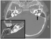
Using mouse models, it has been shown that pendrin is required between embryonic day 16.5 and postnatal day two for normal hearing but not for maintenance of hearing [Choi et al 2011a]. Endolymph volume in homozygous null mice (Slc26a4-/-) is increased and tissue mass in areas occupied by type I and II fibrocytes is reduced. Slc26a4-/- mice lack an endocochlear potential, which is normally generated across the basal cell barrier of the stria vascularis by the potassium channel KCNJ10 and localizes to the intermediate cells.

Chapter Notes

Author History

Fatemeh Alasti, PhD; University of Iowa (2011-2017)
Lorraine A Everett, MD; National Institutes of Health (1998-2001)
Eric D Green, MD, PhD; National Institutes of Health (1998-2001)
Yoichiro Iwasa, MD, PhD; University of Iowa (2020-present)
Daryl A Scott, MD, PhD; University of Iowa (1998-2001)
Amanda M Schaefer, MS, LGC; University of Iowa (2020-present)
Val C Sheffield, MD, PhD; University of Iowa School of Medicine (1998-2001)
Richard JH Smith, MD (1998-present)
Guy Van Camp, PhD; University of Antwerp (1998-2017)
Peter Van Hauwe; University of Antwerp (1998-2001)

Revision History

  • 18 June 2020 (rjhs) Revision: added authors Iwasa and Schaefer, citations Ladsous et al [2014] and Suzuki et al [2007]; updated thyroid surveillance recommendations
  • 19 October 2017 (bp) Comprehensive update posted live
  • 29 May 2014 (me) Comprehensive update posted live
  • 20 December 2012 (cd) Revision: clinical testing available for FOXI1 and KCNJ10
  • 22 December 2011 (me) Comprehensive update posted live
  • 2 April 2009 (me) Comprehensive update posted live
  • 2 July 2008 (cd) Revision: deletion/duplication analysis available clinically
  • 31 August 2006 (me) Comprehensive update posted to live Web site
  • 15 July 2004 (rjhs) Revision: use of an interpreter
  • 28 June 2004 (me) Comprehensive update posted to live Web site
  • 2 July 2003 (rjhs) Revisions
  • 1 May 2001 (me) Comprehensive update posted to live Web site
  • 28 September 1998 (pb) Review posted to live Web site
  • 4 April 1998 (rjhs) Original submission (with DFNA3) by RJH Smith, MD; LA Everett, MD; ED Green, MD, PhD; DA Scott, MD, PhD; VC Sheffield, MD, PhD; G Van Camp, PhD; P Van Hauwe

References

Published Guidelines / Consensus Statements

  • American College of Medical Genetics. Genetics evaluation guidelines for the etiologic diagnosis of congenital hearing loss. Genetic evaluation of congenital hearing loss expert panel. Available online. 2002. Accessed 6-27-23.
  • American College of Medical Genetics. Statement on universal newborn hearing screening. Available online. 2000. Accessed 6-27-23.

Literature Cited

  • Anwar S, Riazuddin S, Ahmed ZM, Tasneem S. Ateeq-ul-Jaleel, Khan SY, Griffith AJ, Friedman TB, Riazuddin S. SLC26A4 mutation spectrum associated with DFNB4 deafness and Pendred's syndrome in Pakistanis. J Hum Genet. 2009;54:266–70. [PubMed: 19287372]
  • Azaiez H, Yang T, Prasad S, Sorensen JL, Nishimura CJ, Kimberling WJ, Smith RJ. Genotype-phenotype correlations for SLC26A4-related deafness. Hum Genet. 2007;122:451–7. [PMC free article: PMC10519375] [PubMed: 17690912]
  • Berrettini S, Forli F, Bogazzi F, Neri E, Salvatori L, Casani AP, Franceschini SS. Large vestibular aqueduct syndrome: audiological, radiological, clinical, and genetic features. Am J Otolaryngol. 2005;26:363–71. [PubMed: 16275403]
  • Campbell C, Cucci RA, Prasad S, Green GE, Edeal JB, Galer CE, Karniski LP, Sheffield VC, Smith RJ. Pendred syndrome, DFNB4, and PDS/SLC26A4 identification of eight novel mutations and possible genotype-phenotype correlations. Hum Mutat. 2001;17:403–11. [PubMed: 11317356]
  • Chattaraj P, Munjal T, Honda K, Rendtorff ND, Ratay JS, Muskett JA, Risso DS, Roux I, Gertz EM, Schäffer AA, Friedman TB, Morell RJ, Tranebjærg L, Griffith AJ. A common SLC26A4-linked haplotype underlying non-syndromic hearing loss with enlargement of the vestibular aqueduct. J Med Genet. 2017;54:665–73. [PMC free article: PMC5880640] [PubMed: 28780564]
  • Choi BY, Kim HM, Ito T, Lee KY, Li X, Monahan K, Wen Y, Wilson E, Kurima K, Saunders TL, Petralia RS, Wangemann P, Friedman TB, Griffith AJ. Mouse model of enlarged vestibular aqueducts defines temporal requirement of Slc26a4 expression for hearing acquisition. J Clin Invest. 2011a;121:4516–25. [PMC free article: PMC3204851] [PubMed: 21965328]
  • Choi BY, Madeo AC, King KA, Zalewski CK, Pryor SP, Muskett JA, Nance WE, Butman JA, Brewer CC, Griffith AJ. Segregation of enlarged vestibular aqueducts in families with non-diagnostic SLC26A4 genotypes. J. Med. Genet. 2009a;46:856–61. [PMC free article: PMC2941511] [PubMed: 19578036]
  • Choi BY, Muskett J, King KA, Zalewski CK, Shawker T, Reynolds JC, Butman JA, Brewer CC, Stewart AK, Alper SL, Griffith AJ. Hereditary hearing loss with thyroid abnormalities. Adv Otorhinolaryngol. 2011b;70:43–9. [PubMed: 21358184]
  • Choi BY, Stewart AK, Madeo AC, Pryor SP, Lenhard S, Kittles R, Eisenman D, Kim HJ, Niparko J, Thomsen J, Arnos KS, Nance WE, King KA, Zalewski CK, Brewer CC, Shawker T, Reynolds JC, Butman JA, Karniski LP, Alper SL, Griffith AJ. Hypo-functional SLC26A4 variants associated with nonsyndromic hearing loss and enlargement of the vestibular aqueduct: genotype-phenotype correlation or coincidental polymorphisms? Hum Mutat. 2009b;30:599–608. [PMC free article: PMC2663020] [PubMed: 19204907]
  • Coyle B, Reardon W, Herbrick JA, Tsui LC, Gausden E, Lee J, Coffey R, Grueters A, Grossman A, Phelps PD, Luxon L, Kendall-Taylor P, Scherer SW, Trembath RC. Molecular analysis of the PDS gene in Pendred syndrome. Hum Mol Genet. 1998;7:1105–12. [PubMed: 9618167]
  • Dawson PA, Markovich D. Pathogenetics of the human SLC26 transporters. Curr Med Chem. 2005;12:385–96. [PubMed: 15720248]
  • Freudenthal B, Kulaveerasingam D, Lingappa L, Shah MA, Brueton L, Wassmer E, Ognjanovic M, Dorison N, Reichold M, Bockenhauer D, Kleta R, Zdebik AA. KCNJ10 mutations disrupt function in patients with EAST syndrome. Nephron Physiol. 2011;119:40–8. [PubMed: 21849804]
  • Goldfeld M, Glaser B, Nassir E, Gomori JM, Hazani E, Bishara N. CT of the ear in Pendred syndrome. Radiology. 2005;235:537–40. [PubMed: 15758191]
  • Huang S, Han D, Yuan Y, Wang G, Kang D, Zhang X, Yan X, Meng X, Dong M, Dai P. Extremely discrepant mutation spectrum of SLC26A4 between Chinese patients with isolated Mondini deformity and enlarged vestibular aqueduct. J Transl Med. 2011;9:167. [PMC free article: PMC3204245] [PubMed: 21961810]
  • Huang SJ, Amendola LM, Sternen DL. Variation among DNA banking consent forms: points for clinicians to bank on. J Community Genet. 2022;13:389–97. [PMC free article: PMC9314484] [PubMed: 35834113]
  • Hulander M, Kiernan AE, Blomqvist SR, Carlsson P, Samuelsson EJ, Johansson BR, Steel KP, Enerbäck S. Lack of pendrin expression leads to deafness and expansion of the endolymphatic compartment in inner ears of Foxi1 null mutant mice. Development. 2003;130:2013–25. [PubMed: 12642503]
  • Ito T, Choi BY, King KA, Zalewski CK, Muskett J, Chattaraj P, Shawker T, Reynolds JC, Butman JA, Brewer CC, Wangemann P, Alper SL, Griffith AJ. SLC26A4 genotypes and phenotypes associated with enlargement of the vestibular aqueduct. Cell Physiol Biochem. 2011;28:545–52. [PMC free article: PMC3709178] [PubMed: 22116369]
  • Jonard L, Niasme-Grare M, Bonnet C, Feldmann D, Rouillon I, Loundon N, Calais C, Catros H, David A, Dollfus H, Drouin-Garraud V, Duriez F, Eliot MM, Fellmann F, Francannet C, Gilbert-Dussardier B, Gohler C, Goizet C, Journel H, Mom T, Thuillier-Obstoy MF, Couderc R, Garabédian EN, Denoyelle F, Marlin S. Screening of SLC26A4, FOXI1 and KCNJ10 genes in unilateral hearing impairment with ipsilateral enlarged vestibular aqueduct. Int J Pediatr Otorhinolaryngol. 2010;74:1049–53. [PubMed: 20621367]
  • Kere J. Overview of the SLC26 family and associated diseases. Novartis Found Symp. 2006;273:2–11. [PubMed: 17120758]
  • King KA, Choi BY, Zalewski C, Madeo AC, Manichaikul A, Pryor SP, Ferruggiaro A, Eisenman D, Kim HJ, Niparko J, Thomsen J, Butman JA, Griffith AJ, Brewer CC. SLC26A4 genotype, but not cochlear radiologic structure, is correlated with hearing loss in ears with an enlarged vestibular aqueduct. Laryngoscope. 2010;120:384–9. [PMC free article: PMC2811762] [PubMed: 19998422]
  • Ladsous M, Vlaeminck-Guillem V, Dumur V, Vincent C, Dubrulle F, Dhaenens CM, et al. Analysis of the thyroid phenotype in 42 patients with pendred syndrome and nonsyndromic enlargement of the vestibular aqueduct. Thyroid. 2014;24:639–48. [PubMed: 24224479]
  • Luxon LM, Cohen M, Coffey RA, Phelps PD, Britton KE, Jan H, Trembath RC, Reardon W. Neuro-otological findings in Pendred syndrome. Int J Audiol. 2003;42:82–8. [PubMed: 12641391]
  • Madeo AC, Manichaikul A, Reynolds JC, Sarlis NJ, Pryor SP, Shawker TH, Griffith AJ. Evaluation of the thyroid in patients with hearing loss and enlarged vestibular aqueducts. Arch Otolaryngol Head Neck Surg. 2009;135:670–6. [PMC free article: PMC2941509] [PubMed: 19620588]
  • Merchant SN, Nakajima HH, Halpin C, Nadol JB Jr, Lee DJ, Innis WP, Curtin H, Rosowski JJ. Clinical investigation and mechanism of air-bone gaps in large vestibular aqueduct syndrome. Ann Otol Rhinol Laryngol. 2007;116:532–41. [PMC free article: PMC2585521] [PubMed: 17727085]
  • Napiontek U, Borck G, Müller-Forell W, Pfarr N, Bohnert A, Keilmann A, Pohlenz J. Intrafamilial variability of the deafness and goiter phenotype in Pendred syndrome caused by a T416P mutation in the SLC26A4 gene. J Clin Endocrinol Metab. 2004;89:5347–51. [PubMed: 15531480]
  • Park HJ, Lee SJ, Jin HS, Lee JO, Go SH, Jang HS, Moon SK, Lee SC, Chun YM, Lee HK, Choi JY, Jung SC, Griffith AJ, Koo SK. Genetic basis of hearing loss associated with enlarged vestibular aqueducts in Koreans. Clin Genet. 2005;67:160–5. [PubMed: 15679828]
  • Park HJ, Shaukat S, Liu XZ, Hahn SH, Naz S, Ghosh M, Kim HN, Moon SK, Abe S, Tukamoto K, Riazuddin S, Kabra M, Erdenetungalag R, Radnaabazar J, Khan S, Pandya A, Usami SI, Nance WE, Wilcox ER, Riazuddin S, Griffith AJ. Origins and frequencies of SLC26A4 (PDS) mutations in east and south Asians: global implications for the epidemiology of deafness. J Med Genet. 2003;40:242–8. [PMC free article: PMC1735432] [PubMed: 12676893]
  • Pera A, Villamar M, Viñuela A, Gandía M, Medà C, Moreno F, Hernández-Chico C. A mutational analysis of the SLC26A4 gene in Spanish hearing-impaired families provides new insights into the genetic causes of Pendred syndrome and DFNB4 hearing loss. Eur J Hum Genet. 2008;16:888–96. [PubMed: 18285825]
  • Pryor SP, Madeo AC, Reynolds JC, Sarlis NJ, Arnos KS, Nance WE, Yang Y, Zalewski CK, Brewer CC, Butman JA, Griffith AJ. SLC26A4/PDS genotype-phenotype correlation in hearing loss with enlargement of the vestibular aqueduct (EVA): evidence that Pendred syndrome and non-syndromic EVA are distinct clinical and genetic entities. J Med Genet. 2005;42:159–65. [PMC free article: PMC1735974] [PubMed: 15689455]
  • Reardon W, Coffey R, Chowdhury T, Grossman A, Jan H, Britton K, Kendall-Taylor P, Trembath R. Prevalence, age of onset, and natural history of thyroid disease in Pendred syndrome. J Med Genet. 1999;36:595–8. [PMC free article: PMC1762963] [PubMed: 10465108]
  • Richards S, Aziz N, Bale S, Bick D, Das S, Gastier-Foster J, Grody WW, Hegde M, Lyon E, Spector E, Voelkerding K, Rehm HL, et al. Standards and guidelines for the interpretation of sequence variants: a joint consensus recommendation of the American College of Medical Genetics and Genomics and the Association for Molecular Pathology. Genet Med. 2015;17:405–24. [PMC free article: PMC4544753] [PubMed: 25741868]
  • Rose J, Muskett JA, King KA, Zalewski CK, Chattaraj P, Butman JA, Kenna MA, Chien WW, Brewer CC, Griffith AJ. Hearing loss associated with enlarged vestibular aqueduct and zero or one mutant allele of SLC26A4. Laryngoscope. 2017;127:E238–43. [PMC free article: PMC6224160] [PubMed: 27859305]
  • Scholl UI, Choi M, Liu T, Ramaekers VT, Häusler MG, Grimmer J, Tobe SW, Farhi A, Nelson-Williams C, Lifton RP. Seizures, sensorineural deafness, ataxia, mental retardation, and electrolyte imbalance (SeSAME syndrome) caused by mutations in KCNJ10. Proc Natl Acad Sci U S A. 2009;106:5842–7. [PMC free article: PMC2656559] [PubMed: 19289823]
  • Scott DA, Wang R, Kreman TM, Andrews M, McDonald JM, Bishop JR, Smith RJ, Karniski LP, Sheffield VC. Functional differences of the PDS gene product are associated with phenotypic variation in patients with Pendred syndrome and non-syndromic hearing loss (DFNB4). Hum Mol Genet. 2000;9:1709–15. [PubMed: 10861298]
  • Senou M, Khalifa C, Thimmesch M, Jouret F, Devuyst O, Col V, Audinot JN, Lipnik P, Moreno JC, Van Sande J, Dumont JE, Many MC, Colin IM, Gérard AC. A coherent organization of differentiation proteins is required to maintain an appropriate thyroid function in the Pendred thyroid. J Clin Endocrinol Metab. 2010;95:4021–30. [PubMed: 20501687]
  • Sloan-Heggen CM, Bierer AO, Shearer AE, Kolbe DL, Nishimura CJ, Frees KL, Ephraim SS, Shibata SB, Booth KT, Campbell CA, Ranum PT, Weaver AE, Black-Ziegelbein EA, Wang D, Azaiez H, Smith RJ. Comprehensive genetic testing in the clinical evaluation of 1119 patients with hearing loss. Hum Genet. 2016;135:441–50. [PMC free article: PMC4796320] [PubMed: 26969326]
  • Stinckens C, Huygen PL, Joosten FB, Van Camp G, Otten B, Cremers CW. Fluctuant, progressive hearing loss associated with Menière like vertigo in three patients with the Pendred syndrome. Int J Pediatr Otorhinolaryngol. 2001;61:207–15. [PubMed: 11700190]
  • Sugiura M, Nakashima T, Naganawa S, Otake Y, Mukaida T, Sone M, Sato E. Sudden sensorineural hearing loss associated with inner ear anomaly. Otol Neurotol. 2005a;26:241–6. [PubMed: 15793412]
  • Sugiura M, Sato E, Nakashima T, Sugiura J, Furuhashi A, Yoshino T, Nakayama A, Mori N, Murakami H, Naganawa S. Long-term follow-up in patients with Pendred syndrome: vestibular, auditory and other phenotypes. Eur Arch Otorhinolaryngol. 2005b;262:737–43. [PubMed: 15747138]
  • Suzuki H, Oshima A, Tsukamoto K, Abe S, Kumakawa K, Nagai K, et al. Clinical characteristics and genotype-phenotype correlation of hearing loss patients with SLC26A4 mutations. Acta Otolaryngol. 2007;127:1292–7. [PubMed: 17851929]
  • Tsukamoto K, Suzuki H, Harada D, Namba A, Abe S, Usami S. Distribution and frequencies of PDS (SLC26A4) mutations in Pendred syndrome and nonsyndromic hearing loss associated with enlarged vestibular aqueduct: a unique spectrum of mutations in Japanese. Eur J Hum Genet. 2003;11:916–22. [PubMed: 14508505]
  • Van Hauwe P, Everett LA, Coucke P, Scott DA, Kraft ML, Ris-Stalpers C, Bolder C, Otten B, de Vijlder JJ, Dietrich NL, Ramesh A, Srisailapathy SC, Parving A, Cremers CW, Willems PJ, Smith RJ, Green ED, Van Camp G. Two frequent missense mutations in Pendred syndrome. Hum Mol Genet. 1998;7:1099–104. [PubMed: 9618166]
  • Wangemann P, Itza EM, Albrecht B, Wu T, Jabba SV, Maganti RJ, Lee JH, Everett LA, Wall SM, Royaux IE, Green ED, Marcus DC. Loss of KCNJ10 protein expression abolishes endocochlear potential and causes deafness in Pendred syndrome mouse model. BMC Med. 2004;2:30. [PMC free article: PMC516044] [PubMed: 15320950]
  • Wémeau JL, Kopp P. Pendred syndrome. Best Pract Res Clin Endocrinol Metab. 2017;31:213–24. [PubMed: 28648509]
  • Wu CC, Lu YC, Chen PJ, Yeh PL, Su YN, Hwu WL, Hsu CJ. Phenotypic analyses and mutation screening of the SLC26A4 and FOXI1 genes in 101 Taiwanese families with bilateral nonsyndromic enlarged vestibular aqueduct (DFNB4) or Pendred syndrome. Audiol Neurootol. 2010;15:57–66. [PubMed: 19648736]
  • Wu CC, Yeh TH, Chen PJ, Hsu CJ. Prevalent SLC26A4 mutations in patients with enlarged vestibular aqueduct and/or Mondini dysplasia: a unique spectrum of mutations in Taiwan, including a frequent founder mutation. Laryngoscope. 2005;115:1060–4. [PubMed: 15933521]
  • Yang T, Gurrola JG 2nd, Wu H, Chiu SM, Wangemann P, Snyder PM, Smith RJ. Mutations of KCNJ10 together with mutations of SLC26A4 cause digenic nonsyndromic hearing loss associated with enlarged vestibular aqueduct syndrome. Am J Hum Genet. 2009;84:651–7. [PMC free article: PMC2681005] [PubMed: 19426954]
  • Yang T, Vidarsson H, Rodrigo-Blomqvist S, Rosengren SS, Enerback S, Smith RJ. Transcriptional control of SLC26A4 is involved in Pendred syndrome and nonsyndromic enlargement of vestibular aqueduct (DFNB4). Am J Hum Genet. 2007;80:1055–63. [PMC free article: PMC1867094] [PubMed: 17503324]
Copyright © 1993-2024, University of Washington, Seattle. GeneReviews is a registered trademark of the University of Washington, Seattle. All rights reserved.

GeneReviews® chapters are owned by the University of Washington. Permission is hereby granted to reproduce, distribute, and translate copies of content materials for noncommercial research purposes only, provided that (i) credit for source (http://www.genereviews.org/) and copyright (© 1993-2024 University of Washington) are included with each copy; (ii) a link to the original material is provided whenever the material is published elsewhere on the Web; and (iii) reproducers, distributors, and/or translators comply with the GeneReviews® Copyright Notice and Usage Disclaimer. No further modifications are allowed. For clarity, excerpts of GeneReviews chapters for use in lab reports and clinic notes are a permitted use.

For more information, see the GeneReviews® Copyright Notice and Usage Disclaimer.

For questions regarding permissions or whether a specified use is allowed, contact: ude.wu@tssamda.

Bookshelf ID: NBK1467PMID: 20301640

Views

Tests in GTR by Gene

Related information

  • MedGen
    Related information in MedGen
  • OMIM
    Related OMIM records
  • PMC
    PubMed Central citations
  • PubMed
    Links to PubMed
  • Gene
    Locus Links

Similar articles in PubMed

See reviews...See all...

Recent Activity

Your browsing activity is empty.

Activity recording is turned off.

Turn recording back on

See more...