U.S. flag

An official website of the United States government

NCBI Bookshelf. A service of the National Library of Medicine, National Institutes of Health.

Adam MP, Feldman J, Mirzaa GM, et al., editors. GeneReviews® [Internet]. Seattle (WA): University of Washington, Seattle; 1993-2024.

Cover of GeneReviews®

GeneReviews® [Internet].

Show details

Pachyonychia Congenita

, PhD, , MD, , MD, PhD, FRCPC, , PhD, , DSc, FRSE, , MD, PhD, FRCPI, FRCP, and , MD, PhD.

Author Information and Affiliations

Initial Posting: ; Last Update: November 30, 2017.

Estimated reading time: 28 minutes

Summary

Clinical characteristics.

Pachyonychia congenita (PC) is characterized by hypertrophic nail dystrophy, painful palmoplantar keratoderma and blistering, oral leukokeratosis, pilosebaceous cysts (including steatocystoma and vellus hair cysts), palmoplantar hyperhydrosis, and follicular keratoses on the trunk and extremities.

Diagnosis/testing.

PC is diagnosed by clinical findings and/or by the identification of a heterozygous pathogenic variant in one of the five keratin genes known to cause PC: KRT6A, KRT6B, KRT6C, KRT16, and KRT17.

Management.

Treatment of manifestations: Pain from the palmoplantar keratoderma may be reduced somewhat by limiting friction and trauma to the feet by minimizing walking or standing, reducing hydration of the stratum corneum by using wicking socks and ventilated footwear, selecting comfortable shoes, and maintaining ideal body weight. Foot care includes paring down of hyperkeratotic areas and topical therapies for hyperkeratosis (emollients and lotions containing keratolyics). Care of thickened nails often requires the use of surgical or razor blades or sanders such as a Dremel® tool. Troublesome nails removed surgically frequently grow back in some form. Good oral hygiene and brushing gently with a soft toothbrush can improve thick, white patches on the tongue and oral mucosa. Secondary fungal and bacterial infections that require treatment are common; cysts usually do not require treatment but can be incised and drained if infected or painful. Bottle-fed infants with leukokeratosis may need a soft nipple with an enlarged opening. Rarely, young children with laryngeal thickening/growths need emergency surgery to reestablish the airway; however, surgery may exacerbate the condition.

Prevention of secondary complications: Attention to pre- and post-grooming hygiene to prevent infection; a "bleach bath" regimen using a mild bleach solution can help prevent infections.

Agents/circumstances to avoid: High temperatures and high humidity may worsen the condition.

Genetic counseling.

Pachyonychia congenita is inherited in an autosomal dominant manner. Approximately 30% of cases appear to be caused by a de novo pathogenic variant. A single case of germline mosaicism has been reported. The offspring of an affected individual have a 50% chance of inheriting the disorder. If the pathogenic variant has been identified in an affected family member, prenatal testing for a pregnancy at increased risk is possible.

Diagnosis

Clinical diagnostic criteria for pachyonychia congenita (PC) include the triad of toenail thickening, plantar keratoderma, and plantar pain, which are present in 97% of individuals with genetically confirmed PC by age ten years [International Pachyonychia Research Registry, Eliason et al 2012].

Suggestive Findings

Pachyonychia congenita (PC) should be suspected in individuals with the following clinical features and/or family history findings.

Clinical features

  • Plantar keratoderma including callus with underlying blisters
  • Plantar pain
  • Hypertrophic nail dystrophy, which may be limited to the toenails or to a few toenails or fingernails (See Figure 1 and Table 2.)
  • Pilosebaceous cysts including widespread steatocystomas/steatocysts (benign lesions) and vellus hair cysts which usually develop at puberty and continue throughout adulthood
  • Oral leukokeratosis
  • Follicular keratoses on the trunk and extremities usually present by early childhood
  • Palmoplantar hyperhydrosis (<50%)
  • Natal or prenatal teeth (i.e., present at birth or by age 1 month in some affected individuals)
Figure 1. . Common findings of pachyonychia congenita include: thickened and dystrophic nails (both fingernails and toenails) (a-c); bullae (usually on the pressure points of the heels and soles); hyperkeratosis (d-e); cysts (f); and oral leukokeratosis (g).

Figure 1.

Common findings of pachyonychia congenita include: thickened and dystrophic nails (both fingernails and toenails) (a-c); bullae (usually on the pressure points of the heels and soles); hyperkeratosis (d-e); cysts (f); and oral leukokeratosis (g).

Family history is consistent with autosomal dominant inheritance.

Note: (1) Approximately 70% of individuals with PC, enrolled in the International Pachyonychia Congenita Research Registry, inherited the condition from an affected parent; therefore, lack of a family history of PC does not preclude the diagnosis. (2) If the family history suggests autosomal recessive inheritance, a condition other than PC should be considered (see Differential Diagnosis).

Establishing the Diagnosis

The diagnosis of PC is established in a proband with the triad of toenail thickening, plantar keratoderma, and plantar pain and/or by identification of a heterozygous pathogenic (or likely pathogenic) variant in one of the five genes listed in Table 1.

Note: (1) Histologic, immunohistologic, or electron microscopic examination of the nails or skin from individuals with PC is not helpful in confirming the diagnosis of PC but can be performed to rule out other diagnoses. (2) Per ACMG/AMP variant interpretation guidelines, the terms "pathogenic variants" and "likely pathogenic variants" are synonymous in a clinical setting, meaning that both are considered diagnostic and both can be used for clinical decision making [Richards et al 2015]. Reference to "pathogenic variants" in this section is understood to include any likely pathogenic variants. (3) Identification of a heterozygous variant of uncertain significance in one of the five genes listed in Table 1 does not establish or rule out the diagnosis.

Molecular genetic testing approaches can include serial single-gene testing, use of a multigene panel, and more comprehensive genomic testing.

Serial single-gene testing

  • For individuals who have focal non-epidermolytic palmoplantar keratoderma (FNEPPK), sequence analysis of KRT6C and KRT16 may be considered first.
  • For individuals who have steatocystoma multiplex (SM) or a history of natal teeth, sequence analysis of KRT17 may be considered first.

A multigene panel that includes KRT6A, KRT6B, KRT6C, KRT16, KRT17, and other genes of interest (see Differential Diagnosis) may also be considered. Note: (1) The genes included in the panel and the diagnostic sensitivity of the testing used for each gene varies by laboratory and are likely to change over time. (2) Some multigene panels may include genes not associated with the condition discussed in this GeneReview; thus, clinicians need to determine which multigene panel is most likely to identify the genetic cause of the condition while limiting identification of variants of uncertain significance and pathogenic variants in genes that do not explain the underlying phenotype. (3) In some laboratories, panel options may include a custom laboratory-designed panel and/or custom phenotype-focused exome analysis that includes genes specified by the clinician. (4) Methods used in a panel may include sequence analysis, deletion/duplication analysis, and/or other non-sequencing-based tests.

For an introduction to multigene panels click here. More detailed information for clinicians ordering genetic tests can be found here.

More comprehensive genomic testing (when available) including exome sequencing and genome sequencing may be considered. Such testing may provide or suggest a diagnosis not previously considered (e.g., mutation of a different gene or genes that results in a similar clinical presentation). For an introduction to comprehensive genomic testing click here. More detailed information for clinicians ordering genomic testing can be found here.

Table 1.

Molecular Genetic Testing Used in Pachyonychia Congenita (PC)

Gene 1Proportion of PC
Attributed to
Pathogenic Variants
in Gene 2, 3
Proportion of Pathogenic Variants 4 Detectable by Method
Sequence analysis 5Gene-targeted deletion/duplication analysis 6
KRT6A 304/774 (39%)>99%Unknown 7
KRT6B 70/774 (9%)>99%Unknown 7
KRT6C 22/774 (3%)>99%Unknown 7
KRT16 247/774 (32%)>99%Unknown 7
KRT17 8130/774 (17%)>99%Unknown 7
1.
2.

Pathogenic variants in at least 800 individuals have been reported [International PC Research Registry, Human Intermediate Filament Database].

3.

The numbers in this table refer only to those individuals enrolled in the International PC Research Registry (IPCRR); not all are published but the data are available on the website.

4.

See Molecular Genetics for information on allelic variants detected in this gene.

5.

Sequence analysis detects variants that are benign, likely benign, of uncertain significance, likely pathogenic, or pathogenic. Variants may include small intragenic deletions/insertions and missense, nonsense, and splice site variants; typically, exon or whole-gene deletions/duplications are not detected. For issues to consider in interpretation of sequence analysis results, click here.

6.

Gene-targeted deletion/duplication analysis detects intragenic deletions or duplications. Methods used may include a range of techniques such as quantitative PCR, long-range PCR, multiplex ligation-dependent probe amplification (MLPA), and a gene-targeted microarray designed to detect single-exon deletions or duplications.

7.

No data on gene-targeted del/dup analysis are available.

8.

To date all identified pathogenic variants causing steatocystoma multiplex (SM) are in KRT17. Careful reexamination of those with SM caused by a heterozygous KRT17 pathogenic variant have identified subtle nail changes in some family members. Many individuals with a clinical diagnosis of SM and no nails changes also have no identifiable pathogenic variant in KRT17. Therefore, genetic heterogeneity is likely for SM.

Clinical Characteristics

Clinical Description

In all types of pachyonychia congenita (PC) most characteristics are visible by age ten years [Shah et al 2014] and typically include toenail thickening, plantar keratoderma, and plantar pain (see Suggestive Findings). However, the absence or presence of certain features as well as the age at onset varies according the gene that is mutated (see Table 2 for phenotypic features of PC). The most recent classification of the condition also incorporates the genetic cause (see Nomenclature).

The severity of findings can differ both within a family and among families with the same pathogenic variant.

Table 2.

International Pachyonychia Congenita Research Registry (IPCRR) Data Summary (as of 10 May 2017)

Gene in Which Pathogenic
Variants Were Confirmed
KRT6A KRT6B KRT6C KRT16 KRT17 TOTAL
Number evaluated for finding 1 3047122247130774
Toenails thickened
10 toenails286 (94%)26 (37%)0 (00%)99 (40%)100 (77%)511 (66%)
7-9 toenails9 (03%)14 (20%)0 (00%)52 (21%)11 (08%)86 (11%)
4-6 toenails2 (01%)21 (30%)3 (14%)63 (26%)7 (05%)96 (12%)
1-3 toenails4 (01%)9 (13%)10 (45%)44 (18%)8 (06%)75 (10%)
Total w/toenails thickened 301 (99%) 70 (99%) 13 (59%) 238 (96%) 126 (97%) 748 (97%)
Age
at
onset
Birth - <1 yr264 (88%)10 (14%)1 (08%)45 (19%)92 (73%)412 (54%)
1-4 yrs33 (11%)19 (27%)6 (46%)78 (33%)24 (19%)160 (21%)
5-14 yrs4 (01%)34 (49%)4 (31%)74 (31%)10 (08%)126 (17%)
≥15 yrs0 (00%)7 (10%)2 (15%)43 (18%)1 (01%)53 (07%)
Fingernails thickened
10 fingernails271 (89%)4 (06%)0 (00%)73 (30%)62 (48%)410 (53%)
7-9 fingernails10 (03%)4 (06%)0 (00%)11 (04%)13 (10%)38 (05%)
4-6 fingernails14 (05%)17 (24%)0 (00%)30 (12%)28 (22%)89 (11%)
1-3 fingernails6 (02%)7 (10%)0 (00%)28 (11%)9 (07%)50 (06%)
Total w/fingernails thickened 301 (99%) 32 (45%) 0 (00%) 142 (57%) 112 (86%) 587 (76%)
Age
at
onset
Birth - <1 yr267 (89%)4 (13%)0 (00%)33 (23%)85 (76%)389 (66%)
1-4 yrs30 (10%)8 (25%)0 (00%)42 (30%)19 (17%)99 (17%)
5-14 yrs3 (01%)11 (34%)0 (00%)34 (24%)6 (05%)54 (09%)
≥15 yrs1 (00%)10 (31%)0 (00%)34 (24%)3 (03%)48 (08%)
Plantar keratoderma
Always (never completely goes away)254 (84%)67 (94%)19 (86%)240 (97%)86 (66%)666 (86%)
Sometimes (feet clear up completely at times)7 (02%)1 (01%)0 (00%)1 (00%)14 (11%)23 (03%)
Seldom (feet usually clear of symptoms)5 (02%)0 (00%)0 (00%)0 (00%)4 (03%)9 (01%)
Total w/plantar keratoderma 266 (88%) 68 (96%) 19 (86%) 241 (98%) 104 (80%) 698 (90%)
Age
at
onset
Birth - <1 yr39 (15%)2 (03%)1 (05%)23 (10%)12 (12%)77 (11%)
1-4 yrs152 (57%)23 (34%)9 (47%)130 (54%)35 (34%)349 (50%)
5-14 yrs70 (26%)42 (62%)9 (47%)82 (34%)43 (41%)246 (35%)
≥15 yrs5 (02%)1 (01%)0 (00%)8 (03%)15 (14%)29 (04%)
Plantar pain w/plantar keratoderma 2
Often require medication for pain65 (24%)12 (18%)5 (26%)74 (31%)19 (18%)175 (25%)
Very painful, but do not use medication114 (43%)32 (47%)11 (58%)111 (46%)34 (33%)302 (43%)
Somewhat painful77 (29%)24 (35%)3 (16%)50 (21%)36 (35%)190 (27%)
Total w/plantar keratoderma/pain 256 (96%) 68 (100%) 19 (100%) 235 (98%) 89 (86%) 667 (96%)
Palmar keratoderma
Always (never completely goes away)86 (28%)11 (15%)2 (09%)148 (60%)21 (16%)268 (35%)
Sometimes (hands clear up completely at times)32 (11%)7 (10%)1 (05%)15 (06%)22 (17%)77 (10%)
Seldom (hands usually clear of symptoms)49 (16%)13 (18%)3 (14%)22 (09%)27 (21%)114 (15%)
Total w/palmar keratoderma 167 (55%) 31 (44%) 6 (27%) 185 (75%) 70 (54%) 459 (59%)
Additional findings
Oral leukokeratosis 269 (88%)18 (25%)4 (18%)88 (36%)34 (26%)413 (53%)
Cysts 188 (62%)49 (69%)4 (18%)64 (26%)121 (93%)426 (55%)
Follicular hyperkeratosis 162 (53%)30 (42%)0 (00%)30 (12%)86 (66%)308 (40%)
Natal or prenatal teeth 12 (04%)0 (00%)0 (00%)0 (00%)99 (76%)111 (14%)
1.

Data from 774 individuals enrolled in the International PC Research Registry with genetically confirmed pachyonychia congenita

2.

By age ≤10 years

Hypertrophic nail dystrophy, the predominant clinical feature of PC, is typically noted within the first few months to years of life, though in rare instances it presents later in life. The nail dystrophy appears to fall into two phenotypes:

  • Nails that grow to full length and have an upward slant caused by the prominent distal hyperkeratosis (often with an accentuated curvature of the nail)
  • Nails that have a nail plate that terminates prematurely leaving a gently sloping distal region of hyperkeratosis and exposed distal finger tip

Focal palmoplantar keratoderma usually presents during the first few years of life when a child starts bearing weight and walking. Blisters develop beneath the keratoderma resulting in intense pain. For many individuals, the blisters and constant foot pain are more severe in warmer weather than cooler weather. The pain associated with plantar focal blistering may require the use of crutches, canes, or wheelchairs. Rarely, keratosis palmoplantaris transgrediens (the contiguous extension of hyperkeratosis beyond the palmar and/or plantar skin) is present.

  • Focal non-epidermolytic palmoplantar keratoderma (FNEPPK), defined as keratoderma of varying severity that may occur on the palms and soles with no (or very mild) nail dystrophy, was previously thought to be a distinct entity but is now considered part of the spectrum of PC.

Oral leukokeratosis (thickened white patches on the tongue and cheek) is often present. In babies, oral leukokeratosis can be misdiagnosed as Candida albicans and may cause difficulty in sucking.

Follicular keratosis, usually on the elbows, knees or trunk, occurs in some persons. It is more prevalent in late childhood and teenage years and becomes less problematic in adults.

Pilosebaceous cysts including widespread steatocystomas/steatocysts (benign lesions) and vellus hair cysts. Cysts may increase in number at puberty. Early onset has been reported [Feng et al 2003] and is recorded in the International Pachyonychia Congenita Research Registry (IPCRR).

  • Steatocystoma multiplex (SM), defined as widespread steatocystomas that can develop at puberty with subtle nail involvement but no palmoplantar keratoderma, occurs in association with heterozygous pathogenic variants in KRT17 (see Table 1, footnote 8).

Natal teeth or prenatal teeth. Although some individuals have a few prenatal or natal teeth, this finding is not consistently present even within the same family [Leachman et al 2005]. Natal teeth are usually associated with pathogenic variants in KRT17. Primary and secondary dentition is normal.

Other findings that may occur:

  • Excessive sweating of the palms and soles (palmoplantar hyperhydrosis), observed in approximately 50% of individuals
  • Axillary and inguinal cyst formation
  • Excessive production of waxy material in the ear
  • Severe and unexplained ear pain
  • Hoarseness (laryngeal involvement), reported primarily in young children. Although rare, laryngeal involvement may cause life-threatening respiratory distress requiring intervention.
  • Angular cheilitis (inflammation and fissuring at the angles of the mouth) which is sometimes secondarily infected
  • Paronychia with pronounced edema (and occasional blister formation) under the nails; may exhibit lymphatic extension and may sometimes be caused by infection

Phenotype Correlations by Gene

Leukokeratosis with laryngeal involvement may be seen in infants and children with PC-K6a (see Nomenclature) caused by a heterozygous pathogenic variant in KRT6A.

All cases of "failure to thrive" and poor feeding in infancy have been found to be in individuals with PC-K6a, caused by a heterozygous pathogenic variant in KRT6A.

Focal non-epidermolytic palmoplantar keratoderma (FNEPPK) has been described only in individuals who have a heterozygous pathogenic variant in either KRT6C or KRT16 [Wilson et al 2010, Fu et al 2011].

Steatocystoma multiplex (SM) has been reported only in individuals who have a heterozygous pathogenic variant in KRT17 [Covello et al 1998].

Individuals with a history of natal teeth are more likely to have a heterozygous pathogenic variant in KRT17 (see Table 2).

Genotype-Phenotype Correlations

Based on data on 774 individuals with PC in the IPCRR, clear genotype-phenotype correlations are evident [Fu et al 2011, Spaunhurst et al 2012] (see Table 2).

In the following instances, the phenotype may vary among individuals with the same pathogenic variant:

  • The same KRT17 pathogenic variant in the highly conserved helix initiation motif has been observed in classic PC and in a few individuals with the milder variant SM with few or no nail changes. The modifying factors responsible for this variable expressivity are not known.
  • In a few reports of late-onset PC, pathogenic variants have been identified outside the helix boundary and some have questioned whether the location of the pathogenic variant affects the age at onset. However, the ages in these cases are the expected ages at onset for the particular type of PC and should likely not be referred to as "late-onset."

Nomenclature

Based on data from the International Pachyonychia Congenita Research Registry (IPCRR), the most recent classification for pachyonychia congenita is by mutated gene [McLean et al 2011, Wilson et al 2011]:

  • PC-K6a (caused by pathogenic variants in KRT6A)
  • PC-K6b (caused by pathogenic variants in KRT6B)
  • PC-K6c (caused by pathogenic variants in KRT6C)
  • PC-K16 (caused by pathogenic variants in KRT16)
  • PC-K17 (caused by pathogenic variants in KRT17)

The classification suggested for PC prior to the identification of the genetic basis of the disease was based solely on clinical findings. Historically, the two major subtypes of PC were based on subtle variable phenotypic features (primarily on the presence or absence of pilosebaceous cysts and natal or prenatal teeth) [Leachman et al 2005, Liao et al 2007]:

  • PC-1 (Jadassohn-Lewandowski syndrome)
  • PC-2 (Jackson-Lawler syndrome)

With detailed clinical histories and pathogenic variants identified in an increasing number of people with PC, it became clear that the older classification of PC-1 and PC-2 was not applicable to the broader population of individuals with PC.

Prevalence

The rarity of PC makes it difficult to accurately assess its prevalence.

The International PC Research Registry has identified 774 individuals in 419 families with genetically confirmed PC.

Differential Diagnosis

Onychomycosis. While the hyperkeratotic nail thickening seen in pachyonychia congenita (PC) may be mistaken for onychomycosis, dermatophytic infections do not affect all finger and toenails, particularly at an early age. In the rare conditions of autoimmune endocrinopathy-candidiasis-ectodermal dystrophy (APECED) and systemic mucocutaneous candidosis, all nails may be affected.

Oral leukokeratosis together with nail dystrophy is often an indication of pachyonychia congenita and may be mistaken for Candida albicans (thrush), white sponge nevus, and/or leukoplakia.

Epidermolysis bullosa simplex (EBS) or other palmoplantar keratodermas can result in a similar pattern of plantar blister formation or hyperkeratosis, respectively; however, they do not share the characteristic nail changes of PC.

Note: EBS may be incorrectly diagnosed in young children with PC because they have a greater tendency toward blister formation and lesser tendency toward keratoderma.

Clouston syndrome (hidrotic ectodermal dysplasia 2), an autosomal dominant disorder caused by a heterozygous pathogenic variant in GJB6, the gene encoding the gap junction beta-6 protein, can also mimic PC [Hale et al 2015]. Alopecia does not typically occur in PC, whereas variable alopecia is a relatively common feature of Clouston syndrome.

Nonsyndromic congenital nail disorder 10 (OMIM 614157) without the associated palmoplantar keratoderma or other features of PC can be confused with PC. This is an autosomal recessive disorder caused by biallelic pathogenic variants in FZD6, encoding frizzled-6 [Wilson et al 2013, Kasparis et al 2016].

Familial onychogryphosis without the associated palmoplantar keratoderma or other features of PC can be confused with PC. Individuals who have nail findings only are unlikely to have a pathogenic variant in one of the PC-related genes.

Twenty-nail dystrophy (OMIM 161050) may occur without keratoderma or other associated changes. Autosomal dominant inheritance has been described.

Dyskeratosis congenita (DC) manifests with features overlapping with PC including nail dystrophy, palmoplantar keratoderma (PPK), hyperhidrosis, and oral leukoplakia. Distinctive features include reticulate hyperpigmentation, skin tumors, and hematologic manifestation. To date, pathogenic variants in ACD, CTC1, DKC1, NHP2, NOP10, PARN, RTEL1, TERC, TERT, TINF2, and WRAP53 have been shown to cause DC; mode of inheritance varies by gene.

Palmoplantar keratoderma striata (PPKS1) (OMIM 148700), an autosomal dominant disorder caused by a heterozygous pathogenic variant in DSG1, can be confused with focal non-epidermolytic palmoplantar keratoderma (FNEPPK) / pachyonychia congenita [Lovgren et al 2017]. However, pain is typically either absent or less significant in PPKS1 than in FNEPPK or PC.

Punctate PPK type 1 (OMIM 148600), an autosomal dominant disorder caused by a heterozygous pathogenic variant in AAGAB, can be painful and focal (due to coalescence of lesions) [Pohler et al 2012].

Olmsted syndrome (OMIM 614594) is characterized by painful palmoplantar keratoderma that may occur with additional features including periorificial keratotic plaques and sometimes constricting digital bands on hands and feet that result in spontaneous amputation, mutilating PPK, alopecia, nail dystrophy, and itching of lesions. Olmsted syndrome is caused by a pathogenic variant in TRPV3 [Lin et al 2012; Wilson et al 2015] and normally inherited in an autosomal dominant manner, although recessive inheritance has been reported [Duchatelet et al 2014].

Management

Evaluations Following Initial Diagnosis

To establish the extent of disease and needs in an individual diagnosed with pachyonychia congenita, the following evaluations are recommended:

  • Thorough clinical examination to assess each affected area. These will vary based on the specific gene involved (see Table 2) and may need to be repeated once genetic testing is completed in order to fully understand the phenotype of the affected individual.
  • Consultation with a clinical geneticist and/or genetic counselor

Treatment of Manifestations

Preliminary treatment guidelines have been published [Goldberg et al 2014] and continue to be refined.

The current treatment modalities primarily center on symptomatic relief of pain, hygienic grooming practices including paring of hyperkeratotic areas, treatment of secondary infection when indicated, and use of various walking aids including wheelchairs, crutches, and canes.

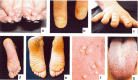
Palmoplantar keratoderma. Frequent grooming of the feet is essential and includes paring down the hyperkeratotic areas. However, trimming too aggressively can greatly increase pain. Some find it helpful to soak the feet prior to the paring. The surface of the skin and the instruments used should be clean to avoid infection. Blisters should be punctured with a sterile needle, the fluid drained, and the blister roof left in place until it dries and is shed away.

Topical therapies to remove the hyperkeratosis:

  • Emollients such as Vaseline® or lanolin-containing products are frequently used. Note: Creams and lotions containing keratolyics such as urea, lactic acid, salicylic acid, or propylene glycol have little effect, with some reporting negative side effects. Occlusive ointments are often poorly tolerated.
  • Oral retinoids, while reducing the keratoderma, do not affect the underlying blistering and fragility of the skin and sometimes increase the pain. Careful regulation of the dosage is necessary [Gruber et al 2012].

Special orthotics or insoles, wicking socks, ventilated footwear or cushioned footwear can help to lessen the pain although pain varies from day to day and at times can be intense even at rest.

Maintaining ideal body weight can be a factor in reducing the hyperkeratosis and pain. Limiting walking or standing can help to reduce trauma and slightly diminish the resulting blisters, callus, and pain.

The origin, nature, and underlying mechanism of plantar pain in individuals with PC is poorly understood. Several recent studies suggest that neuropathic pain treatments may be useful in individuals with PC who experience plantar pain [Pan et al 2016; Wallis et al 2016; Authors, unpublished] (see Therapies Under Investigation).

Nail dystrophy. Thickened nails are not typically painful, but become so when infected or traumatized. An effective tool for very thick nails and for children is a guillotine-type pet nail clipper which places no pressure on the nails. Other tools frequently used are razor or surgical blades or sanders such as a Dremel® tool.

If bacterial or fungal infections occur, systemic antibiotics or antifungals are indicated.

Particularly troublesome nails can be successfully removed surgically; however, few affected individuals have had this procedure and in many cases – regardless of the specific pathogenic variant – the nails have regrown [DeKlotz et al 2017].

Oral leukokeratosis. Good oral hygiene and frequent gentle brushing with a toothbrush can significantly improve the appearance of the thick, white patches on the tongue and oral mucosa; however, if done too vigorously, brushing may also traumatize the mucosa resulting in reactive hyperkeratosis.

Some individuals have reported reduction of the leukokeratosis in response to oral antibiotics, suggesting a possible bacterial contribution; more likely, improvement may be a response to the anti-inflammatory properties of the antibiotics.

Follicular hyperkeratosis. Especially bothersome for children and teens, this finding can be treated with alpha-hydroxy acid creams or lotions or keratolytic emollients; however, these treatments may not be especially effective for PC. The use of emollients such as Vaseline® or lanolin-containing products is reported to be as effective.

Leukokeratosis with laryngeal involvement. Reported only in children with PC-K6a, respiratory insufficiency can on occasion become life threatening, requiring emergent surgical intervention to reestablish the airway. The surgical procedures are repeated as necessary to maintain an open airway; however, surgical procedures to the larynx aimed at improving hoarseness should be avoided as they may tend to worsen the condition.

Cysts. Steatocystoma multiplex and other pilosebaceous cysts can be treated by incision with a number 11 blade and subsequent expression of the contents of the cyst ("incision and drainage"). Oral antibiotics may be indicated in the case of secondary infection. A culture should be obtained if infection is a consideration. Intralesional injection of steroid (e.g., triamcinolone) may reduce inflammation of the area if infection is not suspected. If necessary, cysts can be excised.

Failure to thrive. Poor feeding in infancy has been reported to be ameliorated by the use of a soft nipple with an enlarged opening to reduce the sucking required.

Prevention of Secondary Complications

Infection of the skin and nails following grooming or trauma is the most common secondary complication seen in PC.

  • Pre- and post-grooming hygiene and use of clean instruments minimizes this complication.
  • Antibiotics may be indicated when infection occurs.
  • A simple "bleach bath" regimen using a mild bleach solution can help prevent infections.

Surveillance

In general, individuals with PC have no known associated systemic diseases or predispositions that require routine surveillance.

Agents/Circumstances to Avoid

Some report that higher temperatures and higher humidity worsen the condition.

Evaluation of Relatives at Risk

Molecular genetic testing of at-risk relatives in a family with PC is not indicated because the phenotype is readily observed from a young age and no interventions can prevent the development of manifestations or reduce their severity.

See Genetic Counseling for issues related to testing of at-risk relatives for genetic counseling purposes.

Pregnancy Management

For a pregnant woman with PC, weight gain (increasing stress on the plantar surface) or altered hormonal environment during pregnancy may worsen the painful plantar keratoderma.

Therapies Under Investigation

In 2014, a Phase 1b clinical trial sponsored by PC Project and TransDerm was performed using topical sirolimus. This study included 15 affected individuals and was conducted by Dr Joyce Teng at Stanford University. Background research for this trial was previously published [Hickerson et al 2009].

Short interfering RNA (siRNA) can selectively block expression of a specific K6a-causing pathogenic variant [Hickerson et al 2008, Leachman et al 2008]. The siRNA trial included treatment of a single individual with a specific KRT6A pathogenic variant in a dose-escalation trial of an siRNA directed against the p.Asn171Lys mutated allele [Leachman et al 2010]. The affected individual did not experience any adverse effects from the experimental treatment. The affected person also experienced callus regression on the foot treated with siRNA.

Botulinum toxin has been used to treat pain in several affected individuals, with promising results [Swartling & Vahlquist 2006, Swartling et al 2010, González-Ramos et al 2016]. In addition to these published investigations, a number of other individuals have been treated with botulinum toxin.

Several persons have been treated with statins. The results from studies are mixed. Further research is being conducted and additional animal testing is proposed [Zhao et al 2011].

Other therapies currently under investigation include anti-TNF biologics, duloxetine or duloxetine and a tricyclic combination, gabapentin, topical gabapentin, and capsaicin injections.

Search ClinicalTrials.gov in the US and EU Clinical Trials Register in Europe for access to information on clinical studies for a wide range of diseases and conditions.

Genetic Counseling

Genetic counseling is the process of providing individuals and families with information on the nature, mode(s) of inheritance, and implications of genetic disorders to help them make informed medical and personal decisions. The following section deals with genetic risk assessment and the use of family history and genetic testing to clarify genetic status for family members; it is not meant to address all personal, cultural, or ethical issues that may arise or to substitute for consultation with a genetics professional. —ED.

Mode of Inheritance

Pachyonychia congenita (PC) is inherited in an autosomal dominant manner.

Risk to Family Members

Parents of a proband

  • Up to 70% of individuals diagnosed with pachyonychia congenita have an affected parent.
  • A proband with pachyonychia congenita may have the disorder as the result of a de novo pathogenic variant. The proportion of cases caused by a de novo pathogenic variant is approximately 30%. Of these, some may be novel pathogenic variants while others may be recurrent pathogenic variants identified in other families.
  • Complete clinical examination by a dermatologist to confirm the absence of phenotype is recommended for the evaluation of parents of a proband with an apparent de novo pathogenic variant; if there are no phenotypic signs, no parental genetic testing is necessary.

Sibs of a proband. The risk to the sibs of a proband depends on the genetic status of the proband's parents:

  • If a parent of the proband is affected, the risk to the sibs is 50%.
  • If the parents are clinically unaffected, the risk to the sibs of a proband is low, but slightly greater than that of the general population because of the possibility of germline mosaicism.
    The incidence of germline mosaicism is not known. It is extremely rare: of 774 cases, a single case of germline mosaicism (0.13%) has been reported for PC [Pho et al 2011].

Offspring of a proband. Each child of an individual with pachyonychia congenita has a 50% chance of inheriting the pathogenic variant.

Other family members. The risk to other family members depends on the status of the proband's parents: if a parent is affected, the parent's family members may be affected and at risk of having affected children.

Related Genetic Counseling Issues

Because PC is a very rare disorder, affected individuals and families often feel completely isolated. Connecting affected individuals and families to the PC patient advocacy group (see Resources), which offers many different support services, can often be extremely valuable [Schwartz et al 2013].

Considerations in families with an apparent de novo pathogenic variant. When neither parent of a proband with an autosomal dominant condition has the pathogenic variant identified in the proband or clinical evidence of the disorder, the pathogenic variant is likely de novo. However, non-medical explanations including alternate paternity or maternity (e.g., with assisted reproduction) and undisclosed adoption could also be explored.

Family planning

  • The optimal time for determination of genetic risk and discussion of the availability of prenatal/preimplantation genetic testing is before pregnancy.
  • It is appropriate to offer genetic counseling (including discussion of potential risks to offspring and reproductive options) to young adults who are affected or at risk.

DNA banking. Because it is likely that testing methodology and our understanding of genes, pathogenic mechanisms, and diseases will improve in the future, consideration should be given to banking DNA from probands in whom a molecular diagnosis has not been confirmed (i.e., the causative pathogenic mechanism is unknown). For more information, see Huang et al [2022].

Prenatal Testing and Preimplantation Genetic Testing

Once the pathogenic variant has been identified in an affected family member, prenatal and preimplantation genetic testing are possible.

Resources

GeneReviews staff has selected the following disease-specific and/or umbrella support organizations and/or registries for the benefit of individuals with this disorder and their families. GeneReviews is not responsible for the information provided by other organizations. For information on selection criteria, click here.

  • Pachyonychia Congenita Project and International Pachyonychia Congenita Research Registry (IPCRR)
    PC Project
    P.O. Box 17850
    Holladay UT 84117
    Phone: 801-987-8758
    Email: info@pachyonychia.org
  • National Institute of Arthritis and Musculoskeletal and Skin Diseases

Molecular Genetics

Information in the Molecular Genetics and OMIM tables may differ from that elsewhere in the GeneReview: tables may contain more recent information. —ED.

Table B.

OMIM Entries for Pachyonychia Congenita (View All in OMIM)

148041KERATIN 6A, TYPE II; KRT6A
148042KERATIN 6B, TYPE II; KRT6B
148067KERATIN 16, TYPE I; KRT16
148069KERATIN 17, TYPE I; KRT17
167200PACHYONYCHIA CONGENITA 1; PC1
167210PACHYONYCHIA CONGENITA 2; PC2
612315KERATIN 6C, TYPE II; KRT6C
615726PACHYONYCHIA CONGENITA 3; PC3
615728PACHYONYCHIA CONGENITA 4; PC4
615735PALMOPLANTAR KERATODERMA, NONEPIDERMOLYTIC, FOCAL OR DIFFUSE; PPKNEFD

Molecular Pathogenesis

Keratins form a cytoskeletal intermediate filament network within all epithelial cells. Epithelia in different body regions utilize a range of different keratins. Keratins associated with PC are constitutively expressed in the nail, palmoplantar skin, oral mucosa, and hair. Thus, pathogenic variants in the genes encoding these keratins lead to pathology in these major body sites.

Pathogenic variants in at least 400 families have been reported to date [Human Intermediate Filament Database, Szeverenyi et al 2008, Wilson et al 2011, Wilson et al 2014, IPCRR]. A number of the pathogenic variants are recurrent but others are family specific. More than 100 different pathogenic variants have been identified. The majority are found in or near the helix initiation motif in the 1A domain or the helix termination motif at the end of the 2B domain (Figure 2), consistent with the location of pathogenic variants in most other keratin disorders [Wilson et al 2011]. A genotype/phenotype correlation is observed in the keratin disorder epidermolysis bullosa simplex (EBS), in which the more severe pathogenic variants occur in the helix boundary domains and those causing a milder phenotype occur within or outside these regions. To date this has not been observed in PC; possibly, pathogenic variants in these less conserved regions in KRT6A, KRT6B, KRT6C, KRT16, or KRT17 are in general not severe enough to produce a clinical phenotype.

Figure 2. . Schematic diagram showing the basic protein structure of a keratin filament.

Figure 2.

Schematic diagram showing the basic protein structure of a keratin filament. The α-helical rod domain is divided into four domains: 1A, 1B, 2A, and 2B, connected by non-helical linkers L1, L12, and L2. At the ends of the rod domain are the helix (more...)

KRT6A

Gene structure. The cDNA comprises 2450 bp in nine exons. For a detailed summary of gene and protein information, see Table A, Gene.

Pathogenic variants. The majority of pathogenic variants are heterozygous missense variants; in some individuals, small in-frame deletions/insertions and splice site and nonsense variants have been reported. Most pathogenic variants occur in the highly conserved helix boundary motif domains located at either end of the alpha-helical keratin rod domain. There are a number of recurrent pathogenic variants; the major ones for PC-K6a, located at Asn171, are either a single amino acid deletion c.516_518del or a missense variant affecting the neighboring amino acid residue Asn172 (Table 3).

Table 3.

Selected KRT6A Pathogenic Variants

DNA Nucleotide Change
(Alias 1)
Predicted Protein ChangeReference Sequences
c.511A>Gp.Asn171Asp NM_005554​.3
NP_005545​.1
c.511A>Tp.Asn171Tyr
c.512A>Gp.Asn171Ser
c.513C>Ap.Asn171Lys
c.516_518del
(514_516delAAC)
p.Asn172del

Variants listed in the table have been provided by the authors. GeneReviews staff have not independently verified the classification of variants.

GeneReviews follows the standard naming conventions of the Human Genome Variation Society (varnomen​.hgvs.org). See Quick Reference for an explanation of nomenclature.

1.

Variant designation that does not conform to current naming conventions

Normal gene product. The protein, keratin, type II cytoskeletal 6A (K6a keratin), consists of 564 amino acids. Keratins form a cytoskeletal intermediate filament network within all epithelial cells.

Abnormal gene product. Pathogenic variants cause disruption of the cytoskeleton resulting in keratin filament aggregation leading to collapse of the cytoskeleton and cell fragility. The highly conserved helix boundary domains where the majority of pathogenic variants occur are critical during normal keratin filament assembly.

KRT6B

Gene structure. The cDNA comprises 2331 bp in nine exons (reference sequence NM_005555.3). For a detailed summary of gene and protein information, see Table A, Gene.

Pathogenic variants. The pathogenic variants reported to date are heterozygous missense variants or small in-frame deletions in either the highly conserved helix initiation or helix termination domains.

Normal gene product. The protein, keratin, type II cytoskeletal 6B (K6b keratin), consists of 564 amino acids. Keratins form a cytoskeletal network within all epithelial cells.

Abnormal gene product. Pathogenic variants cause disruption of the cytoskeleton resulting in keratin filament aggregation leading to collapse of the cytoskeleton and cell fragility. The highly conserved helix boundary domains where the majority of pathogenic variants occur are critical during normal keratin filament assembly.

KRT6C

Gene structure. The cDNA comprises 2345 bp in nine exons (reference sequence NM_173086.4). For a detailed summary of gene and protein information, see Table A, Gene.

Pathogenic variants. The pathogenic variants reported to date are heterozygous missense variants or small in-frame deletions in regions encoding either the highly conserved helix initiation or helix termination domains.

Normal gene product. The protein, keratin, type II cytoskeletal 6C (K6c keratin), consists of 564 amino acids (NP_775109.2). Keratins form a cytoskeletal network within all epithelial cells.

Abnormal gene product. Pathogenic variants cause disruption of the cytoskeleton resulting in keratin filament aggregation leading to collapse of the cytoskeleton and cell fragility. The highly conserved helix boundary domains where the majority of pathogenic variants occur are critical during normal keratin filament assembly.

KRT16

Gene structure. The cDNA comprises 1720 bp in eight exons (reference sequence NM_005557.3). For a detailed summary of gene and protein information, see Table A, Gene.

Pathogenic variants. The majority of pathogenic variants are heterozygous missense variants; in some individuals, small in-frame deletions and nonsense variants have been reported. Most pathogenic variants occur in the highly conserved helix boundary motif domains located at either end of the alpha-helical keratin rod domain.

Normal gene product. The protein, keratin, type I cytoskeletal 16 (K16), consists of 473 amino acids. Keratins form a cytoskeletal network within all epithelial cells.

Abnormal gene product. Pathogenic variants cause disruption of the cytoskeleton resulting in keratin filament aggregation leading to collapse of the cytoskeleton and cell fragility. The highly conserved helix boundary domains where the majority of pathogenic variants occur are critical during normal keratin filament assembly.

KRT17

Gene structure. The cDNA comprises 1574 bp in eight exons. For a detailed summary of gene and protein information, see Table A, Gene.

Pathogenic variants. The majority of pathogenic variants are heterozygous missense variants; in some individuals, small in-frame deletions have been reported. The majority of pathogenic variants in KRT17 occur in the helix initiation motif, in which several recurrent pathogenic variants have been observed, particularly c.275A>G.

Table 4.

Selected KRT17 Pathogenic Variants

DNA Nucleotide ChangePredicted Protein ChangeReference Sequences
c.275A>Gp.Asn92Ser NM_000422​.2
NP_000413​.1

Variants listed in the table have been provided by the authors. GeneReviews staff have not independently verified the classification of variants.

GeneReviews follows the standard naming conventions of the Human Genome Variation Society (varnomen​.hgvs.org). See Quick Reference for an explanation of nomenclature.

Normal gene product. The protein, keratin, type I cytoskeletal 17 (K17), consists of 432 amino acids. Keratins form a cytoskeletal network within all epithelial cells.

Abnormal gene product. Pathogenic variants cause disruption of the cytoskeleton resulting in keratin filament aggregation leading to collapse of the cytoskeleton and cell fragility. The highly conserved helix boundary domains where the majority of pathogenic variants occur are critical during normal keratin filament assembly.

Chapter Notes

Author Notes

TransDerm, Inc is a therapeutic company dedicated to finding treatment for rare skin disorders.

Acknowledgments

We thank the many affected individuals and the clinicians without whose input our work into the genetic basis of PC would not have been possible. Thanks to Holly Evans of PC Project for helping with IPCRR data preparation. Research into the genetic basis and treatment of keratin disorders in the WHIM lab has been supported by The Wellcome Trust, DEBRA UK, and The PC Project.

Author History

C David Hansen, MD (2011-present)
Peter R Hull, MD, PhD, FRCPC (2011-present)
Roger L Kaspar, PhD (2006-present)
Sancy A Leachman, MD, PhD; University of Utah (2006-2014)
WH Irwin McLean, DSc, FRSE (2006-present)
Leonard M Milstone, MD; Yale University (2011-2017)
Edel O'Toole, MD, PhD, FRCPI, FRCP (2014-present)
Mary E Schwartz, LLD; PC Project (2006-2017)
Frances JD Smith, PhD (2006-present)
Eli Sprecher, MD, PhD (2011-present)
Maurice van Steensel, MD, PhD; University Hospital Maastricht (2014-2017)

Revision History

  • 30 November 2017 (ma) Comprehensive update posted live
  • 24 July 2014 (me) Comprehensive updated posted live
  • 1 December 2011 (me) Comprehensive update posted live
  • 25 June 2009 (me) Comprehensive update posted live
  • 6 December 2007 (cd) Revision: clarification of PC phenotypes
  • 27 January 2006 (me) Review posted live
  • 14 July 2005 (rlk) Original submission

References

Literature Cited

  • Covello SP, Smith FJD, Sillevis Smitt JH, Paller AS, Munro CS, Jonkman MF, Uitto J, McLean WHI. Keratin 17 mutations cause either steatocystoma multiplex or pachyonychia congenita type 2. Br J Dermatol. 1998;139:475–80. [PubMed: 9767294]
  • DeKlotz CMC, Schwartz ME, Milstone LM. Nail removal in pachyonychia congenita: patient-reported survey outcomes. J Am Acad Dermatol. 2017;76:990–2. [PubMed: 28411774]
  • Duchatelet S, Guibbal L, de Veer S, Fraitag S, Nitschké P, Zarhrate M, Bodemer C, Hovnanian A. Olmsted syndrome with erythromelalgia caused by recessive transient receptor potential vanilloid 3 mutations. Br J Dermatol. 2014;171:675–8. [PubMed: 24606194]
  • Eliason MJ, Leachman SA, Feng BJ, Schwartz ME, Hansen CD. A review of the clinical phenotype of 254 patients with genetically confirmed pachyonychia congenita. J Am Acad Dermatol. 2012;67:680–6. [PubMed: 22264670]
  • Feng YG, Xiao SX, Ren XR, Wang WQ, Liu A, Pan M. Keratin 17 mutation in pachyonychia congenita type 2 with early onset sebaceous cysts. Br J Dermatol. 2003;148:452–5. [PubMed: 12653736]
  • Fu T, Leachman SA, Wilson NJ, Smith FJD, Schwartz ME, Tang JY. Genotype-phenotype correlations among pachyonychia congenita patients with K16 mutations. J Invest Dermatol. 2011;131:1025–8. [PMC free article: PMC3775566] [PubMed: 21160496]
  • Goldberg I, Fruchter D, Meilick A, Schwartz ME, Sprecher E. Best treatment practices for pachyonychia congenita. JEADV. 2014;28:279–85. [PubMed: 23363249]
  • González-Ramos J, Sendagorta-Cudós E, González-López G, Mayor-Ibarguren A, Feltes-Ochoa R, Herranz-Pinto P. Efficacy of botulinum toxin in pachyonychia congenita type 1: report of two new cases. Dermatol Ther. 2016;29:32–6. [PubMed: 26445325]
  • Gruber R, Edlinger M, Kaspar RL, Hansen CD, Leachman S, Milstone LM, Smith FJ, Sidoroff A, Fritsch PO, Schmuth M. An appraisal of oral retinoids in the treatment of pachyonychia congenita. J Am Acad Dermatol. 2012;66:e193–9. [PubMed: 21601946]
  • Hale GI, Wilson NJ, Smith FJ, Wylie G, Schwartz ME, Zamiri M. Mutations in GJB6 causing phenotype resembling pachyonychia congenita. Br J Dermatol. 2015;172:1447–9. [PubMed: 25385569]
  • Hickerson RP, Leake D, Pho LN, Leachman SA, Kaspar RL. Rapamycin selectively inhibits expression of an inducible keratin (K6a) in human keratinocytes and improves symptoms in pachyonychia congenita patients. J Dermatol Sci. 2009;56:82–8. [PubMed: 19699613]
  • Hickerson RP, Smith FJD, Reeves RE, Contag CH, Leake D, Leachman SA, Milstone LM, McLean WHI, Kaspar RL. Single-nucleotide-specific siRNA targeting in a dominant-negative skin model. J Invest Dermatol. 2008;128:594–605. [PubMed: 17914454]
  • Huang SJ, Amendola LM, Sternen DL. Variation among DNA banking consent forms: points for clinicians to bank on. J Community Genet. 2022;13:389–97. [PMC free article: PMC9314484] [PubMed: 35834113]
  • Kasparis C, Reid D, Wilson NJ, Okur V, Cole C, Hansen CD, Bosse K, Betz RC, Khan M, Smith FJD. Isolated recessive nail dysplasia caused by FZD6 mutations: report of three families and review of the literature. Clin Exp Dermatol. 2016;41:884–9. [PMC free article: PMC5132090] [PubMed: 27786367]
  • Leachman SA, Hickerson RP, Hull PR, Smith FJD, Milstone LM, Lane EB, Bale SJ, Roop DR, McLean WHI, Kaspar RL. Therapeutic siRNAs for dominant genetic skin disorders including pachyonychia congenita. J Dermatol Sci. 2008;51:151–7. [PMC free article: PMC2587483] [PubMed: 18495438]
  • Leachman SA, Hickerson RP, Schwartz ME, Bullough EE, Hutcherson SL, Boucher KM, Hansen CD, Eliason MJ, Srivatsa GS, Kornbrust DJ, Smith FJD, McLean WHI, Milstone LM, Kaspar RL. First-in-human mutation-targeted siRNA phase Ib trial of an inherited skin disorder. Mol Ther. 2010;18:442–6. [PMC free article: PMC2839285] [PubMed: 19935778]
  • Leachman SA, Kaspar RL, Fleckman P, Florell SR, Smith FJD, McLean WHI, Lunny DP, Milstone LM, van Steensel MA, Munro CS, O'Toole EA, Celebi JT, Kansky A, Lane EB. Clinical and pathological features of pachyonychia congenita. J Investig Dermatol Symp Proc. 2005;10:3–17. [PubMed: 16250204]
  • Liao H, Sayers JM, Wilson NJ, Irvine AD, Mellerio JE, Baselga E, Bayliss SJ, Uliana V, Fimiani M, Lane EB, McLean WHI, Leachman SA, Smith FJD. A spectrum of mutations in keratins K6a, K16 and K17 causing pachyonychia congenita. J Dermatol Sci. 2007;48:199–205. [PubMed: 17719747]
  • Lin Z, Chen Q, Lee M, Cao X, Zhang J, Ma D, Chen L, Hu X, Wang H, Wang X, Zhang P, Liu X, Guan L, Tang Y, Yang H, Tu P, Bu D, Zhu X, Wang K, Li R, Yang Y. Exome sequencing reveals mutations in TRPV3 as a cause of Olmsted syndrome. Am J Hum Genet. 2012;90:558–64. [PMC free article: PMC3309189] [PubMed: 22405088]
  • Lovgren ML, McAleer MA, Irvine AD, Wilson NJ, Tavadia S, Schwartz ME, Cole C, Sandilands A, Smith FJD, Zamiri M. Mutations in desmoglein 1 cause diverse inherited palmoplantar keratoderma phenotypes: implications for genetic screening. Br J Dermatol. 2017;176:1345–50. [PMC free article: PMC5485079] [PubMed: 27534273]
  • McLean WHI, Hansen CD, Eliason M, Smith FJD. The phenotypic and molecular genetic features of pachyonychia congenita. J Invest Dermatol. 2011;131:1015–7. [PubMed: 21430705]
  • Pan B, Byrnes K, Schwartz M, Hansen CD, Campbell CM, Krupiczojc M, Caterina MJ, Polydefkis M. Peripheral neuropathic changes in pachyonychia congenita. Pain. 2016;157:2843–53. [PubMed: 27776012]
  • Pho LN, Smith FJD, Konecki D, Bale S, McLean WHI, Cohen B, Eliason M, Leachman SA. Paternal germ cell mosaicism in autosomal dominant pachyonychia congenita. Arch Dermatol. 2011;147:1077–80. [PubMed: 21576551]
  • Pohler E, Mamai O, Hirst J, Zamiri M, Horn H, Nomura T, Irvine AD, Moran B, Wilson NJ, Smith FJD, Goh CS, Sandilands A, Cole C, Barton GJ, Evans AT, Shimizu H, Akiyama M, Suehiro M, Konohana I, Shboul M, Teissier S, Boussofara L, Denguezli M, Saad A, Gribaa M, Dopping-Hepenstal PJ, McGrath JA, Brown SJ, Goudie DR, Reversade B, Munro CS, McLean WHI. Haploinsufficiency for AAGAB causes clinically heterogeneous forms of punctate palmoplantar keratoderma. Nat Genet. 2012;44:1272–6. [PMC free article: PMC3836166] [PubMed: 23064416]
  • Richards S, Aziz N, Bale S, Bick D, Das S, Gastier-Foster J, Grody WW, Hegde M, Lyon E, Spector E, Voelkerding K, Rehm HL, et al. Standards and guidelines for the interpretation of sequence variants: a joint consensus recommendation of the American College of Medical Genetics and Genomics and the Association for Molecular Pathology. Genet Med. 2015;17:405–24. [PMC free article: PMC4544753] [PubMed: 25741868]
  • Schwartz ME, Zimmerman GM, Smith FJD, Sprecher E. Pachyonychia Congenita Project: a partnership of patient and medical professional. J Derm Nurses Association. 2013;5:42–47.
  • Shah S, Boen M, Kenner-Bell B, Schwartz M, Rademaker A, Paller AS. Pachyonychia congenita in pediatric patients: natural history, features, and impact. JAMA Dermatol. 2014;150:146–53. [PubMed: 24132595]
  • Spaunhurst KM, Hogendorf AM, Smith FJD, Lingala B, Schwartz ME, Cywinska-Bernas A, Zeman KJ, Tang JY. Pachyonychia congenita patients with mutations in KRT6A have more extensive disease compared with patients who have mutations in KRT16. Br J Dermatol. 2012;166:875–8. [PubMed: 22098151]
  • Szeverenyi I, Cassidy AJ, Chung CW, Lee BT, Common JE, Ogg SC, Chen H, Sim SY, Goh WL, Ng KW, Simpson JA, Chee LL, Eng GH, Li B, Lunny DP, Chuon D, Venkatesh A, Khoo KH, McLean WHI, Lim YP, Lane EB. The human intermediate filament database: comprehensive information on a gene family involved in many human diseases. Hum Mutat. 2008;29:351–60. [PubMed: 18033728]
  • Swartling C, Karlqvist M, Hymnelius K, Weis J, Vahlquist A. Botulinum toxin in the treatment of sweat-worsened foot problems in patients with epidermolysis bullosa simplex and pachyonychia congenita. Br J Dermatol. 2010;163:1072–6. [PubMed: 20618323]
  • Swartling C, Vahlquist A. Treatment of pachyonychia congenita with plantar injections of botulinum toxin. Br J Dermatol. 2006;154:763–5. [PubMed: 16536826]
  • Wallis T, Poole CD, Hoggart B. Can skin disease cause neuropathic pain? A study in pachyonychia congenita. Clin Exp Dermatol. 2016;41:26–33. [PubMed: 26358843]
  • Wilson NJ, Cole C, Milstone LM, Kiszewski AE, Hansen CD, O'Toole EA, Schwartz ME, McLean WHI, Smith FJD. Expanding the phenotypic spectrum of Olmsted syndrome. J Invest Dermatol. 2015;135:2879–83. [PMC free article: PMC4652067] [PubMed: 26067147]
  • Wilson NJ, Hansen CD, Azkur D, Kocabas CN, Metin A, Coskun Z, Schwartz ME, Hull PR, McLean WHI, Smith FJD. Recessive mutations in the gene encoding frizzled 6 cause twenty nail dystrophy--expanding the differential diagnosis for pachyonychia congenita. J Dermatol Sci. 2013;70:58–60. [PubMed: 23374899]
  • Wilson NJ, Leachman SA, Hansen CD, McMullan AC, Milstone LM, Schwartz ME, McLean WHI, Hull PR, Smith FJD. A large mutational study in pachyonychia congenita. J Invest Dermatol. 2011;131:1018–24. [PubMed: 21326300]
  • Wilson NJ, Messenger AG, Leachman SA, O'Toole EA, Lane EB, McLean WHI, Smith FJD. Keratin K6c mutations cause focal palmoplantar keratoderma. J Invest Dermatol. 2010;130:425–9. [PubMed: 19609311]
  • Wilson NJ, O'Toole E, Milstone LM, Hansen CD, Shepherd AA, Al-Asadi E, Schwartz ME, McLean WHI, Sprecher E, Smith FJD. The molecular genetic analysis of the expanding pachyonychia congenita case collection. Br J Dermatol. 2014;171:343–55. [PMC free article: PMC4282083] [PubMed: 24611874]
  • Zhao Y, Gartner U, Smith FJD, McLean WHI. Statins downregulate K6a promoter activity: a possible therapeutic avenue for pachyonychia congenita. J Invest Dermatol. 2011;131:1045–52. [PubMed: 21390048]
Copyright © 1993-2024, University of Washington, Seattle. GeneReviews is a registered trademark of the University of Washington, Seattle. All rights reserved.

GeneReviews® chapters are owned by the University of Washington. Permission is hereby granted to reproduce, distribute, and translate copies of content materials for noncommercial research purposes only, provided that (i) credit for source (http://www.genereviews.org/) and copyright (© 1993-2024 University of Washington) are included with each copy; (ii) a link to the original material is provided whenever the material is published elsewhere on the Web; and (iii) reproducers, distributors, and/or translators comply with the GeneReviews® Copyright Notice and Usage Disclaimer. No further modifications are allowed. For clarity, excerpts of GeneReviews chapters for use in lab reports and clinic notes are a permitted use.

For more information, see the GeneReviews® Copyright Notice and Usage Disclaimer.

For questions regarding permissions or whether a specified use is allowed, contact: ude.wu@tssamda.

Bookshelf ID: NBK1280PMID: 20301457

Views

Tests in GTR by Gene

Related information

  • MedGen
    Related information in MedGen
  • OMIM
    Related OMIM records
  • PMC
    PubMed Central citations
  • PubMed
    Links to PubMed
  • Gene
    Locus Links

Similar articles in PubMed

See reviews...See all...

Recent Activity

Your browsing activity is empty.

Activity recording is turned off.

Turn recording back on

See more...