U.S. flag

An official website of the United States government

NCBI Bookshelf. A service of the National Library of Medicine, National Institutes of Health.

Adam MP, Feldman J, Mirzaa GM, et al., editors. GeneReviews® [Internet]. Seattle (WA): University of Washington, Seattle; 1993-2024.

Cover of GeneReviews®

GeneReviews® [Internet].

Show details

Neurofibromatosis 1

Synonyms: NF1, Von Recklinghausen Disease, Von Recklinghausen's Neurofibromatosis

, MD, PhD.

Author Information and Affiliations

Initial Posting: ; Last Update: April 21, 2022.

Estimated reading time: 1 hour

Summary

Clinical characteristics.

Neurofibromatosis 1 (NF1) is a multisystem disorder characterized by multiple café au lait macules, intertriginous freckling, multiple cutaneous neurofibromas, and learning disability or behavior problems. About half of people with NF1 have plexiform neurofibromas, but most are internal and not suspected clinically. Plexiform neurofibromas can cause pain, neurologic deficits, and abnormalities of involved or adjacent structures. Less common but potentially more serious manifestations include optic nerve and other central nervous system gliomas, malignant peripheral nerve sheath tumors, scoliosis, tibial dysplasia, vasculopathy, and gastrointestinal, endocrine, or pulmonary disease.

Diagnosis/testing.

The diagnosis of NF1 is established in a proband with two or more of the characteristic clinical features or one characteristic clinical feature and a heterozygous NF1 pathogenic variant.

Management.

Treatment of manifestations: Referral to specialists for treatment of abnormalities of the eye, central or peripheral nervous system, cardiovascular system, lungs, endocrine system, spine, or long bones; surgical removal of disfiguring or uncomfortable discrete cutaneous or subcutaneous neurofibromas. Surgical treatment of diffuse or large plexiform neurofibromas is possible but may be associated with damage to involved nerves or adjacent tissues and stimulate growth of residual tumor. Complete surgical excision, when possible, of malignant peripheral nerve sheath tumors is the treatment of choice; chemotherapy may be beneficial in some individuals. Treatment of optic gliomas is generally unnecessary as they are usually asymptomatic and clinically stable. Dystrophic scoliosis often requires surgical management, whereas nondystrophic scoliosis can usually be treated conservatively. Individualized developmental and educational interventions may be beneficial, and methylphenidate treatment often benefits individuals with attention-deficit/hyperactivity disorder.

Surveillance: Annual physical examination by a physician familiar with the disorder; ophthalmologic examination annually in children, and regularly but less frequently in adults; developmental assessment of children; regular blood pressure monitoring; MRI for identification and follow up of clinically suspected intracranial or other tumors that are not apparent on physical examination. Begin annual mammography in women at age 30 years with consideration of annual breast MRI in women between ages 30 and 50 years. Individuals with NF1 whole-gene deletions, large or growing plexiform neurofibromas or intracranial tumors, symptomatic vascular disease, progressive osseous lesions, or other serious disease manifestations require more frequent targeted follow up.

Genetic counseling.

NF1 is inherited in an autosomal dominant manner. Approximately half of affected individuals have NF1 as the result of a de novo NF1 disease-causing variant. Each child of an individual with NF1 has a 50% chance of inheriting the disease-causing variant. Penetrance is close to 100%; thus, a child who inherits an NF1-causing variant is expected to develop features of NF1, but the features may be considerably more (or less) severe in an affected child than in his or her affected parent. Prenatal testing for a pregnancy at increased risk and preimplantation genetic testing are possible if the disease-causing variant in a family is known.

Diagnosis

Suggestive Findings

Neurofibromatosis 1 (NF1) should be suspected in individuals who have any one of the following clinical features:

  • Six or more café au lait macules (CALMs; Figure 1) >5 mm in greatest diameter in prepubertal individuals and >15 mm in greatest diameter in postpubertal individuals
  • Freckling in the axillary or inguinal regions
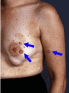
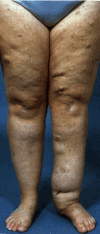
  • Two or more neurofibromas (Figure 2) of any type or one plexiform neurofibroma (Figure 3)
  • Optic pathway glioma
  • Two or more Lisch nodules identified by slit lamp examination or two or more choroidal abnormalities (bright, patchy nodules imaged by optical coherence tomography/near-infrared reflectance imaging)
  • A distinctive osseous lesion such as sphenoid dysplasia, anterolateral bowing of the tibia, or pseudarthrosis of a long bone
  • A parent who meets the diagnostic criteria for NF1
Figure 1.

Figure 1.

Café au lait macules

Figure 2.

Figure 2.

Neurofibromas

Figure 3.

Figure 3.

Plexiform neurofibroma

Establishing the Diagnosis

The diagnosis of NF1 is established in a proband with two or more of the features described in Suggestive Findings [Legius et al 2021].

Note:

1.

About half of individuals with Legius syndrome (see Differential Diagnosis) have skin pigmentary features that meet the diagnostic criteria of NF1 (i.e., ≥6 CALMs and axillary or inguinal freckling), but individuals with Legius syndrome do not have neurofibromas, optic pathway gliomas, Lisch nodules, or typical osseous lesions of NF1 [Legius et al 2021]. Moreover, Legius syndrome is much rarer than NF1, and children with Legius syndrome almost always have an affected parent with only CALMs and intertriginous freckling (see Differential Diagnosis).

2.

CALMs, intertriginous freckling, cutaneous neurofibromas, and Lisch nodules are usually bilateral in NF1. A diagnosis of mosaic NF1 should be considered if such lesions are present only on one side or in one segment of the body.

3.

Sphenoid wing dysplasia is not a separate criterion in those with ipsilateral orbital plexiform neurofibromas.

4.

A germline NF1 pathogenic variant must be identified for genetic testing to serve as a criterion for diagnosis [Legius et al 2021]. The criterion is not met by identification of an NF1 variant only in tumor tissue or identification of a germline likely pathogenic variant or variant of uncertain significance.

5.

Negative NF1 molecular testing does not rule out a diagnosis of NF1 [Accetturo et al 2020]. Some individuals diagnosed with NF1 based on clinical criteria do not have a pathogenic variant detectable by current technology. Many clinical features of NF1 increase in frequency with age, and some individuals who have unequivocal NF1 as adults cannot be diagnosed in early childhood, before these features become apparent.

If the phenotypic findings suggest the diagnosis of NF1, single-gene testing may be considered. Sequence analysis of NF1 genomic DNA (gDNA) and/or cDNA (complementary DNA, copied from mRNA) is performed in association with gene-targeted deletion analysis. Because of the frequency of pathogenic variants that affect splicing (22%-30%, more than 1/3 of which are not detected by gDNA sequencing of protein-coding regions), methods that include cDNA sequencing have higher detection rates than methods based solely on analysis of gDNA (Table 1).

Note:

1.

If an NF1 variant is not detected, sequence analysis and deletion/duplication analysis of SPRED1 (see Differential Diagnosis) may be considered in individuals with only pigmentary features of NF1.

2.

Chromosomal microarray analysis (CMA) may be performed instead of sequence analysis to detect NF1 whole-gene deletions if the NF1 microdeletion phenotype is suspected clinically (see Genotype-Phenotype Correlations).

3.

A karyotype may be considered to look for a translocation or complex cytogenetic abnormality if a clinical diagnosis of NF1 is certain, but no pathogenic variant is found on sequence analysis of NF1 gDNA or cDNA and gene-targeted deletion analysis.

If the phenotype is indistinguishable from other disorders characterized by hyperpigmentation, tumors, and/or other overlapping features, a multigene panel that includes NF1, SPRED1, and other genes of interest (see Differential Diagnosis) may be considered. A rasopathy multigene panel is usually most appropriate. Note: (1) The genes included in the panel and the diagnostic sensitivity of the testing used for each gene vary by laboratory and are likely to change over time. (2) Many multigene panels include genes not associated with the condition discussed in this GeneReview; thus, clinicians need to determine which multigene panel is most likely to identify the genetic cause of the condition while limiting identification of variants of uncertain significance and pathogenic variants in genes that do not explain the underlying phenotype. (3) In some laboratories, panel options may include a custom laboratory-designed panel and/or custom phenotype-focused exome analysis that includes genes specified by the clinician. (4) Methods used in a panel may include sequence analysis, deletion/duplication analysis, and/or other non-sequencing-based tests.

For an introduction to multigene panels click here. More detailed information for clinicians ordering genetic tests can be found here.

If the phenotype is nonspecific (e.g., developmental delay and hypotonia in a young child) comprehensive genomic testing (exome sequencing or genome sequencing) may be considered. Note: Identification of one or more characteristic clinical features (see Suggestive Findings) is required to establish the diagnosis in an individual with an NF1 pathogenic variant.

For an introduction to comprehensive genomic testing click here. More detailed information for clinicians ordering genomic testing can be found here.

Table 1.

Molecular Genetic Testing Used in Neurofibromatosis 1

Gene 1MethodProportion of Probands with a Disease-Causing Variant 2 Detectable by Method
NF1 cDNA & gDNA sequence analysis 3, 4>95% 5
gDNA sequence analysis 3~60%-90% 6
Gene-targeted deletion/duplication analysis 4~13% 7, 8
CMA 9~5%-11% 7, 8
Karyotype<1% 8
1.
2.

See Molecular Genetics for information on variants detected in this gene.

3.

Sequence analysis detects variants that are benign, likely benign, of uncertain significance, likely pathogenic, or pathogenic. Variants may include small intragenic deletions/insertions and missense, nonsense, or splice site variants. Deletion/duplication analysis is often included in NF1 molecular analysis; If exome sequencing is used, confirm that deletion/duplcaiton analysis is included (usually performed by read-depth analysis of next generation sequencing data). For issues to consider in interpretation of sequence analysis results, click here.

4.

Gene-targeted deletion/duplication analysis detects intragenic deletions or duplications. If deletion/duplication analysis is not performed with sequence analysis (e.g., read-depth analysis), alternate methods may include quantitative PCR, multiplex ligation-dependent probe amplification (MLPA), or FISH.

5.
6.
7.

Whole-gene deletions occur in 5%-11% of individuals with NF1 [Kehrer-Sawatzki et al 2020].

8.
9.

Chromosomal microarray analysis (CMA) uses oligonucleotide or SNP arrays to detect genome-wide large deletions/duplications (including NF1) that may not be detected by sequence analysis. The ability to determine the size of the deletion/duplication depends on the type of microarray used and the density of probes in the 17q11.2 region. CMA designs in current clinical use target the 17q11.2 region.

Clinical Characteristics

Clinical Description

Neurofibromatosis 1 (NF1) is an extremely variable multisystem disease; the progression and severity may differ throughout life in an affected individual as well as in affected family members with the same NF1 pathogenic variant [Gutmann et al 2017, Monroe et al 2017, Gianluca et al 2020]. The cardinal clinical manifestations of NF1 include multiple café au lait macules, intertriginous freckling, multiple cutaneous neurofibromas, subcutaneous or deep nodular neurofibromas, plexiform neurofibromas, and characteristic ocular signs. Problems with learning, behavior, and social adaptation are unusually common among people with NF1. Optic or non-optic gliomas occur much more often than expected, but most of these tumors exhibit a benign course. Individuals with NF1, especially those with plexiform or deep nodular neurofibromas in large numbers or of large size, are at high risk of developing malignant peripheral nerve sheath tumors, which tend to occur at a much younger age and have a worse prognosis than in the general population. Women with NF1 are at increased risk of developing breast cancer and have complications of pregnancy more often than expected. Hypertension is frequent in people with NF1, and NF1 vasculopathy may cause stroke or other cardiovascular complications in affected children and young adults. Vertebral or tibial dysplasia can cause major disability in some individuals, and NF1-associated gastrointestinal, endocrine, or pulmonary disease – although less frequent – may be quite serious.

Table 2.

Neurofibromatosis 1: Frequency of Select Features

Feature% of Persons w/Feature 1Typical Age of OnsetComment
Café au lait macules>99%Infancy & childhood↑ in number & size during 1st few yrs of life; macules fade in older persons.
Intertrigenous freckling85%Infancy & early childhoodFrequency ↑ w/age during childhood.
Lisch nodules>95%Early childhoodFrequency ↑ w/age during childhood.
Choroidal abnormalities82%-98%Early childhood↑ in number & size during childhood
Optic pathway glioma15%-20%Birth - 6 yrsFrequency lower in adults
Non-optic glioma2%-5%Any ageFrequency lower in children than adults
Cutaneous
neurofibromas
99%Adolescence-adulthoodInfrequent in childhood; variably ↑ in size & number throughout life
Nodular neurofibromas (subcutaneous or deep)~15%AdolescenceFrequency shown is on clinical exam; frequency is 2-3x higher on whole-body MRI.
Plexiform
neurofibroma(s)
~30%Infancy (sometimes congenital) or childhoodFrequency shown is on clinical exam; frequency is ~50% on whole-body MRI.
Malignant peripheral
nerve sheath tumor
8%-13%Adolescence - adulthoodCross-sectional prevalence 2%-5% after mid-childhood
Intellectual disability4%-8%ChildhoodPersists throughout life
Learning difficulties50%-60%ChildhoodPersist throughout life
Behavior issues30%-67%Childhood
Seizures6%-7%Any age
Long bone dysplasia2%Infancy (congenital)
Dystrophic scoliosis5%6-10 yrsRapidly progressive scoliosis due to vertebral dysplasia
Nondystrophic scoliosis5%AdolescenceMilder scoliosis w/o vertebral anomalies
Osteoporosis~20%Mid-adulthoodOsteopenia is frequent at all ages; osteoporosis occurs earlier than in general population but is rare in children & uncommon in young adults
Hypertension≥15%-20%Any agePrevalence greater in adults than children
1.

Many of the features listed in this table have different frequencies at different ages. The table gives life-time cumulative incidence figures that may be higher, and sometimes much higher, than the prevalence at any given age. Most frequencies in this table are from Ferner & Gutmann [2013] or DeBella et al [2000]. References for more recently defined features are given in the discussion of individual features below.

Cutaneous Features

Café au lait macules (CALMs). Typically, CALMs in individuals with NF1 are ovoid in shape with well-defined borders, uniform in color (a little darker than the background pigmentation of the individual's skin), and about 1-3 cm in size; however, they may be smaller or much larger, lighter or darker, or irregular in shape [Ozarslan et al 2021, Albaghdadi et al 2022]. The pigmentation may also be irregular, with freckling or a more deeply pigmented smaller CALM within a larger, more typically colored lesion. They usually appear in infancy and early childhood, and once established remain stable in number and size (except for growth of the skin itself). They may be difficult to see in older adults with NF1 because of the wrinkling and diffuse pigmentation that often occur with aging.

CALMs are flat and flush with the surrounding skin; if the skin of the lesion is raised or has an unusually soft or irregular texture in comparison to the surrounding skin, an underlying plexiform neurofibroma is likely. The darker pigmentation of CALMs may be difficult to see in people with very fair skin or very dark skin, where the color of the lesions is similar to that of the rest of the skin. A Wood's light is useful in such individuals to demonstrate the pigmented macules. CALMs are not seen on the palms or soles in people with NF1 but can occur almost anywhere else on the body.

Freckling. Clusters of freckles are frequent in sun-exposed areas and may also be seen diffusely over the trunk, proximal extremities, and neck in people with NF1. Individuals with NF1 also develop freckles in areas where skin rubs against skin – in the axilla, groin, and under the breasts in women [Ozarslan et al 2021, Albaghdadi et al 2022].

Neurofibromas. See Other Tumors.

Other skin findings. Juvenile xanthogranuloma and nevus anemicus are more common than expected in people with NF1 and may be useful in supporting the diagnosis in young children who do not meet the standard diagnostic criteria [Miraglia et al 2020, Ozarslan et al 2021]. Juvenile xanthogranulomas are small, tan- or orange-colored papules that may occur in clusters. Nevus anemicus is an irregularly shaped macule that is paler than surrounding skin and does not get red when rubbed, as the skin surrounding it does.

Ocular Findings

Lisch nodules are innocuous iris hamartomas that can be demonstrated on slit lamp examination in almost all adults with NF1, but in fewer than half of children with NF1 younger than age five years. They can be distinguished from iris freckles by their three-dimensional nodular appearance.

Choroidal freckling cannot be seen on standard ophthalmologic examination but can be visualized by scanning laser ophthalmoscopy with infrared or near-infrared light, infrared reflectance imaging, or optical coherence tomography [Vagge et al 2016, Moramarco et al 2018]. The lesions, which are Schwann cell proliferations arrayed in concentric rings around an axon, occur in the majority of people with NF1 of all ages and increase in prevalence, number, and size with age during childhood [Touzé et al 2021].

Other findings frequently seen on ophthalmologic examination of individuals with NF1 include microvascular abnormalities of the retina [Parrozzani et al 2018, Moramarco et al 2019] and hyperpigmented spots of the fundus [Moramarco et al 2021].

Optic pathway gliomas in individuals with NF1 are usually asymptomatic and remain so throughout life [Di Nicola & Viola 2020, Shofty et al 2020]. The clinical course in individuals with optic pathway gliomas tends to be milder in individuals with NF1 than in those who do not have NF1. Optic pathway gliomas in NF1 are frequently stable for many years or only very slowly progressive [Sellmer et al 2018, Kinori et al 2021]. In addition, the majority of optic pathway gliomas appear to regress spontaneously – their prevalence declines from approximately 20% in young children to less than 5% in older adults with NF1 [Sellmer et al 2018]. Symptomatic optic pathway gliomas in individuals with NF1 usually present before age six years with loss of visual acuity, proptosis, or strabismus, but these tumors may not become symptomatic until later in childhood or adulthood [Friedrich & Nuding 2016, Kinori et al 2021].

Increased tortuosity of the optic nerve can be seen on brain MRI in children with NF1, but optic nerve tortuosity is not associated with the occurrence of optic pathway glioma among individuals with NF1 [Ji et al 2013].

Other Tumors

Neurofibromas are benign Schwann cell tumors that can affect virtually any nerve in the body [Brena et al 2020, Serra et al 2020, Ozarslan et al 2021]. Cutaneous neurofibromas are discrete, well-circumscribed masses, usually ranging in size from 1-2 mm to a few centimeters. Their consistency varies from soft to rubbery to firm. They may be sessile or pedunculated, and the involved skin may be the same color and tone as adjacent uninvolved skin or may be pinker or browner or bluer. Most are asymptomatic, but they may itch or be tender to touch. Cutaneous neurofibromas are rare in children but present in almost all adults with NF1.

Subcutaneous neurofibromas lie under the skin (the skin can be moved over them). Most feel rubbery and are nodular, but they may be diffuse, with indistinct borders, and of soft or heterogeneous consistency. The skin overlying a superficial diffuse neurofibroma may exhibit unusual pigmentation or hair patterning. Subcutaneous neurofibromas may be isolated or occur in clusters or continuously like beads on a string along a nerve. Most are small, but subcutaneous neurofibromas can grow to be 5 cm in diameter or more. They may be tender and are sometimes painful. Subcutaneous neurofibromas are uncommon in children but present in about 15% of adults with NF1 on clinical examination.

Cutaneous and subcutaneous neurofibromas continue to develop throughout life, although the rate of appearance may vary greatly from year to year. The total number of neurofibromas seen on clinical examination in adults with NF1 varies from a few to hundreds or even thousands. Some women experience a rapid increase in the number and size of neurofibromas during pregnancy, but this does not appear to produce a persistent increase in the tumor burden in comparison to those of child-bearing age with NF1 who have not been pregnant [Well et al 2020].

Plexiform neurofibromas. About half of people with NF1 have plexiform neurofibromas. Most of these tumors are internal, and thus not apparent on clinical examination. They can, however, be seen on MRI (see Imaging). Plexiform neurofibromas tend to grow in childhood and adolescence and then remain stable throughout adulthood [Nguyen et al 2012]. Although most plexiform neurofibromas are asymptomatic, they may cause pain, grow to cause disfigurement, produce overgrowth or erosion of adjacent tissue, or impinge on the function of nerves and other structures.

Superficial diffuse plexiform neurofibromas are soft and irregular; they are often associated with thickening, hypertrophy, and/or hyperpigmentation of the associated skin. More extensive diffuse plexiform neurofibromas may have a characteristic “bag of worms” feel on palpation, indicating involvement of multiple nerves and branches. Plexiform neurofibromas may also be firm and nodular, occurring singly or extending for some distance or even along the entire extent of a nerve, producing a “beads on a string” feel on palpation. Deeper plexiform neurofibromas that are not apparent on clinical examination may also be diffuse or nodular, and singular or clustered on any nerve, nerve root, or nerve plexus.

Malignant peripheral nerve sheath tumors (MPNST) are the most common malignant neoplasms associated with NF1. In comparison to the general population, MPNST tend to occur at a younger age and be associated with a poorer prognosis in people with NF1 [Martin et al 2020, Sharma et al 2021]. Most, if not all, MPNST arise in preexisting diffuse or nodular plexiform neurofibromas. The most frequent clinical sign of malignant change is persistent pain, either as a new symptom or as exacerbation of existing pain. This pain may be accompanied by rapid growth or change in texture of the tumor clinically or on MRI.

Individuals with NF1 who have a type 1 whole-gene deletion or benign subcutaneous neurofibromas, or whose burden of benign internal plexiform neurofibromas is high, appear to be at greater risk of developing MPNST than people with NF1 who do not have these features [Nguyen et al 2014]. Individuals with atypical neurofibromas also appear to be at unusually high risk of developing MPNST [Higham et al 2018].

Brain tumors. Non-optic gliomas in people with NF1 are usually asymptomatic, and most are discovered as incidental findings on head MRI done as a routine screen or for other indications. These are usually low-grade tumors that grow slowly or not at all over many years [Sellmer et al 2017], although symptomatic and/or high-grade brain tumors are seen occasionally [Byrne et al 2017, Glombova et al 2019].

At least 20% of people with NF1 who have one non-optic glioma have two or more of these tumors [Sellmer et al 2017, Glombova et al 2019]. Second central nervous system (CNS) gliomas occur in 17%-20% of individuals with NF1 who have optic pathway gliomas [Sharif et al 2006, Sellmer et al 2018].

Breast cancer. Women with NF1 are at substantially increased risk of developing breast cancer before age 50 years [Uusitalo et al 2016] and of dying of breast cancer [Evans et al 2020]. The cumulative risk of developing contralateral breast cancer is greater than expected among women with NF1 [Evans et al 2020]. Breast cancers in women with NF1 are more likely to be HER2-positive and to have other unfavorable tumor markers [Evans et al 2020].

Hematologic malignancies. Although still rare, juvenile myelomonocytic leukemia (JMML) is hundreds of times more frequent in children with NF1 than in other children [Niemeyer & Flotho 2019]. The usual clinical features at presentation are splenomegaly, hepatomegaly, and leukemic infiltrates of the lung in association with a peripheral blood smear that shows myelocytes, meta-myelocytes, and sometimes nucleated red cells. Juvenile xanthogranulomas may be seen in children with NF1 and JMML but do not appear to be more frequent than expected in other children with NF1 [Liy-Wong et al 2017]. It is not clear whether lymphoreticular malignancies occur more frequently than expected in adults with NF1 [Bergqvist et al 2021].

Additional tumors. A variety of other tumors may also be seen more often than expected in individuals with NF1, including rhabdomyosarcomas [Crucis et al 2015], pheochromocytomas [Gruber et al 2017], paragangliomas [Gruber et al 2017], gastrointestinal stromal tumors [Nishida et al 2016], and glomus tumors [Kumar et al 2014]. People with NF1 may also be at increased risk for some other cancers [Seminog & Goldacre 2013, Varan et al 2016, Landry et al 2021].

Other Neurologic Manifestations

Motor function. Hypotonia and impairments in coordination, balance, and fine motor function are frequent in children with NF1 [Iannuzzi et al 2016, Haas-Lude et al 2018, Pardej et al 2022]. Children with NF1 also have less muscle strength than unaffected children of the same age, sex, and weight [Summers et al 2015].

Intellectual and learning disabilities. Deficits in visual-spatial performance are most common in individuals with NF1, but specific learning disorders and problems with executive function, memory, and language are also frequent [Vogel et al 2017]. The average IQ of people with NF1 is ~1 SD lower than individuals in the general population, and frank intellectual disability (IQ <70) occurs in 4%-8% of individuals with NF1, a frequency about twice that in the general population [Vogel et al 2017, Al-Farsi et al 2022]. Intellectual disability is more frequent among individuals whose NF1 is caused by a whole-gene deletion.

Behavioral issues include problems with social competence and attention. Both children and adults with NF1 report increased social difficulties such as isolation and reduced peer acceptance and exhibit fewer social skills and prosocial behaviors [Chisholm et al 2018, Payne et al 2020]. Attention-deficit/hyperactivity disorder is present in 30%-50% of children and adolescents with NF1 [Vogel et al 2017, Chisholm et al 2018, Domon-Archambault et al 2018] and may persist into adulthood [Mautner et al 2015]. Symptoms of autism are frequent in children with NF1, and 25% of children with NF1 meet standard diagnostic criteria for autism spectrum disorder [Vogel et al 2017, Chisholm et al 2018, Domon-Archambault et al 2018, Payne et al 2020, Chisholm et al 2022]. Autistic symptoms appear to be less common among adults than children with NF1 [Morris et al 2016]. Sleep disturbance is frequent in individuals with NF1 at all ages [Domon-Archambault et al 2018, Fjermestad et al 2018]. Psychiatric diseases such as mood disorders, anxiety disorders, and emerging personality disorders may also occur more often than expected among adults with NF1 [Domon-Archambault et al 2018, Kenborg et al 2021].

Polyneuropathy. A few percent of people with NF1 develop a diffuse polyneuropathy, often (though not always) in association with multiple nerve root tumors [Barnett et al 2019, Bayat & Bayat 2020]. NF1 polyneuropathy may be asymptomatic or produce sensory deficits, pain, or itching. The risk for MPNST appears to be higher in individuals who have polyneuropathy than in those who do not.

Seizures occur in about 5% of individuals with NF1, with a slightly higher prevalence in adults than in children [Bernardo et al 2020, Sorrentino et al 2021]. The seizures may be generalized but more often are focal, occurring in association with a brain tumor, area of infarction, or mesial temporal sclerosis [Pecoraro et al 2017, Bernardo et al 2020, Sorrentino et al 2021]. Neurodevelopmental abnormalities are more common in individuals with NF1 and epilepsy [Sorrentino et al 2021].

The approach to treating epilepsy in individuals with NF1 is similar to that used in those who do not have NF1 [Bernardo et al 2020, Sorrentino et al 2021]. Control of focal seizures may require the use of more than one anti-seizure drug or surgical removal of the affected part of the brain.

Headaches occur in at least half of individuals with NF1 and are more frequent in adults than children [Fjermestad et al 2018, Hirabaru & Matsuo 2018, Kongkriangkai et al 2019]. Migraine headaches are most common in NF1, but other kinds of headaches may occur. NF1-associated lesions are often seen on head MRI in people with headaches, but the frequency is similar to that found in those without headaches [Afridi et al 2015].

Neuroimaging. Hyperintense lesions (unidentified bright objects [UBOs] or focal areas of high signal intensity [FASI]) are seen on T2-weighted brain MRI in more than half of children with NF1 [Sellmer et al 2018]. These may occur in the optic tracts, basal ganglia, brain stem, cerebellum, or cortex, and they usually show no evidence of a mass effect. Typical UBOs are not seen on T1-weighted MRI imaging or on CT scan. UBOs correspond pathologically to areas of spongiform myelinopathy [DiPaolo et al 1995]. They peak in number and size at about age seven years and then tend to regress, but some persist into adulthood [Sellmer et al 2018, Calvez et al 2020]. The presence of UBOs does not appear to be related to the occurrence of seizures in children with NF1 [Hsieh et al 2011]. Some studies have suggested that the presence, number, volume, location, or disappearance of UBOs over time correlates with learning disabilities or behavioral abnormalities in children with NF1, but findings have not been consistent across investigations [Payne et al 2014, Roy et al 2015, Parmeggiani et al 2018, Eby et al 2019, Baudou et al 2020].

Enlargement of the corpus callosum is seen in some children with NF1 and has been associated with learning disabilities [Pride et al 2010, Aydin et al 2016].

MRI is the imaging method of choice for demonstrating optic gliomas, brain tumors, and neurofibromas of cranial, spinal, or peripheral nerves, as well as diffuse or nodular neurofibromas anywhere in the body. Positron emission tomography is useful in recognizing MPNST [Nishida et al 2021], and high-resolution ultrasound examination can be used to characterize dermal and superficial plexiform neurofibromas [Winter et al 2020].

Musculoskeletal Features

Long bone, sphenoid wing, or vertebral dysplasia. Osseous dysplasia may occur as a primary abnormality in individuals with NF1 (typical of long-bone dysplasia), or in association with an adjacent plexiform neurofibroma or dural ectasia (vertebral or sphenoid wing dysplasia). Healing of fractured or defective bone in any of these focal lesions is often unsatisfactory [Elefteriou et al 2009]. Surgical treatment of osseous dysplasia and its associated deformities of the long bones, craniofacies, and spine is frequently difficult and best accomplished by experienced specialists [Mladenov et al 2020].

Dysplasia of the long bones, most often the tibia and fibula, is an infrequent but characteristic feature of NF1 [Elefteriou et al 2009]. The lesion is congenital and almost always unilateral. It usually presents in infancy with anteriolateral bowing of the lower leg. Early recognition of tibial dysplasia permits bracing, which may prevent fracture. The initial radiographic changes are narrowing of the medullary canal with cortical thickening at the apex of the bowing [Stevenson et al 2007].

Sphenoid wing dysplasia typically presents with asymmetry of the orbits, but it is sometimes found incidentally on cranial imaging. Sphenoid wing dysplasia is often static but may be progressive, occasionally disrupting the integrity of the orbit and producing pulsating enophthalmos [Chauvel-Picard et al 2020].

Vertebral dysplasia usually presents as dystrophic scoliosis, which typically develops between ages six and eight years, much earlier than common adolescent scoliosis. Dystrophic scoliosis, which is characterized by acute angulation of the spine over a short segment, may be rapidly progressive within a few months after becoming apparent [Kaspiris et al 2022].

Nondystrophic scoliosis, which is not usually associated with vertebral abnormalities in individuals with NF1, resembles common adolescent scoliosis in its age at onset and more benign course [Elefteriou et al 2009].

Osteoporosis. Generalized osteopenia is more common than expected in people with NF1 [Rodari et al 2018, Filopanti et al 2019, Jalabert et al 2021, Kaspiris et al 2022], and fractures occur more often than expected [Heervä et al 2012]. Adults with NF1 develop osteoporosis more frequently and at a younger age than in the general population [Filopanti et al 2019, Kaspiris et al 2022]. The pathogenesis of these bony changes is not fully understood, but individuals with NF1 often have lower-than-expected serum 25-hydroxy vitamin D concentrations, elevated serum parathyroid hormone levels, and evidence of increased bone resorption [Riccardi et al 2020, Tezol et al 2021, Kaspiris et al 2022]. The function of both osteoblasts and osteoclasts appears to be abnormal in bone from people with NF1 [Riccardi et al 2020, Kaspiris et al 2022].

Vascular Involvement

Arterial hypertension occurs in at least 15%-20% of individuals with NF1 [Dubov et al 2016, Sivasubramanian & Meyers 2021]. It may develop at any age but is more frequent in adults than children. Often no specific cause is found, but hypertension may be caused by renal artery stenosis or mid-aortic syndrome, usually as a manifestation of NF1 vasculopathy, especially in children [Celik et al 2021, Sivasubramanian & Meyers 2021]. Hydronephrosis and other structural abnormalities of the urinary tract are frequent in individuals with NF1-related hypertension but are also seen in those without hypertension [Dubov et al 2016, Celik et al 2021].

Although much more frequent in people with NF1 than in the general population, pheochromocytomas or paragangliomas are found in fewer than 1% of adults with NF1. These tumors are usually asymptomatic, but they can cause arterial hypertension [Al-Sharefi et al 2019].

Pulmonary hypertension is a rare but very serious complication of NF1 in older adults [Jutant et al 2018, Jutant et al 2020]. It is usually associated with parenchymal lung disease, which itself may be a manifestation of NF1 vasculopathy affecting vessels in the lungs [Jutant et al 2018].

Stroke is more common and often occurs at a younger age among people with NF1 than in the general population [Terry et al 2016]. Anatomically variant stenotic or ectatic cerebral arteries and intracranial aneurysms occur more frequently in individuals with NF1 than in the general population [Bekiesińska-Figatowska et al 2014, D'Arco et al 2014, Barreto-Duarte et al 2021]. The internal carotid, middle cerebral, or anterior cerebral artery are most often affected. Small telangiectatic vessels form around the stenotic area and appear as a "puff of smoke" (moya moya) on cerebral angiography. Moya moya vasculopathy develops about three times more often than expected in children with NF1 after cranial irradiation for primary brain tumor [Murphy et al 2015].

Cardiac Issues

Congenital anomalies of the circulatory system were observed 3.35 times (95% confidence interval 1.64-6.83x) more often than expected among children with NF1 in a study performed through Finnish population-based registries [Leppävirta et al 2018]. Pulmonary valve stenosis and mitral valve anomalies are the most frequent cardiac defects seen in individuals with NF1 [Lin et al 2000, Pinna et al 2019]. Congenital heart defects and hypertrophic cardiomyopathy may be especially frequent among persons with NF1 whole-gene deletions [Nguyen et al 2013, Pinna et al 2019]. Intracardiac neurofibromas may also occur [Nguyen et al 2013].

Pulmonary Disease

NF1-associated diffuse lung disease occurs in 10%-20% of adults [Jutant et al 2018, Alves Júnior et al 2019]. Symptoms are usually nonspecific and may include dyspnea on exertion, shortness of breath, chronic cough, or chest pain. Symptoms do not usually appear until the third or fourth decade of life, although characteristic signs may be found on imaging studies in children with NF1 [Spinnato et al 2019]. Chest CT is the method of choice for identifying NF1-associated diffuse lung disease, which typically is characterized by upper-lobe cystic and bullous disease and basilar interstitial disease. NF1 pulmonary disease is often poorly responsive to available medical treatments.

Growth

Individuals with NF1 tend to be below average in height and above average in head circumference for age [Zessis et al 2018]. However, very few individuals with NF1 have height more than 3 SD below the mean or head circumference more than 4 SD above the mean.

In contrast, individuals with whole-gene NF1 deletions have overgrowth (especially in height) between ages two and six years [Ning et al 2016, Kehrer-Sawatzki et al 2020]. The clinical features in some of these individuals resemble those of Weaver syndrome (see Genotype-Phenotype Correlations).

Pubertal development is usually normal, although decreased pubertal growth velocity occurs in both girls and boys [Zessis et al 2018]. However, delayed puberty is common [Virdis et al 2003], and precocious puberty and/or growth hormone excess may also occur in children with NF1, especially in those with tumors of the optic chiasm [Cambiaso et al 2017, Hannah-Shmouni & Stratakis 2019].

Life Expectancy

The median life expectancy of individuals with NF1 is at least eight years lower than in the general population [Evans et al 2011, Wilding et al 2012]. Even higher excess mortality has been identified in females [Uusitalo et al 2015]. Malignancy (MPNST) and vasculopathy are the most important causes of early death in individuals with NF1 [Evans et al 2011, Masocco et al 2011, Uusitalo et al 2015].

Quality of Life

Quality of life assessments are lower in both children and adults with NF1 than in comparison groups [Vranceanu et al 2015]. Cosmetic, medical, social, and behavioral features of NF1 all may compromise the quality of life in people with NF1, and clinical depression may impair their ability to function effectively [Domon-Archambault et al 2018]. A population-based Danish registry study found that people with NF1 were more than twice as likely to be hospitalized and to have more frequent and longer hospitalizations at all ages in comparison to the general population [Kenborg et al 2020].

NF1 Phenotypic Variants

Mosaic NF1 (i.e., somatic mosaicism for an NF1 pathogenic variant) may present as clinical features of NF1 localized to one or more segments of the body or as typical (generalized) NF1 [Ejerskov et al 2021]. Mosaic NF1 is usually milder than typical NF1 involving the same pathogenic variant, and some adults with mosaic NF1 have no clinical features of NF1 [Kluwe et al 2020, Yang et al 2020]. Mosaic NF1 involving just a single body segment is sometimes called "segmental NF1," but the term "localized mosaic NF1" is preferred because it is more informative and because non-mosaic NF1 may sometimes involve just one part of the body by chance, especially in young children. Adults with mosaic NF1 may have children with typical (i.e., non-mosaic) NF1 [Legius & Brems 2020] (see Genetic Counseling).

NF1-Noonan syndrome phenotype occurs in approximately 12% of individuals with NF1. The features may include ocular hypertelorism, downslanted palpebral fissures, ptosis, low-set ears, webbed neck, pectus anomaly, and pulmonic stenosis. Affected family members with NF1 may or may not have concomitant features of Noonan syndrome [Chen et al 2014, Ekvall et al 2014]. The NF1-Noonan syndrome phenotype is genetically heterogeneous. Some individuals have disease-causing variants of both NF1 and PTPN11 (the gene most often involved in Noonan syndrome) [D'Amico et al 2021]. In other individuals, only an NF1 variant is identified [De Luca et al 2005]. Watson syndrome, an overlapping phenotype characterized by pulmonary valvular stenosis, CALMs, short stature, and mild intellectual disability, is caused by NF1 variants [Allanson et al 1991].

Familial spinal neurofibromatosis is characterized by neurofibromas of every spinal nerve root but few (if any) cutaneous manifestations of NF1 [Bettegowda et al 2021]. Despite the name, this phenotype may also occur in simplex cases [Ruggieri et al 2015].

Multiple spinal ganglioneuromas (rather than neurofibromas) and multiple subcutaneous tumors were reported in an adult with an NF1 pathogenic variant whose clinical features were not diagnostic of NF1 [Bacci et al 2010].

Multiple lipomas have been observed in association with a pathogenic variant of NF1 but no other clinical features of NF1 [Ramirez et al 2021].

Symptomatic optic glioma was reported in a male with an NF1 pathogenic variant and no other diagnostic features of NF1 at age 21 years [Buske et al 1999].

Encephalocraniocutaneous lipomatosis was reported in one child with more than five CALMs and an NF1 pathogenic variant [Legius et al 1995].

Genotype-Phenotype Correlations

Several allele-phenotype correlations have been observed in NF1:

  • 1.4-Mb (type 1) deletion of the entire NF1 gene are associated with larger numbers and earlier appearance of cutaneous and plexiform neurofibromas, a higher risk of developing MPNST, more frequent and more severe cognitive abnormalities, somatic overgrowth, and large hands and feet [Bettegowda et al 2021, Kehrer-Sawatzki & Cooper 2021, Pacot et al 2021, Well et al 2021]. A recurrent pattern of dysmorphic features that includes coarse facial appearance, flat forehead, ocular hypertelorism, broad nasal tip, low-set ears, and broad neck is often observed among adolescents and adults [Kehrer-Sawatzki & Cooper 2021].
  • An unusually severe phenotype with frequent plexiform or spinal neurofibromas, optic pathway gliomas, malignant neoplasms, and skeletal abnormalities has been observed in adults with missense variants of one of five codons between 844 and 848 [NM_000267.3] that code for the cysteine-serine rich domain of neurofibromin [Koczkowska et al 2018, Bettegowda et al 2021].
  • p.Met992del is associated with typical pigmentary features of NF1 but no cutaneous, subcutaneous, or surface plexiform neurofibromas [Bettegowda et al 2021]. One quarter of individuals with this variant lack the clinical features needed to meet NF1 diagnostic criteria, but brain tumors outside of the optic pathways, cognitive or learning disabilities, and Noonan syndrome features occur at frequencies similar to those seen in other individuals with NF1.
  • p.Arg1038Gly has been associated with mild pigmentary features of NF1 but a paucity of neurofibromas and frequent NF1-Noonan syndrome features [Trevisson et al 2019].
  • Missense variants affecting p.Met1149 have been associated with a mild phenotype characterized by pigmentary features, frequent learning problems, and features of NF1-Noonan syndrome [Koczkowska et al 2020]
  • Missense variants affecting p.Arg1276 have been associated with a higher-than-expected frequency of cardiovascular malformations, especially pulmonic stenosis, NF1-Noonan phenotype, and symptomatic spinal neurofibromas [Koczkowska et al 2020].
  • Missense variants affecting p.Lys1423 have been associated with higher-than-expected frequencies of plexiform neurofibromas, as well as of learning problems, cardiovascular malformations (especially pulmonic stenosis), and NF1-Noonan phenotype [Koczkowska et al 2020].
  • Several missense variants affecting p.Arg1809 are associated with multiple CALMs but absence of cutaneous neurofibromas or clinically apparent plexiform neurofibromas, although learning disabilities, short stature, and pulmonic stenosis often occur [Pinna et al 2015, Rojnueangnit et al 2015, Bettegowda et al 2021].

Penetrance

Pedigree studies demonstrate that the penetrance of NF1 is nearly complete after childhood [Rasmussen & Friedman 2000], but molecular testing has documented incomplete penetrance of pathogenic NF1 variants in a small number of individuals [Bettegowda et al 2021].

Nomenclature

NF1 was previously referred to as peripheral neurofibromatosis, to distinguish it from NF2 (central neurofibromatosis) – although CNS involvement may also occur in NF1.

"Neurofibromatosis" without further specification is sometimes used in the literature to refer to NF1, but this usage is confusing because other authors employ the term "neurofibromatosis" to designate a group of conditions that includes (in addition to NF1) Legius syndrome, NF2, and schwannomatosis.

Prevalence

A Finnish register-based total population study estimated the prevalence of NF1 at 1:2,052 between ages 0 and 74 years [Kallionpää et al 2018]. The prevalence was highest in the youngest cohort and lower in older cohorts, reflecting the increased mortality (see Life Expectancy). The incidence at birth in this study was estimated at 1:1,871 [Uusitalo et al 2015], making NF1 one of the most common autosomal dominant genetic disorders. The prevalence and birth incidence were higher than previous estimates [Evans et al 2010] – likely the result of the diagnosis of more mildly affected individuals rather than differences in the actual incidence and prevalence in the populations studied.

Almost half of all affected individuals have the disorder as the result of de novo variants. If the Finnish estimate of the incidence of NF1 at birth is correct and fertility in affected individuals is reduced by half [Huson et al 1989], the mutation rate for NF1 would be >1:8000, among the highest known for any gene in humans. The cause of the unusually high mutation rate is unknown.

Differential Diagnosis

More than 100 genetic conditions and multiple congenital anomaly syndromes that include café au lait macules (CALMs) or other individual features of neurofibromatosis 1 (NF1) have been described, but few of these disorders are ever confused with NF1. The conditions to consider in the differential diagnosis of NF1 are summarized in Table 3.

Table 3.

Genes of Interest in the Differential Diagnosis of Neurofibromatosis 1

Gene(s)DisorderMOIClinical Characteristics / Comment
AKT1 1 Proteus syndrome See footnote 1.Hamartomatous overgrowth of multiple tissues, connective tissue nevi, epidermal nevi, & hyperostoses
BRAF
MAP2K1
PTPN11
RAF1
Noonan syndrome with multiple lentigines (previously referred to as LEOPARD syndrome)ADMultiple lentigines, ocular hypertelorism, deafness, & congenital heart disease
BRAF
KRAS
LZTR1
MAP2K1
NRAS
PTPN11
RAF1
RIT1
SOS1
Noonan syndrome (NS)AD
(AR) 2
Short stature, congenital heart defect, neck webbing, & characteristic facies. Persons w/NF1 may have NS-like facial features. Facial features of NS change w/age. Features found irrespective of age: low-set, posteriorly rotated ears w/fleshy helices, vivid blue or blue-green irides, hypertelorism, downslanted palpebral fissures, epicanthal folds, & ptosis.
GNAS 3Fibrous dysplasia/McCune-Albright syndrome (FD/MAS)See footnote 3.Large CALMs w/irregular margins & polyostotic fibrous dysplasia
KIT
SNAI2
Piebald trait (OMIM 172800)ADAreas of cutaneous pigmentation & depigmentation w/hyperpigmented borders of the unpigmented areas, & white forelock
LZTR1
SMARCB1
Schwannomatosis ADPredisposition to develop multiple schwannomas & (less often) meningiomas. Most common presenting feature: localized or diffuse pain or asymptomatic mass.
MLH1
MSH2
MSH6
PMS2
Constitutional mismatch repair deficiency (CMMRD; see Lynch Syndrome.)ARRare childhood cancer predisposition syndrome. Affected persons often have colorectal cancer or cancer of the small intestine prior to 2nd decade of life. Cutaneous phenotype is remarkably similar to NF1. CMMRD is distinguishable from NF1 in that the parents are often consanguineous & 1 or both parents often have clinical findings &/or family history of Lynch syndrome due to heterozygous pathogenic variant at one of these loci. Typically, neither parent has clinical findings consistent w/NF1.
NF2 Neurofibromatosis 2 (NF2)ADBilateral vestibular schwannomas, schwannomas of other cranial & peripheral nerves, cutaneous schwannomas, meningiomas, & juvenile posterior subcapsular cataract
PDGFRB Infantile myofibromatosis (OMIM 228550)ADMultiple tumors of the skin, subcutaneous tissues, skeletal muscle, bones, & viscera
SPRED1 Legius syndrome ADMultiple CALMs w/o neurofibromas, other tumors, or Lisch nodules. Addl features of Legius: freckling, lipomas, macrocephaly, & learning disabilities / ADHD / DDs. ~8% of children w/≥6 CALMs & no other features of NF1 have Legius syndrome. 5

AD = autosomal dominant; ADHD = attention-deficit/hyperactivity disorder; AR = autosomal recessive; CALMs = café au lait macules; DD = developmental delay; MOI = mode of inheritance; NS = Noonan syndrome

1.

A somatic mosaic AKT1 pathogenic variant has been identified in more than 90% of individuals meeting Proteus syndrome diagnostic criteria. There are no confirmed occurrences of vertical transmission or sib recurrence.

2.

NS is most often inherited in an AD manner. NS caused by pathogenic variants in LZTR1 can be inherited in either an AD or an AR manner.

3.

FD/MAS is the result of early embryonic postzygotic somatic activating mutation of GNAS. There are no verified instances of vertical transmission of FD/MAS.

5.

Clinically distinguishing Legius syndrome from NF1 may be impossible in a young child because neurofibromas and Lisch nodules do not usually arise until later in childhood or adolescence in those with NF1. Examination of the parents for signs of Legius syndrome or NF1 may distinguish the two conditions, but in simplex cases, reevaluation of the individual after adolescence or molecular testing may be necessary to establish the diagnosis [Legius et al 2021].

Other disorders with features of NF1

  • Isolated familial multiple CALMs (OMIM 114030). Among 253 individuals with familial multiple CALMs but no other clinical features of NF1, 86.6% had disease-causing variants of NF1, 7.1% had disease-causing variants of SPRED1, and 6.3% did not have an NF1 or SPRED1 disease-causing variant identified [Messiaen et al 2009].
  • Multiple orbital neurofibromas, painful peripheral nerve tumors, distinctive face, and marfanoid habitus [Babovic-Vuksanovic et al 2012]

Management

The American Academy of Pediatrics and American College of Medical Genetics and Genomics (ACMG) have published management guidelines for children with NF1 [Miller et al 2019], and the ACMG has published management guidelines for affected adults [Stewart et al 2018]. Similar recommendations have been made by the NF France Network [Bergqvist et al 2020] and by other experts [Ly & Blakeley 2019, Baudou & Chaix 2020]. Referral to a neurofibromatosis clinic staffed by a variety of medical specialists with experience and particular interest in NF1 may benefit many individuals [Toledano-Alhadef et al 2020].

Evaluations Following Initial Diagnosis

To establish the extent of disease and needs in an individual diagnosed with neurofibromatosis 1 (NF1), the evaluations summarized in Table 4 (if not performed as part of the evaluation that led to the diagnosis) are recommended.

Table 4.

Recommended Evaluations Following Initial Diagnosis in Individuals with Neurofibromatosis 1

System/ConcernEvaluationComment
Integument Clinical assessment for skin findings of neurofibromas &/or plexiform neurofibromas
Eyes Ophthalmologic eval incl fundoscopy, slit lamp exam of the irides, infrared reflectance imaging or optical coherence tomography of the fundus, & vision assessment
Neurologic Neurologic exam; assessment for seizures, headaches, & painNote: Routine use of brain MRI in asymptomatic persons is controversial. 1
Development Developmental assessment
Psychiatric Neuropsychiatric assessment
Skeletal Clinical assessment for asymmetry, long bone dysplasia, sphenoid wing dysplasia, vertebral dysplasia &/or scoliosis, & recurrent fractures
Vascular Blood pressureEvaluate those w/hypertension for renovascular disease or midaortic syndrome, hydronephrosis or other structural renal anomalies, & pheochromocytoma or paraganglioneuroma [Sivasubramanian & Meyers 2021].
Cardiac History & clinical exam for signs/symptoms of congenital heart defects &/or cardiomyopathy
Growth Plot height, weight, & head circumference on age-appropriate charts.
Genetic
counseling
By genetics professionals 2To inform affected persons & their families re nature, MOI, & implications of NF1 to facilitate medical & personal decision making
1.

Proponents state that brain MRI is useful to identify structural anomalies of the brain or skull, tumors, or vascular disease before it becomes clinically apparent. Those who oppose head MRI in asymptomatic individuals point to the uncertain clinical significance of unidentified bright objects (UBOs), increased cost, requirement for sedation in small children, and findings resulting in repeating imaging for reassurance.

2.

Medical geneticist, certified genetic counselor, certified advanced genetic nurse

Treatment of Manifestations

Table 5.

Treatment of Manifestations in Individuals with Neurofibromatosis 1

Manifestation/ConcernTreatmentConsiderations/Other
Optic pathway
gliomas (OPG)
  • Monitoring by brain MRI & mgmt per ophthalmologist & oncologist w/experience in NF1
  • Chemotherapy for progressive OPG, although the results are mixed
  • Surgical treatment is usually reserved for cosmetic palliation in a blind eye.
  • Children w/NF1 & low-grade progressive gliomas (most of which were OPG) had better survival w/carboplatin & vincristine than children w/o NF1 who had similar tumors [Ater et al 2016].
  • Radiotherapy is usually avoided due to ↑ risk of inducing malignancy or moya moya vasculopathy in the exposed field.
Cutaneous /
subcutaneous
neurofibromas
Surgical removal, laser, or electrocautery for neurofibromas that are disfiguring or in locations that result in discomfort (e.g., at belt or collar lines)Laser ablation is rapid & effective for removing large numbers of neurofibromas w/satisfactory cosmetic results [Méni et al 2015].
Plexiform
neurofibromas
  • Assess size, extent, & monitor growth w/MRI.
  • Monitor for pain, neurologic deficit, &/or tumor growth (which suggests MPNST).
  • Exam by MRI, PET, or PET/CT when MPNST is suspected; definitive diagnosis of MPNST requires biopsy.
  • Selumetinib, a MEK inhibitor, is FDA-approved for treatment of NF1-related inoperable plexiform neurofibromas. 1, 2
  • Surgical removal of small superficial plexiform neurofibromas may be possible.
  • Surgical treatment of larger tumors is often unsatisfactory because of involvement w/nerves & tendency to grow back.
  • Radiotherapy is contraindicated due to risk of inducing MPNST.
Malignant peripheral nerve sheath tumors (MPNST)
  • Mgmt per surgical &/or medical oncologists familiar w/NF1
  • Complete surgical excision, when possible, is the only treatment that offers the possibility of cure [Valentin et al 2016].
  • Adjuvant chemotherapy or radiotherapy may be helpful in some [Valentin et al 2016].
Treatments for MPNST involving MEK inhibitors, immunotherapy, &/or radiation therapy are currently being evaluated in clinical trials [Marjanska et al 2020].
Brain tumors Monitoring by brain MRI & mgmt per oncologist w/experience in NF1
  • Brain stem & cerebellar gliomas in those w/NF1 are usually less aggressive than in persons w/o NF1 [Sellmer et al 2017].
  • Avoid radiation therapy, as MPNST or other gliomas may develop w/in treatment field [Kleinerman 2009, Madden et al 2014]. Transformation of a pilocytic astrocytoma to a more malignant brain tumor may occur after radiation therapy in persons w/NF1 [Krishnatry et al 2016].
Breast cancer Although more aggressive breast cancer is reported in NF1, women w/NF1 should be treated in the same manner as others w/similar pathology & tumor markers.Avoiding radiotherapy, if possible, is reasonable.
Hematologic
malignancies
Mgmt per oncologist w/experience in treatment of NF1
Pain Pain treatment:
  • Depends on nature, severity, & degree to which it interferes w/ADL [Bellampalli & Khanna 2019];
  • Is empiric & similar to that in those w/o NF1.
Persons w/intractable pain that interferes w/ADL despite conventional treatment should be referred to pain specialist.
Developmental delay /
Intellectual disability
Mgmt per developmental specialist [Miller et al 2019]
Behavioral/
psychiatric
manifestations
Methylphenidate treatment may be beneficial in children w/ADHD [Lion-François et al 2014].
Seizures Mgmt per neurologist w/experience in treatment of epilepsy
Headaches Mgmt per neurologist w/experience in treatment of headaches
Long bone dysplasia
  • Assess by x-ray when suspected on clinical exam.
  • Consider CT or 3D-CT reconstructions when surgical treatment is planned.
Surgical treatment of tibial pseudarthrosis is difficult & often unsatisfactory [Paley 2019].
Sphenoid wing
dysplasia w/or w/o
assoc plexiform
neurofibroma
Mgmt per craniofacial team w/experience in treatment of NF1Mgmt recommendations for orbital/periorbital plexiform neurofibroma in children w/NF1 have been made by a multidisciplinary expert task force [Avery et al 2017].
Vertebral dysplasia /
Dystrophic scoliosis
Mgmt per orthopedist & spine specialist w/experience in treatment of NF1Often requires surgical mgmt, which may be complex & difficult [Jia et al 2021]
Scoliosis
(nondystrophic)
Mgmt per orthopedistTreatment similar to that of idiopathic scoliosis.
Osteopenia /
Recurrent fractures
Hypovitaminosis D is common in persons w/NF1 of all ages.
Hypertension Treatment per nephrologist &/or cardiologist based on cause of hypertension [Sivasubramanian & Meyers 2021]
Stroke / Moya
moya vasculopathy
  • MR angiography to assess NF1 vasculopathy [D'Arco et al 2014]
  • Treatment per neurologist & vascular surgeon
Cardiac
manifestations
Mgmt per cardiologist & cardiac surgeon
Growth deficiency /
Pubertal delay
Mgmt per pediatric endocrinologist
Precocious puberty Mgmt per endocrinologist

ADHD = attention-deficit/hyperactivity disorder; ADL = activities of daily living; MPNST = malignant peripheral nerve sheath tumor

1.
2.

Treatments for plexiform neurofibromas involving other MEK inhibitors are currently being evaluated in clinical trials [Marjanska et al 2020, Solares et al 2021].

Imaging

MRI is the method of choice for demonstrating the size and extent of plexiform neurofibromas [Ahlawat et al 2016] and for monitoring their growth over time [Nguyen et al 2012]. MRI is also useful in characterizing optic pathway gliomas, other brain tumors, structural abnormalities of the brain, and signs of cerebrovascular disease in people with NF1 [Lin et al 2011, Prada et al 2015, Blanchard et al 2016, Sellmer et al 2017, Sellmer et al 2018]. MR angiography is valuable in assessing NF1 vasculopathy [D'Arco et al 2014]. Conventional radiographic studies can demonstrate the skeletal anomalies that occur in people with NF1 [Patel & Stacy 2012], but CT imaging or three-dimensional CT reconstructions may be necessary when surgical treatment of bony lesions is being planned. PET and CT/PET can help to distinguish benign and malignant peripheral nerve sheath tumors [Chirindel et al 2015, Salamon et al 2015, Van Der Gucht et al 2016], but definitive differentiation can only be made by histologic examination of the tumor. CT/PET appears to be useful in guiding percutaneous biopsies of peripheral nerve sheath tumors suspected of being malignant [Brahmi et al 2015].

Surveillance

Surveillance recommendations for children and adults with NF1 have been published by ACMG [Stewart et al 2018, Miller et al 2019]. The evaluations summarized in Table 6a (routine surveillance of children with NF1), and Table 6b (routine surveillance of affected adults) are based on these recommendations.

Table 6a.

Recommended Surveillance for Children with Neurofibromatosis 1

System/ConcernEvaluationFrequency
Eyes Ophthalmologic examAnnually until adolescence or as recommended by ophthalmologist; exam as needed in older children
Tumors Physical exam for neurofibromas, new or changing plexiform neurofibromas, & other signs/symptoms of malignancy by a clinical provider familiar w/the patientAnnually 1
Neurologic Neurologic assessment for neurologic deficit, seizures, headaches, & painAnnually 1. Note: brain MRI only as indicated based on clinically apparent signs or symptoms
Neurodevelopment
  • Developmental assessment by screening questionnaire
  • Neuropsychiatric assessment
As needed
Skeletal Clinical assessment for asymmetry & scoliosisAnnually throughout childhood until growth is complete 1
Assess for increased fractures.Annually
Cardiovascular
  • Blood pressure assessment
  • Clinical assessment for cardiac disease; assess for signs & symptoms of vascular complications
  • Monitoring of known cardiac &/or vascular disease per cardiologist/vascular specialist
Annually & prior to surgical procedures 1
Growth
deficiency
Assess height & head circumference on NF1 specific growth charts.Annually throughout childhood
Endocrine
manifestations
Assess pubertal development.Annually throughout early childhood
1.

Persons with NF1 whole-gene deletions, large or growing plexiform neurofibromas or intracranial tumors, symptomatic vascular disease, progressive osseous lesions, or other serious disease manifestations require more frequent targeted follow up.

Table 6b.

Recommended Surveillance for Adults with Neurofibromatosis 1

System/ConcernEvaluationFrequency
Tumors Physical exam for neurofibromas, new or changing plexiform neurofibromas, & other signs/symptoms of malignancy by a clinical provider familiar w/the patientAnnually
Breast cancer (in
women w/NF1)
Mammography 1Annually beginning at age 30 yrs
Contrast-enhanced breast MRI 1Consider annually btwn ages 30 & 50 yrs.
Neurologic Neurologic assessment for neurologic deficit, headaches, seizures, sleep disturbance, & painAnnually. Note: brain MRI only as indicated based on clinically apparent signs or symptoms
Neuropsychiatric Assess for cognitive issues & depression.Annually or as needed
Skeletal Clinical assessment for scoliosis & osteoporosisAnnually
Serum vitamin D levelsAs needed
Cardiovascular
  • Blood pressure assessment
  • Clinical assessment for cardiac disease; assess for signs & symptoms of vascular complications
  • Monitoring known cardiac &/or vascular disease per cardiologist / vascular specialist
Annually
1.

The efficacy and cost-effectiveness of such screening have not yet been demonstrated [Howell et al 2017].

Agents/Circumstances to Avoid

Activity restrictions may be required in those with tibial dysplasia or dystrophic scoliosis if recommended by orthopedic specialist.

Radiotherapy of individuals with NF1 appears to be associated with a high risk of developing malignant peripheral nerve sheath tumors within the field of treatment [Evans et al 2002, Sharif et al 2006, Tsang et al 2017].

Evaluation of Relatives at Risk

See Genetic Counseling for issues related to testing of at-risk relatives for genetic counseling purposes.

Pregnancy Management

Although most pregnancies in individuals with NF1 are normal, preterm delivery, delivery by cæsarean section, hypertension, and placental abruptions are more common than expected [Chetty et al 2011, Terry et al 2013, Leppävirta et al 2018]. Although many individuals with NF1 report a rapid increase in the number and size of neurofibromas during pregnancy, no ongoing difference in cutaneous or plexiform neurofibroma volume was observed on whole-body MRI between 13 persons with NF1 who had a pregnancy and 13 age-matched nulligravid persons with NF1 over an observation period that averaged 4.7 years and spanned the pregnancy [Well et al 2020].

Therapies Under Investigation

Therapy of NF1-related MPNST by interfering with various critical cell signaling pathways is under active investigation in preclinical models and early clinical trials [Brosseau et al 2020, Foiadelli et al 2020].

Many preclinical and clinical investigations of NF1-related gliomas are underway [Packer et al 2020, Packer & Vezina 2020].

Gene therapy to correct the primary disease-causing NF1 variant is also being studied in model systems [Cui et al 2020, Leier et al 2020].

Clinical studies are in progress to assess treatments for NF1-associated leukemia, cutaneous neurofibromas, pain, constipation, and hypertension, as well as for cognitive, learning, behavioral, social, and motor impairments.

See NIH ClinicalTrials.gov in the US and EU Clinical Trials Register in Europe for list of current clinical trials for NF1.

Genetic Counseling

Genetic counseling is the process of providing individuals and families with information on the nature, mode(s) of inheritance, and implications of genetic disorders to help them make informed medical and personal decisions. The following section deals with genetic risk assessment and the use of family history and genetic testing to clarify genetic status for family members; it is not meant to address all personal, cultural, or ethical issues that may arise or to substitute for consultation with a genetics professional. —ED.

Mode of Inheritance

Neurofibromatosis 1 (NF1) is inherited in an autosomal dominant manner.

Risk to Family Members

Parents of a proband

  • Approximately 50% of individuals diagnosed with NF1 have an affected parent.
  • Approximately 50% of individuals diagnosed with NF1 have the disorder as the result of a de novo NF1 pathogenic variant.
  • Recommendations for the evaluation of both parents of a proband with an apparent de novo pathogenic variant (i.e., neither parent is known to have NF1) include:
    • Medical history and physical examination with particular attention to dermal and other features of NF1; and
    • Ophthalmologic examination (including slit lamp examination and infrared reflectance imaging or optical coherence tomography) to look for Lisch nodules, choroidal freckling, or other ophthalmologic signs of NF1.
  • If neither parent of an individual with NF1 has features that meet the clinical diagnostic criteria for NF1 [Legius et al 2021] after detailed medical history, physical examination, and ophthalmologic examination, the proband most likely has NF1 as the result of a de novo pathogenic variant. Alternatively, the proband may have NF1 as the result of a disease-causing variant inherited from a parent who is mosaic or, rarely, from a heterozygous parent with incomplete penetrance. If the disease-causing variant has been identified in a child with NF1, targeted molecular testing of the parents can be performed to look for mosaicism and determine if a parent is heterozygous (but apparently unaffected due to incomplete penetrance).
    Note: Parental somatic and germline mosaicism may be present even if there are no clinical signs of NF1 and no evidence of the proband’s disease-causing NF1 variant on standard molecular testing of either parent’s leukocyte DNA [Lázaro et al 1995, Bottillo et al 2010, Trevisson et al 2014, Yang et al 2020].
  • The family history may appear to be negative because of failure to recognize NF1 in family members or early death of a parent before the recognition of signs or symptoms. Therefore, an apparently negative family history cannot be confirmed unless both parents have undergone detailed clinical examination for signs of NF1.
  • Note: An individual in whom NF1 appears to have arisen as the result of de novo mutation may have somatic mosaicism associated with segmental or unusually mild manifestations of NF1 [Messiaen et al 2011, García-Romero et al 2016, Kluwe et al 2020]. The risk of a parent with mosaicism for an NF1 pathogenic variant transmitting the disorder to his or her child is less than 50%, but if the pathogenic variant is transmitted, it will be present in every cell in the child's body and the child may be much more severely affected.

Sibs of a proband. The risk to the sibs of the proband depends on the clinical status of the proband's parents.

  • If a parent is affected, the risk to the sibs is 50%; a sib who inherits an NF1 pathogenic variant will develop features of NF1, but the features may be considerably more (or less) severe in an affected sib than in the proband.
  • If a parent of a proband has mosaicism for a disease-causing NF1 variant, the risk to sibs of having typical (usually more severe) NF1 is less than 50% and probably depends on the frequency of the NF1 variant in parental germ cells. The frequency of the NF1 variant in parental germ cells cannot be predicted from the frequency of the variant in parental blood or other somatic tissues.
  • If neither parent of an individual with NF1 meets the clinical diagnostic criteria for NF1 after careful medical history, physical examination, and ophthalmologic examination, the risk to the sibs of the affected individual of having NF1 is low but greater than that of the general population because of the possibility of parental germline mosaicism. Germline mosaicism for an NF1 pathogenic variant has been demonstrated by molecular testing in a few families in which affected sibs were born to unaffected parents [Lázaro et al 1995, Bottillo et al 2010, Trevisson et al 2014, Yang et al 2020].

Offspring of a proband

  • Each child of an individual with NF1 has a 50% chance of inheriting the NF1 pathogenic variant.
  • Penetrance is close to 100%; thus, a child who inherits an NF1 pathogenic variant is expected to develop features of NF1, but the features may be considerably more (or less) severe in an affected child than in the child's affected parent.

Other family members. The risk to other family members depends on the status of the proband's parents: if a proband's parent is affected, other members of his or her family may be at risk.

Related Genetic Counseling Issues

Possibility of multiple de novo pathogenic variants in a single family. Upadhyaya et al [2003] reported the occurrence of three different NF1 pathogenic variants in one family and advised caution in assuming that the same pathogenic variant is present in all members of an affected family. Two different NF1 pathogenic variants have been reported in other families [Klose et al 1999, Pacot et al 2019].

Considerations in families with an apparent de novo pathogenic variant. When neither parent of a proband with an autosomal dominant condition has the pathogenic variant identified in the proband or clinical evidence of the disorder, the pathogenic variant is likely de novo. However, non-medical explanations including alternate paternity or maternity (e.g., with assisted reproduction) and undisclosed adoption may also be considered.

Family planning

  • The optimal time for determination of genetic risk and discussion of the availability of prenatal/preimplantation genetic testing is before pregnancy.
  • Genetic counseling (including discussion of potential risks to offspring and reproductive options) should be offered to young adults who are affected or at risk.

Prenatal Testing and Preimplantation Genetic Testing

Molecular genetic testing. Once the NF1 pathogenic variant has been identified in an affected family member, prenatal testing for a pregnancy at increased risk and preimplantation genetic testing are possible.

Ultrasound examination. Prenatal diagnosis of exceptionally severe NF1 by prenatal ultrasound examination has been reported [McEwing et al 2006], but ultrasound examination is unlikely to be informative in most instances.

Differences in perspective may exist among medical professionals and within families regarding the use of prenatal testing. While most centers would consider use of prenatal testing to be a personal decision, discussion of these issues may be helpful.

Resources

GeneReviews staff has selected the following disease-specific and/or umbrella support organizations and/or registries for the benefit of individuals with this disorder and their families. GeneReviews is not responsible for the information provided by other organizations. For information on selection criteria, click here.

Molecular Genetics

Information in the Molecular Genetics and OMIM tables may differ from that elsewhere in the GeneReview: tables may contain more recent information. —ED.

Table A.

Neurofibromatosis 1: Genes and Databases

GeneChromosome LocusProteinLocus-Specific DatabasesHGMDClinVar
NF1 17q11​.2 Neurofibromin NF1 database NF1 NF1

Data are compiled from the following standard references: gene from HGNC; chromosome locus from OMIM; protein from UniProt. For a description of databases (Locus Specific, HGMD, ClinVar) to which links are provided, click here.

Table B.

OMIM Entries for Neurofibromatosis 1 (View All in OMIM)

162200NEUROFIBROMATOSIS, TYPE I; NF1
613113NEUROFIBROMIN 1; NF1

Molecular Pathogenesis

NF1 encodes neurofibromin, which activates ras GTPase, thereby controlling cellular proliferation and acting as a tumor suppressor [Rad & Tee 2016, Bergoug et al 2020]. Neurofibromin has other functions as well, including involvement in somatic cell division and regulation of adenylyl-cyclase activity and intracellular cyclic-AMP generation [Mo et al 2022].

NF1 is large, and more than 3000 different disease-causing variants have been described [Bettegowda et al 2021]. Many pathogenic variants have been observed repeatedly, but none has been found in more than a very small proportion of families studied. Most disease-causing variants are novel, and about half are de novo. Most of the germline NF1 pathogenic variants described appear to cause severe truncation of the gene product, often by altering mRNA splicing.

Mechanism of disease causation. Loss of function

NF1-specific laboratory technical considerations. Splice variants may not be demonstrable by standard gDNA sequencing alone.

Table 7.

Notable NF1 Pathogenic Variants

Reference SequencesDNA Nucleotide ChangePredicted Protein ChangeComment [Reference]
NM_000267​.3
NP_000258​.1
c.2970_2972delAATp.Met992delSee Genotype-Phenotype Correlations.
c.3112A>Gp.Arg1038Gly
c.3445A>Gp.Met1149Val
c.3446T>Cp.Met1149Thr
c.3447G>Ap.Met1149Ile
c.3447G>Cp.Met1149Ile
c.3447G>Tp.Met1149Ile
c.3826C>Gp.Arg1276Gly
c.3826_3827delinsGAp.Arg1276Glu
c.3827G>Ap.Arg1276Gln
c.3827G>Cp.Arg1276Pro
c.3827G>Tp.Arg1276Leu
c.4267A>Cp.Lys1423Gln
c.4267A>Gp.Lys1423Glu
c.4268A>Cp.Lys1423Thr
c.4268A>Tp.Lys1423Met
c.5425C>Ap.Arg1809Ser
c.5425C>Tp.Arg1809Cys
c.5426G>Cp.Arg1809Pro
c.5426G>Tp.Arg1809Leu
NG_009018​.1 1.4-Mb deletion 1--

Variants listed in the table have been provided by the author. GeneReviews staff have not independently verified the classification of variants.

GeneReviews follows the standard naming conventions of the Human Genome Variation Society (varnomen​.hgvs.org). See Quick Reference for an explanation of nomenclature.

1.

Type 1 deletion of the entire NF1 gene

Cancer and Benign Tumors

Several kinds of tumors that occur with increased frequency among persons with NF1 may exhibit somatic (but not germline) NF1 variants of one or both alleles in individuals who do not have clinical features of NF1 [D'Angelo et al 2019, Eoli et al 2019, Dunnett-Kane et al 2020, Fisher et al 2021]. Examples of sporadic tumors with NF1 somatic variants include malignant peripheral nerve sheath tumors, pheochromocytomas, juvenile myelomonocytic leukemia, gliomas, and breast cancer. Somatic NF1 pathogenic variants may also be found in liposarcomas, lung adenocarcinomas, ovarian carcinomas, colorectal carcinomas, bladder transitional cell carcinoma, neuroblastomas, melanomas, and adult acute myeloid leukemia, all of which are uncommon in individuals with NF1 [Philpott et al 2017, Dunnett-Kane et al 2020]. Cancers associated with somatic NF1 variants often have different mutational spectra than those seen in individuals with germline NF1 variants.

Chapter Notes

Revision History

  • 21 April 2022 (sw) Comprehensive update posted live
  • 6 June 2019 (ha) Revision: ACMG patient management guidelines for children with NF1 [Miller et al 2019] and for affected adults [Stewart et al 2018] added
  • 17 May 2018 (ma) Revision: treatment of and surveillance for NF1-related breast cancer added to Management
  • 11 January 2018 (ha) Revision: to diagnostic criteria; Wimmer et al 2017 added
  • 2 November 2017 (ha) Comprehensive update posted live
  • 4 September 2014 (me) Comprehensive update posted live
  • 3 May 2012 (me) Comprehensive update posted live
  • 2 June 2009 (me) Comprehensive update posted live
  • 31 January 2007 (me) Comprehensive update posted live
  • 5 October 2004 (me) Comprehensive update posted live
  • 30 September 2002 (me) Comprehensive update posted live
  • 2 October 1998 (pb) Review posted live
  • Spring 1996 (jmf) Original submission

References

Literature Cited

  • Accetturo M, Bartolomeo N, Stella A. In-silico analysis of NF1 missense variants in clinvar: translating variant predictions into variant interpretation and classification. Int J Mol Sci. 2020;21:721. [PMC free article: PMC7037781] [PubMed: 31979111]
  • Afridi SK, Leschziner GD, Ferner RE. Prevalence and clinical presentation of headache in a National Neurofibromatosis 1 Service and impact on quality of life. Am J Med Genet A. 2015;167A:2282–5. [PubMed: 26044068]
  • Ahlawat S, Fayad LM, Khan MS, Bredella MA, Harris GJ, Evans DG, Farschtschi S, Jacobs MA, Chhabra A, Salamon JM, Wenzel R, Mautner VF, Dombi E, Cai W, Plotkin SR, Blakeley JO. Whole Body MRI Committee for the REiNS International Collaboration; REiNS International Collaboration Members 2016. Current whole-body MRI applications in the neurofibromatoses: NF1, NF2, and schwannomatosis. Neurology. 2016;87:S31–9. [PMC free article: PMC5578359] [PubMed: 27527647]
  • Albaghdadi M, Thibodeau ML, Lara-Corrales I. Updated approach to patients with multiple café au lait macules. Dermatol Clin. 2022;40:9–23. [PubMed: 34799039]
  • Al-Farsi FAH, Al-Alyani OBS, Al-Kumzari A, Al-Saadi T. Systemic review and meta-analysis of the intellectual integrity of children with neurofibromatosis type 1. World Neurosurg. 2022;157:69–74. [PubMed: 34648986]
  • Allanson JE, Upadhyaya M, Watson GH, Partington M, MacKenzie A, Lahey D, MacLeod H, Sarfarazi M, Broadhead W, Harper PS, et al. Watson syndrome: is it a subtype of type 1 neurofibromatosis? J Med Genet. 1991;28:752–6. [PMC free article: PMC1017110] [PubMed: 1770531]
  • Al-Sharefi A, Javaid U, Perros P, Ealing J, Truran P, Nag S, Kamaruddin S, Abouglila K, Cains F, Lewis L, James RA. Clinical presentation and outcomes of phaeochromocytomas/paragangliomas in neurofibromatosis type 1. Eur Endocrinol. 2019;15:95–100. [PMC free article: PMC6785954] [PubMed: 31616500]
  • Alves Júnior SF, Zanetti G, Alves de Melo AS, Souza AS Jr, Souza LS, de Souza Portes Meirelles G, Irion KL, Hochhegger B, Marchiori E. Neurofibromatosis type 1: State-of-the-art review with emphasis on pulmonary involvement. Respir Med. 2019;149:9–15. [PubMed: 30885426]
  • Anderson MK, Johnson M, Thornburg L, Halford Z. A review of selumetinib in the treatment of neurofibromatosis type 1-related plexiform neurofibromas. Ann Pharmacother. 2022;56:716–26. [PubMed: 34541874]
  • Ater JL, Xia C, Mazewski CM, Booth TN, Freyer DR, Packer RJ, Sposto R, Vezina G, Pollack IF. Nonrandomized comparison of neurofibromatosis type 1 and non-neurofibromatosis type 1 children who received carboplatin and vincristine for progressive low-grade glioma: a report from the Children's Oncology Group. Cancer. 2016;122:1928–36. [PMC free article: PMC4892942] [PubMed: 27061921]
  • Avery RA, Katowitz JA, Fisher MJ, Heidary G, Dombi E, Packer RJ, Widemann BC, et al. Orbital/periorbital plexiform neurofibromas in children with neurofibromatosis type 1: multidisciplinary recommendations for care. Ophthalmology. 2017;124:123–32. [PMC free article: PMC5173440] [PubMed: 27817916]
  • Aydin S, Kurtcan S, Alkan A, Guler S, Filiz M, Yilmaz TF, Sahin TU, Aralasmak A. Relationship between the corpus callosum and neurocognitive disabilities in children with NF-1: diffusion tensor imaging features. Clin Imaging. 2016;40:1092–5. [PubMed: 27423006]
  • Babovic-Vuksanovic D, Messiaen L, Nagel C, Brems H, Scheithauer B, Denayer E, Mao R, Sciot R, Janowski KM, Schuhmann MU, Claes K, Beert E, Garrity JA, Spinner RJ, Stemmer-Rachamimov A, Gavrilova R, Van Calenbergh F, Mautner V, Legius E. Multiple orbital neurofibromas, painful peripheral nerve tumors, distinctive face and marfanoid habitus: a new syndrome. Eur J Hum Genet. 2012;20:618–25. [PMC free article: PMC3355267] [PubMed: 22258529]
  • Bacci C, Sestini R, Ammannati F, Bianchini E, Palladino T, Carella M, Melchionda S, Zelante L, Papi L. Multiple spinal ganglioneuromas in a patient harboring a pathogenic NF1 mutation. Clin Genet. 2010;77:293–7. [PubMed: 19863548]
  • Barnett C, Alon T, Abraham A, Kim RH, McCuaig JM, Kongkham P, Maurice C, Suppiah S, Zadeh G, Bril V. Evidence of small-fiber neuropathy in neurofibromatosis type 1. Muscle Nerve. 2019;60:673–8. [PubMed: 31465118]
  • Barreto-Duarte B, Andrade-Gomes FH, Arriaga MB, Araújo-Pereira M, Cubillos-Angulo JM, Andrade BB. Association between neurofibromatosis type 1 and cerebrovascular diseases in children: a systematic review. PLoS One. 2021;16:e0241096. [PMC free article: PMC7781472] [PubMed: 33395412]
  • Baudou E, Chaix Y. The value of screening tests in children with neurofibromatosis type 1 (NF1). Childs Nerv Syst. 2020;36:2311–19. [PubMed: 32524181]
  • Baudou E, Nemmi F, Biotteau M, Maziero S, Assaiante C, Cignetti F, Vaugoyeau M, Audic F, Peran P, Chaix Y. Are morphological and structural MRI characteristics related to specific cognitive impairments in neurofibromatosis type 1 (NF1) children? Eur J Paediatr Neurol. 2020;28:89–100. [PubMed: 32893091]
  • Bayat M, Bayat A. Neurological manifestations of neurofibromatosis: a review. Neurol Sci. 2020;41:2685–90. [PubMed: 32358705]
  • Bekiesińska-Figatowska M, Brągoszewska H, Duczkowski M, Romaniuk-Doroszewska A, Szkudlińska-Pawlak S, Duczkowska A, Mądzik J, Kowalska B, Pęczkowski P. Circle of Willis abnormalities in children with neurofibromatosis type 1. Neurol Neurochir Pol. 2014;48:15–20. [PubMed: 24636765]
  • Bellampalli SS, Khanna R. Towards a neurobiological understanding of pain in neurofibromatosis type 1: mechanisms and implications for treatment. Pain. 2019;160:1007–18. [PMC free article: PMC6478401] [PubMed: 31009417]
  • Bergoug M, Doudeau M, Godin F, Mosrin C, Vallée B, Bénédetti H. Neurofibromin structure, functions and regulation. Cells. 2020;9:2365. [PMC free article: PMC7692384] [PubMed: 33121128]
  • Bergqvist C, Hemery F, Jannic A, Ferkal S, Wolkenstein P. Lymphoproliferative malignancies in patients with neurofibromatosis 1. Orphanet J Rare Dis. 2021;16:230. [PMC free article: PMC8136208] [PubMed: 34011343]
  • Bergqvist C, Servy A, Valeyrie-Allanore L, Ferkal S, Combemale P, Wolkenstein P, et al. Neurofibromatosis 1 French national guidelines based on an extensive literature review since 1966. Orphanet J Rare Dis. 2020;15:37. [PMC free article: PMC6998847] [PubMed: 32014052]
  • Bernardo P, Cinalli G, Santoro C. Epilepsy in NF1: a systematic review of the literature. Childs Nerv Syst. 2020;36:2333–50. [PubMed: 32613422]
  • Bettegowda C, Upadhayaya M, Evans DG, Kim A, Mathios D, Hanemann CO, et al. Genotype-phenotype correlations in neurofibromatosis and their potential clinical use. Neurology. 2021;97:S91–S98. [PMC free article: PMC8594005] [PubMed: 34230207]
  • Bianchessi D, Ibba MC, Saletti V, Blasa S, Langella T, Paterra R, Cagnoli GA, Melloni G, Scuvera G, Natacci F, Cesaretti C, Finocchiaro G, Eoli M. Simultaneous Detection of NF1, SPRED1, LZTR1, and NF2 gene mutations by targeted NGS in an Italian cohort of suspected NF1 patients. Genes (Basel). 2020;11:671. [PMC free article: PMC7349720] [PubMed: 32575496]
  • Blanchard G, Lafforgue MP, Lion-François L, Kemlin I, Rodriguez D, Castelnau P, Carneiro M, Meyer P, Rivier F, Barbarot S, Chaix Y, et al. Systematic MRI in NF1 children under six years of age for the diagnosis of optic pathway gliomas. Study and outcome of a French cohort. Eur J Paediatr Neurol. 2016;20:275–81. [PubMed: 26774135]
  • Bottillo I, Torrente I, Lanari V, Pinna V, Giustini S, Divona L, De Luca A, Dallapiccola B. Germline mosaicism in neurofibromatosis type 1 due to a paternally derived multi-exon deletion. Am J Med Genet A. 2010;152A:1467–73. [PubMed: 20503322]
  • Brahmi M, Thiesse P, Ranchere D, Mognetti T, Pinson S, Renard C, Decouvelaere AV, Blay JY, Combemale P. Diagnostic accuracy of PET/CT-guided percutaneous biopsies for malignant peripheral nerve sheath tumors in neurofibromatosis type 1 patients. PLoS One. 2015;10:e0138386. [PMC free article: PMC4596851] [PubMed: 26445379]
  • Brena M, Besagni F, Hernandez-Martin A, Tadini G. Clinical features of NF1 in the skin. In: Multidisciplinary Approach to Neurofibromatosis Type 1, in: Gianluca T, Legius E, Brems H (eds). Multidisciplinary approach to neurofibromatosis type 1. Cham, Switzerland : Springer, [2020]. pp. 46-70.
  • Brosseau JP, Liao CP, Le LQ. Translating current basic research into future therapies for neurofibromatosis type 1. Br J Cancer. 2020;123:178–86. [PMC free article: PMC7374719] [PubMed: 32439933]
  • Buske A, Gewies A, Lehmann R, Rüther K, Algermissen B, Nürnberg P, Tinschert S. Recurrent NF1 gene mutation in a patient with oligosymptomatic neurofibromatosis type 1 (NF1). Am J Med Genet. 1999;86:328–30. [PubMed: 10494088]
  • Byrne S, Connor S, Lascelles K, Siddiqui A, Hargrave D, Ferner RE. Clinical presentation and prognostic indicators in 100 adults and children with neurofibromatosis 1 associated non-optic pathway brain gliomas. J Neurooncol. 2017;133:609–14. [PMC free article: PMC5537330] [PubMed: 28593402]
  • Calì F, Chiavetta V, Ruggeri G, Piccione M, Selicorni A, Palazzo D, Bonsignore M, Cereda A, Elia M, Failla P, Figura MG, Fiumara A, Maitz S, Luana Mandarà GM, Mattina T, Ragalmuto A, Romano C, Ruggieri M, Salluzzo R, Saporoso A, Schepis C, Sorge G, Spanò M, Tortorella G, Romano V. Mutation spectrum of NF1 gene in Italian patients with neurofibromatosis type 1 using Ion Torrent PGM™ platform. Eur J Med Genet. 2017;60:93–9. [PubMed: 27838393]
  • Calvez S, Levy R, Calvez R, Roux CJ, Grévent D, Purcell Y, Beccaria K, Blauwblomme T, Grill J, Dufour C, Bourdeaut F, Doz F, Robert MP, Boddaert N, Dangouloff-Ros V. Focal areas of high signal intensity in children with neurofibromatosis type 1: expected evolution on MRI. AJNR Am J Neuroradiol. 2020;41:1733–9. [PMC free article: PMC7583105] [PubMed: 32816766]
  • Cambiaso P, Galassi S, Palmiero M, Mastronuzzi A, Del Bufalo F, Capolino R, Cacchione A, Buonuomo PS, Gonfiantini MV, Bartuli A, Cappa M, Macchiaiolo M. Growth hormone excess in children with neurofibromatosis type-1 and optic glioma. Am J Med Genet A. 2017;173:2353–8. [PubMed: 28631895]
  • Celik B, Aksoy OY, Bastug F, Poyrazoglu HG. Renal manifestations in children with neurofibromatosis type 1. Eur J Pediatr. 2021;180:3477–82. [PubMed: 34091747]
  • Chauvel-Picard J, Lion-Francois L, Beuriat PA, Paulus C, Szathmari A, Mottolese C, Gleizal A, Di Rocco F. Craniofacial bone alterations in patients with neurofibromatosis type 1. Childs Nerv Syst. 2020;36:2391–9. [PubMed: 32583151]
  • Chen PC, Yin J, Yu HW, Yuan T, Fernandez M, Yung CK, Trinh QM, Peltekova VD, Reid JG, Tworog-Dube E, Morgan MB, Muzny DM, Stein L, McPherson JD, Roberts AE, Gibbs RA, Neel BG, Kucherlapati R. Next-generation sequencing identifies rare variants associated with Noonan syndrome. Proc Natl Acad Sci U S A. 2014;111:11473–8. [PMC free article: PMC4128129] [PubMed: 25049390]
  • Chetty SP, Shaffer BL, Norton ME. Management of pregnancy in women with genetic disorders: Part 2: Inborn errors of metabolism, cystic fibrosis, neurofibromatosis type 1, and turner syndrome in pregnancy. Obstet Gynecol Surv. 2011;66:765–76. [PubMed: 22192461]
  • Chirindel A, Chaudhry M, Blakeley JO, Wahl R. 18F-FDG PET/CT qualitative and quantitative evaluation in neurofibromatosis type 1 patients for detection of malignant transformation: comparison of early to delayed imaging with and without liver activity normalization. J Nucl Med. 2015;56:379–85. [PubMed: 25655626]
  • Chisholm AK, Anderson VA, Pride NA, Malarbi S, North KN, Payne JM. Social function and autism spectrum disorder in children and adults with neurofibromatosis type 1: a systematic review and meta-analysis. Neuropsychol Rev. 2018;28:317–40. [PubMed: 30097761]
  • Chisholm AK, Haebich KM, Pride NA, Walsh KS, Lami F, Ure A, Maloof T, Brignell A, Rouel M, Granader Y, Maier A, Barton B, Darke H, Dabscheck G, Anderson VA, Williams K, North KN, Payne JM. Delineating the autistic phenotype in children with neurofibromatosis type 1. Mol Autism. 2022;13:3. [PMC free article: PMC8729013] [PubMed: 34983638]
  • Crucis A, Richer W, Brugières L, Bergeron C, Marie-Cardine A, Stephan JL, Girard P, Corradini N, Munzer M, Lacour B, Minard-Colin V, Sarnacki S, Ranchere-Vince D, Orbach D, Bourdeaut F. Rhabdomyosarcomas in children with neurofibromatosis type I: a national historical cohort. Pediatr Blood Cancer. 2015;62:1733–8. [PubMed: 25893277]
  • Cui XW, Ren JY, Gu YH, Li QF, Wang ZC. NF1, neurofibromin and gene therapy: prospects of next-generation therapy. Curr Gene Ther. 2020;20:100–8. [PubMed: 32767931]
  • D'Amico A, Rosano C, Pannone L, Pinna V, Assunto A, Motta M, Ugga L, Daniele P, Mandile R, Mariniello L, Siano MA, Santoro C, Piluso G, Martinelli S, Strisciuglio P, De Luca A, Tartaglia M, Melis D. Clinical variability of neurofibromatosis 1: A modifying role of cooccurring PTPN11 variants and atypical brain MRI findings. Clin Genet. 2021;100:563–72. [PubMed: 34346503]
  • D'Angelo F, Ceccarelli M, Tala A, Garofano L, Zhang J, Frattini V, Caruso FP, Lewis G, Alfaro KD, Bauchet L, Berzero G, Cachia D, Cangiano M, Capelle L, de Groot J, DiMeco F, Ducray F, Farah W, Finocchiaro G, Goutagny S, Kamiya-Matsuoka C, Lavarino C, Loiseau H, Lorgis V, Marras CE, McCutcheon I, Nam DH, Ronchi S, Saletti V, Seizeur R, Slopis J, Suñol M, Vandenbos F, Varlet P, Vidaud D, Watts C, Tabar V, Reuss DE, Kim SK, Meyronet D, Mokhtari K, Salvador H, Bhat KP, Eoli M, Sanson M, Lasorella A, Iavarone A. The molecular landscape of glioma in patients with neurofibromatosis 1. Nat Med. 2019;25:176–87. [PMC free article: PMC6857804] [PubMed: 30531922]
  • D'Arco F, D'Amico A, Caranci F, Di Paolo N, Melis D, Brunetti A. Cerebrovascular stenosis in neurofibromatosis type 1 and utility of magnetic resonance angiography: our experience and literature review. Radiol Med. 2014;119:415–21. [PubMed: 24297593]
  • De Luca A, Bottillo I, Sarkozy A, Carta C, Neri C, Bellacchio E, Schirinzi A, Conti E, Zampino G, Battaglia A, Majore S, Rinaldi MM, Carella M, Marino B, Pizzuti A, Digilio MC, Tartaglia M, Dallapiccola B. NF1 gene mutations represent the major molecular event underlying neurofibromatosis-Noonan syndrome. Am J Hum Genet. 2005;77:1092–101. [PMC free article: PMC1285166] [PubMed: 16380919]
  • DeBella K, Poskitt K, Szudek J, Friedman JM. Use of "unidentified bright objects" on MRI for diagnosis of neurofibromatosis 1 in children. Neurology. 2000;54:1646–51. [PubMed: 10762507]
  • Di Nicola M, Viola F. Ocular manifestations of neurofibromatosis type 1. In: Tadini G, Legius E, Brems H, eds. Multidisciplinary Approach to Neurofibromatosis Type 1. Cham, Switzerland: Springer; 2020:71-84.
  • DiPaolo DP, Zimmerman RA, Rorke LB, Zackai EH, Bilaniuk LT, Yachnis AT. Neurofibromatosis type 1: pathologic substrate of high-signal-intensity foci in the brain. Radiology. 1995;195:721–4. [PubMed: 7754001]
  • Domon-Archambault V, Gagnon L, Benoît A, Perreault S. Psychosocial features of neurofibromatosis type 1 in children and adolescents. J Child Neurol. 2018;33:225–32. [PubMed: 29318935]
  • Dubov T, Toledano-Alhadef H, Chernin G, Constantini S, Cleper R, Ben-Shachar S. High prevalence of elevated blood pressure among children with neurofibromatosis type 1. Pediatr Nephrol. 2016;31:131–6. [PubMed: 26314566]
  • Dunnett-Kane V, Burkitt-Wright E, Blackhall FH, Malliri A, Evans DG, Lindsay CR. Germline and sporadic cancers driven by the RAS pathway: parallels and contrasts. Ann Oncol. 2020;31:873–83. [PMC free article: PMC7322396] [PubMed: 32240795]
  • Eby NS, Griffith JL, Gutmann DH, Morris SM. Adaptive functioning in children with neurofibromatosis type 1: relationship to cognition, behavior, and magnetic resonance imaging. Dev Med Child Neurol. 2019;61:972–8. [PubMed: 30659594]
  • Ejerskov C, Raundahl M, Gregersen PA, Handrup MM. Clinical features and disease severity in patients with mosaic neurofibromatosis type 1: a single-center study and literature review. Orphanet J Rare Dis. 2021;16:180. [PMC free article: PMC8048193] [PubMed: 33853649]
  • Ekvall S, Sjörs K, Jonzon A, Vihinen M, Annerén G, Bondeson ML. Novel association of neurofibromatosis type 1-causing mutations in families with neurofibromatosis-Noonan syndrome. Am J Med Genet A. 2014;164A:579–87. [PubMed: 24357598]
  • Elefteriou F, Kolanczyk M, Schindeler A, Viskochil DH, Hock JM, Schorry EK, Crawford AH, Friedman JM, Little D, Peltonen J, Carey JC, Feldman D, Yu X, Armstrong L, Birch P, Kendler DL, Mundlos S, Yang FC, Agiostratidou G, Hunter-Schaedle K, Stevenson DA. Skeletal abnormalities in neurofibromatosis type 1: approaches to therapeutic options. Am J Med Genet A. 2009;149A:2327–38. [PubMed: 19764036]
  • Eoli M, Saletti V, Finocchiaro G. Neurological malignancies in neurofibromatosis type 1. Curr Opin Oncol. 2019;31:554–61. [PubMed: 31436563]
  • Evans DG, Baser ME, McGaughran J, Sharif S, Howard E, Moran A. Malignant peripheral nerve sheath tumours in neurofibromatosis 1. J Med Genet. 2002;39:311–4. [PMC free article: PMC1735122] [PubMed: 12011145]
  • Evans DG, Howard E, Giblin C, Clancy T, Spencer H, Huson SM, Lalloo F. Birth incidence and prevalence of tumor-prone syndromes: estimates from a UK family genetic register service. Am J Med Genet A. 2010;152A:327–32. [PubMed: 20082463]
  • Evans DG, Kallionpää RA, Clementi M, Trevisson E, Mautner VF, Howell SJ, Lewis L, Zehou O, Peltonen S, Brunello A, Harkness EF, Wolkenstein P, Peltonen J. Breast cancer in neurofibromatosis 1: survival and risk of contralateral breast cancer in a five country cohort study. Genet Med. 2020;22:398–406. [PMC free article: PMC7000349] [PubMed: 31495828]
  • Evans DG, O'Hara C, Wilding A, Ingham SL, Howard E, Dawson J, Moran A, Scott-Kitching V, Holt F, Huson SM. Mortality in neurofibromatosis 1: in North West England: an assessment of actuarial survival in a region of the UK since 1989. Eur J Hum Genet. 2011;19:1187–91. [PMC free article: PMC3198144] [PubMed: 21694737]
  • Ferner RE, Gutmann DH. Neurofibromatosis type 1 (NF1): diagnosis and management. Handb Clin Neurol. 2013;115:939–55. [PubMed: 23931823]
  • Filopanti M, Verga U, Ulivieri FM, Giavoli C, Rodari G, Arosio M, Natacci F, Spada A. Trabecular bone score (TBS) and bone metabolism in patients affected with type 1 neurofibromatosis (NF1). Calcif Tissue Int. 2019;104:207–13. [PubMed: 30421324]
  • Fisher MJ, Jones DTW, Li Y, Guo X, Sonawane PS, Waanders AJ, Phillips JJ, Weiss WA, Resnick AC, Gosline S, Banerjee J, Guinney J, Gnekow A, Kandels D, Foreman NK, Korshunov A, Ryzhova M, Massimi L, Gururangan S, Kieran MW, Wang Z, Fouladi M, Sato M, Øra I, Holm S, Markham SJ, Beck P, Jäger N, Wittmann A, Sommerkamp AC, Sahm F, Pfister SM, Gutmann DH. Integrated molecular and clinical analysis of low-grade gliomas in children with neurofibromatosis type 1 (NF1). Acta Neuropathol. 2021;141:605–17. [PubMed: 33585982]
  • Fjermestad KW, Nyhus L, Kanavin ØJ, Heiberg A, Hoxmark LB. Health survey of adults with neurofibromatosis 1 compared to population study controls. J Genet Couns. 2018;27:1102–10. [PubMed: 29429039]
  • Foiadelli T, Naso M, Licari A, Orsini A, Magistrali M, Trabatti C, Luzzi S, Mosconi M, Savasta S, Marseglia GL. Advanced pharmacological therapies for neurofibromatosis type 1-related tumors. Acta Biomed. 2020;91:101–14. [PMC free article: PMC7975824] [PubMed: 32608378]
  • Friedrich RE, Nuding MA. Optic pathway glioma and cerebral focal abnormal signal intensity in patients with neurofibromatosis type 1: characteristics, treatment choices and follow-up in 134 affected individuals and a brief review of the literature. Anticancer Res. 2016;36:4095–121. [PubMed: 27466519]
  • Galvin R, Watson AL, Largaespada DA, Ratner N, Osum S, Moertel CL. Neurofibromatosis in the era of precision medicine: development of MEK inhibitors and recent successes with selumetinib. Curr Oncol Rep. 2021;23:45. [PMC free article: PMC10859964] [PubMed: 33721151]
  • García-Romero MT, Parkin P, Lara-Corrales I. Mosaic Neurofibromatosis Type 1: A Systematic Review. Pediatr Dermatol. 2016;33:9–17. [PubMed: 26338194]
  • Gianluca T, Legius E, Brems H (eds). Multidisciplinary approach to neurofibromatosis type 1 Cham, Switzerland : Springer, [2020] 313 pages.
  • Giugliano T, Santoro C, Torella A, Del Vecchio Blanco F, Grandone A, Onore ME, Melone MAB, Straccia G, Melis D, Piccolo V, Limongelli G, Buono S, Perrotta S, Nigro V, Piluso G. Clinical and genetic findings in children with neurofibromatosis type 1, Legius syndrome, and other related neurocutaneous disorders. Genes (Basel). 2019;10:580. [PMC free article: PMC6722641] [PubMed: 31370276]
  • Glombova M, Petrak B, Lisy J, Zamecnik J, Sumerauer D, Liby P. Brain gliomas, hydrocephalus and idiopathic aqueduct stenosis in children with neurofibromatosis type 1. Brain Dev. 2019;41:678–90. [PubMed: 31000370]
  • Gross AM, Dombi E, Widemann BC. Current status of MEK inhibitors in the treatment of plexiform neurofibromas. Childs Nerv Syst. 2020;36:2443–52. [PubMed: 32607696]
  • Gruber LM, Erickson D, Babovic-Vuksanovic D, Thompson GB, Young WF Jr, Bancos I. Pheochromocytoma and paraganglioma in patients with neurofibromatosis type 1. Clin Endocrinol (Oxf). 2017;86:141–9. [PubMed: 27460956]
  • Gutmann DH, Ferner RE, Listernick RH, Korf BR, Wolters PL, Johnson KJ. Neurofibromatosis type 1. Nat Rev Dis Primers. 2017;3:17004. [PubMed: 28230061]
  • Haas-Lude K, Heimgärtner M, Winter S, Mautner VF, Krägeloh-Mann I, Lidzba K. Motor dysfunction in NF1: Mediated by attention deficit or inherent to the disorder? Eur J Paediatr Neurol. 2018;22:164–9. [PubMed: 29111114]
  • Hannah-Shmouni F, Stratakis CA. Growth hormone excess in neurofibromatosis 1. Genet Med. 2019;21:1254–5. [PubMed: 30283094]
  • Heervä E, Huilaja L, Leinonen P, Peltonen S, Peltonen J. Follow-up of six patients with neurofibromatosis 1-related osteoporosis treated with alendronate for 23 months. Calcif Tissue Int. 2014;94:608–12. [PubMed: 24390519]
  • Heervä E, Koffert A, Jokinen E, Kuorilehto T, Peltonen S, Aro HT, Peltonen J. A controlled register-based study of 460 neurofibromatosis 1 patients: increased fracture risk in children and adults over 41 years of age. J Bone Miner Res. 2012;27:2333–7. [PubMed: 22692994]
  • Higham CS, Dombi E, Rogiers A, Bhaumik S, Pans S, Connor SEJ, Miettinen M, Sciot R, Tirabosco R, Brems H, Baldwin A, Legius E, Widemann BC, Ferner RE. The characteristics of 76 atypical neurofibromas as precursors to neurofibromatosis 1 associated malignant peripheral nerve sheath tumors. Neuro Oncol. 2018;20:818–25. [PMC free article: PMC5961015] [PubMed: 29409029]
  • Hirabaru K, Matsuo M. Neurological comorbidity in children with neurofibromatosis type 1. Pediatr Int. 2018;60:70–5. [PubMed: 28796925]
  • Howell SJ, Hockenhull K, Salih Z, Evans DG. Increased risk of breast cancer in neurofibromatosis type 1: current insights. Breast Cancer (Dove Med Press). 2017;9:531–6. [PMC free article: PMC5573065] [PubMed: 28860858]
  • Hsieh HY, Fung HC, Wang CJ, Chin SC, Wu T. Epileptic seizures in neurofibromatosis type 1 are related to intracranial tumors but not to neurofibromatosis bright objects. Seizure. 2011;20:606–11. [PubMed: 21621428]
  • Huson SM, Compston DA, Clark P, Harper PS. A genetic study of von Recklinghausen neurofibromatosis in south east Wales. I. Prevalence, fitness, mutation rate, and effect of parental transmission on severity. J Med Genet. 1989;26:704–11. [PMC free article: PMC1015740] [PubMed: 2511318]
  • Iannuzzi S, Albaret JM, Chignac C, Faure-Marie N, Barry I, Karsenty C, Chaix Y. Motor impairment in children with Neurofibromatosis type 1: effect of the comorbidity with language disorders. Brain Dev. 2016;38:181–7. [PubMed: 26321374]
  • Jalabert M, Ferkal S, Souberbielle JC, Sbidian E, Mageau A, Eymard F, Le Corvoisier P, Allanore L, Chevalier X, Wolkenstein P, Guignard S. Bone status according to neurofibromatosis type 1 phenotype: a descriptive study of 60 women in France. Calcif Tissue Int. 2021;108:738–45. [PubMed: 33558959]
  • Ji J, Shimony J, Gao F, McKinstry RC, Gutmann DH. Optic nerve tortuosity in children with neurofibromatosis type 1. Pediatr Radiol. 2013;43:1336–43. [PMC free article: PMC3904184] [PubMed: 23636538]
  • Jia F, Wang G, Sun J, Liu X. Combined anterior-posterior versus posterior-only spinal fusion in treating dystrophic neurofibromatosis scoliosis with modern instrumentation: a systematic review and meta-analysis. Clin Spine Surg. 2021;34:132–42. [PubMed: 32969869]
  • Jutant EM, Girerd B, Jaïs X, Savale L, O'Connell C, Perros F, Sitbon O, Humbert M, Montani D. Pulmonary hypertension associated with neurofibromatosis type 1. Eur Respir Rev. 2018;27:180053. [PMC free article: PMC9488681] [PubMed: 30158278]
  • Jutant EM, Jaïs X, Girerd B, Savale L, Ghigna MR, Perros F, Mignard X, Jevnikar M, Bourlier D, Prevot G, Tromeur C, Bauer F, Bergot E, Dauphin C, Favrolt N, Traclet J, Soumagne T, De Groote P, Chabanne C, Magro P, Bertoletti L, Gueffet JP, Chaouat A, Goupil F, Moceri P, Borie R, Fadel E, Wolkenstein P, Brillet PY, Simonneau G, Sitbon O, Humbert M, Montani D. Phenotype and outcomes of pulmonary hypertension associated with neurofibromatosis type 1. Am J Respir Crit Care Med. 2020;202:843–52. [PubMed: 32437637]
  • Kallionpää RA, Uusitalo E, Leppävirta J, Pöyhönen M, Peltonen S, Peltonen J. Prevalence of neurofibromatosis type 1 in the Finnish population. Genet Med. 2018;20:1082–6. [PubMed: 29215653]
  • Kaspiris A, Savvidou OD, Vasiliadis ES, Hadjimichael AC, Melissaridou D, Iliopoulou-Kosmadaki S, Iliopoulos ID, Papadimitriou E, Chronopoulos E. Current aspects on the pathophysiology of bone metabolic defects during progression of scoliosis in neurofibromatosis type 1. J Clin Med. 2022;11:444. [PMC free article: PMC8781800] [PubMed: 35054138]
  • Kehrer-Sawatzki H, Cooper DN. Classification of NF1 microdeletions and its importance for establishing genotype/phenotype correlations in patients with NF1 microdeletions. Hum Genet. 2021;140:1635–49. [PMC free article: PMC8553723] [PubMed: 34535841]
  • Kehrer-Sawatzki H, Kluwe L, Salamon J, Well L, Farschtschi S, Rosenbaum T, Mautner VF. Clinical characterization of children and adolescents with NF1 microdeletions. Childs Nerv Syst. 2020;36:2297–310. [PMC free article: PMC7575500] [PubMed: 32533297]
  • Kenborg L, Andersen EW, Duun-Henriksen AK, Jepsen JRM, Doser K, Dalton SO, Bidstrup PE, Krøyer A, Frederiksen LE, Johansen C, Østergaard JR, Hove H, Sørensen SA, Riccardi VM, Mulvihill JJ, Winther JF. Psychiatric disorders in individuals with neurofibromatosis 1 in Denmark: A nationwide register-based cohort study. Am J Med Genet A. 2021;185:3706–16. [PubMed: 34327813]
  • Kenborg L, Duun-Henriksen AK, Dalton SO, Bidstrup PE, Doser K, Rugbjerg K, Pedersen C, Krøyer A, Johansen C, Andersen KK, Østergaard JR, Hove H, Sørensen SA, Riccardi VM, Mulvihill JJ, Winther JF. Multisystem burden of neurofibromatosis 1 in Denmark: registry- and population-based rates of hospitalizations over the life span. Genet Med. 2020;22:1069–78. [PubMed: 32107470]
  • Kinori M, Armarnik S, Listernick R, Charrow J, Zeid JL. Neurofibromatosis type 1-associated optic pathway glioma in children: a follow-up of 10 years or more. Am J Ophthalmol. 2021;221:91–6. [PubMed: 32283094]
  • Kleinerman RA. Radiation-sensitive genetically susceptible pediatric sub-populations. Pediatr Radiol. 2009;39 Suppl 1:S27–31. [PMC free article: PMC2656401] [PubMed: 19083227]
  • Klose A, Peters H, Hoffmeyer S, Buske A, Lüder A, Hess D, Lehmann R, Nürnberg P, Tinschert S. Two independent mutations in a family with neurofibromatosis type 1 (NF1). Am J Med Genet. 1999;83:6–12. [PubMed: 10076878]
  • Kluwe L, Friedrich RE, Farschtschi SC, Hagel C, Kehrer-Sawatzki H, Mautner VF. Null phenotype of neurofibromatosis type 1 in a carrier of a heterozygous atypical NF1 deletion due to mosaicism. Hum Mutat. 2020;41:1226–31. [PubMed: 32248581]
  • Koczkowska M, Callens T, Chen Y, Gomes A, Hicks AD, Sharp A, Johns E, Uhas KA, Armstrong L, Bosanko KA, Babovic-Vuksanovic D, Baker L, Basel DG, Bengala M, Bennett JT, Chambers C, Clarkson LK, Clementi M, Cortés FM, Cunningham M, D'Agostino MD, Delatycki MB, Digilio MC, Dosa L, Esposito S, Fox S, Freckmann ML, Fauth C, Giugliano T, Giustini S, Goetsch A, Goldberg Y, Greenwood RS, Griffis C, Gripp KW, Gupta P, Haan E, Hachen RK, Haygarth TL, Hernández-Chico C, Hodge K, Hopkin RJ, Hudgins L, Janssens S, Keller K, Kelly-Mancuso G, Kochhar A, Korf BR, Lewis AM, Liebelt J, Lichty A, Listernick RH, Lyons MJ, Maystadt I, Martinez Ojeda M, McDougall C, McGregor LK, Melis D, Mendelsohn N, Nowaczyk MJM, Ortenberg J, Panzer K, Pappas JG, Pierpont ME, Piluso G, Pinna V, Pivnick EK, Pond DA, Powell CM, Rogers C, Ruhrman Shahar N, Rutledge SL, Saletti V, Sandaradura SA, Santoro C, Schatz UA, Schreiber A, Scott DA, Sellars EA, Sheffer R, Siqveland E, Slopis JM, Smith R, Spalice A, Stockton DW, Streff H, Theos A, Tomlinson GE, Tran G, Trapane PL, Trevisson E, Ullrich NJ, Van den Ende J, Schrier Vergano SA, Wallace SE, Wangler MF, Weaver DD, Yohay KH, Zackai E, Zonana J, Zurcher V, Claes KBM, Eoli M, Martin Y, Wimmer K, De Luca A, Legius E, Messiaen LM. Clinical spectrum of individuals with pathogenic NF1 missense variants affecting p.Met1149, p.Arg1276, and p.Lys1423: genotype-phenotype study in neurofibromatosis type 1. Hum Mutat. 2020;41:299–315. [PMC free article: PMC6973139] [PubMed: 31595648]
  • Koczkowska M, Chen Y, Callens T, Gomes A, Sharp A, Johnson S, Hsiao MC, Chen Z, Balasubramanian M, Barnett CP, Becker TA, Ben-Shachar S, Bertola DR, Blakeley JO, Burkitt-Wright EMM, Callaway A, Crenshaw M, Cunha KS, Cunningham M, D'Agostino MD, Dahan K, De Luca A, Destrée A, Dhamija R, Eoli M, Evans DGR, Galvin-Parton P, George-Abraham JK, Gripp KW, Guevara-Campos J, Hanchard NA, Hernández-Chico C, Immken L, Janssens S, Jones KJ, Keena BA, Kochhar A, Liebelt J, Martir-Negron A, Mahoney MJ, Maystadt I, McDougall C, McEntagart M, Mendelsohn N, Miller DT, Mortier G, Morton J, Pappas J, Plotkin SR, Pond D, Rosenbaum K, Rubin K, Russell L, Rutledge LS, Saletti V, Schonberg R, Schreiber A, Seidel M, Siqveland E, Stockton DW, Trevisson E, Ullrich NJ, Upadhyaya M, van Minkelen R, Verhelst H, Wallace MR, Yap YS, Zackai E, Zonana J, Zurcher V, Claes K, Martin Y, Korf BR, Legius E, Messiaen LM. Genotype-phenotype correlation in NF1: evidence for a more severe phenotype associated with missense mutations affecting NF1 codons 844-848. Am J Hum Genet. 2018;102:69–87. [PMC free article: PMC5777934] [PubMed: 29290338]
  • Kongkriangkai AM, King C, Martin LJ, Wakefield E, Prada CE, Kelly-Mancuso G, Schorry EK. Substantial pain burden in frequency, intensity, interference and chronicity among children and adults with neurofibromatosis Type 1. Am J Med Genet A. 2019;179:602–7. [PMC free article: PMC6451830] [PubMed: 30737893]
  • Krishnatry R, Zhukova N, Guerreiro Stucklin AS, Pole JD, Mistry M, Fried I, Ramaswamy V, Bartels U, Huang A, Laperriere N, Dirks P, Nathan PC, Greenberg M, Malkin D, Hawkins C, Bandopadhayay P, Kieran MW, Manley PE, Bouffet E, Tabori U. Clinical and treatment factors determining long-term outcomes for adult survivors of childhood low-grade glioma: a population-based study. Cancer. 2016;122:1261–9. [PubMed: 26970559]
  • Kumar MG, Emnett RJ, Bayliss SJ, Gutmann DH. Glomus tumors in individuals with neurofibromatosis type 1. J Am Acad Dermatol. 2014;71:44–8. [PubMed: 24685357]
  • Landry JP, Schertz KL, Chiang YJ, Bhalla AD, Yi M, Keung EZ, Scally CP, Feig BW, Hunt KK, Roland CL, Guadagnolo A, Bishop AJ, Lazar AJ, Slopis JM, McCutcheon IE, Torres KE. Comparison of cancer prevalence in patients with neurofibromatosis type 1 at an academic cancer center vs in the general population from 1985 to 2020. JAMA Netw Open. 2021;4:e210945. [PMC free article: PMC7974640] [PubMed: 33734413]
  • Lázaro C, Gaona A, Lynch M, Kruyer H, Ravella A, Estivill X. Molecular characterization of the breakpoints of a 12-kb deletion in the NF1 gene in a family showing germ-line mosaicism. Am J Hum Genet. 1995;57:1044–9. [PMC free article: PMC1801366] [PubMed: 7485153]
  • Legius E, Brems H. Genetic basis of neurofibromatosis type 1 and related conditions, including mosaicism. Childs Nerv Syst. 2020;36:2285–95. [PubMed: 32601904]
  • Legius E, Messiaen L, Wolkenstein P, Pancza P, Avery RA, Berman Y, Blakeley J, Babovic-Vuksanovic D, Cunha KS, Ferner R, Fisher MJ, Friedman JM, Gutmann DH, Kehrer-Sawatzki H, Korf BR, Mautner VF, Peltonen S, Rauen KA, Riccardi V, Schorry E, Stemmer-Rachamimov A, Stevenson DA, Tadini G, Ullrich NJ, Viskochil D, Wimmer K, Yohay K, Huson SM, Evans DG, Plotkin SR, et al. Revised diagnostic criteria for neurofibromatosis type 1 and Legius syndrome: an international consensus recommendation. Genet Med. 2021;23:1506–13. [PMC free article: PMC8354850] [PubMed: 34012067]
  • Legius E, Wu R, Eyssen M, Marynen P, Fryns JP, Cassiman JJ. Encephalocraniocutaneous lipomatosis with a mutation in the NF1 gene. J Med Genet. 1995;32:316–9. [PMC free article: PMC1050386] [PubMed: 7643367]
  • Leier A, Bedwell DM, Chen AT, Dickson G, Keeling KM, Kesterson RA, Korf BR, Marquez Lago TT, Müller UF, Popplewell L, Zhou J, Wallis D. Mutation-directed therapeutics for neurofibromatosis type i. Mol Ther Nucleic Acids. 2020;20:739–53. [PMC free article: PMC7225739] [PubMed: 32408052]
  • Leppävirta J, Kallionpää RA, Uusitalo E, Vahlberg T, Pöyhönen M, Peltonen J, Peltonen S. Congenital anomalies in neurofibromatosis 1: a retrospective register-based total population study. Orphanet J Rare Dis. 2018;13:5. [PMC free article: PMC5769274] [PubMed: 29335026]
  • Lin AE, Birch PH, Korf BR, Tenconi R, Niimura M, Poyhonen M, Armfield Uhas K, Sigorini M, Virdis R, Romano C, Bonioli E, Wolkenstein P, Pivnick EK, Lawrence M, Friedman JM. Cardiovascular malformations and other cardiovascular abnormalities in neurofibromatosis 1. Am J Med Genet. 2000;95:108–17. [PubMed: 11078559]
  • Lin N, Baird L, Koss M, Kopecky KE, Gone E, Ullrich NJ, Scott RM, Smith ER. Discovery of asymptomatic moyamoya arteriopathy in pediatric syndromic populations: radiographic and clinical progression. Neurosurg Focus. 2011;31:E6. [PubMed: 22133171]
  • Lion-François L, Gueyffier F, Mercier C, Gérard D, Herbillon V, Kemlin I, Rodriguez D, Ginhoux T, Peyric E, Coutinho V, Bréant V, des Portes V, Pinson S, Combemale P, Kassaï B, et al. The effect of methylphenidate on neurofibromatosis type 1: a randomised, double-blind, placebo-controlled, crossover trial. Orphanet J Rare Dis. 2014;9:142. [PMC free article: PMC4172829] [PubMed: 25205361]
  • Liy-Wong C, Mohammed J, Carleton A, Pope E, Parkin P, Lara-Corrales I. The relationship between neurofibromatosis type 1, juvenile xanthogranuloma, and malignancy: A retrospective case-control study. J Am Acad Dermatol. 2017;76:1084–7. [PubMed: 28189268]
  • Ly KI, Blakeley JO. The diagnosis and management of neurofibromatosis type 1. Med Clin North Am. 2019;103:1035–54. [PubMed: 31582003]
  • Madden JR, Rush SZ, Stence N, Foreman NK, Liu AK. Radiation-induced gliomas in 2 pediatric patients with neurofibromatosis type 1: case study and summary of the literature. J Pediatr Hematol Oncol. 2014;36:e105–8. [PubMed: 24136023]
  • Marjanska A, Galazka P, Wysocki M, Styczynski J. New frontiers in therapy of peripheral nerve sheath tumors in patients with neurofibromatosis type 1: latest evidence and clinical implications. Anticancer Res. 2020;40:1817–31. [PubMed: 32234870]
  • Martin E, Coert JH, Flucke UE, Slooff WM, van de Sande MAJ, van Noesel MM, Grünhagen DJ, Wijnen MHWA, Verhoef C. Neurofibromatosis-associated malignant peripheral nerve sheath tumors in children have a worse prognosis: a nationwide cohort study. Pediatr Blood Cancer. 2020;67:e28138. [PubMed: 31889416]
  • Maruoka R, Takenouchi T, Torii C, Shimizu A, Misu K, Higasa K, Matsuda F, Ota A, Tanito K, Kuramochi A, Arima Y, Otsuka F, Yoshida Y, Moriyama K, Niimura M, Saya H, Kosaki K. The use of next-generation sequencing in molecular diagnosis of neurofibromatosis type 1: a validation study. Genet Test Mol Biomarkers. 2014;18:722–35. [PMC free article: PMC4216997] [PubMed: 25325900]
  • Masocco M, Kodra Y, Vichi M, Conti S, Kanieff M, Pace M, Frova L, Taruscio D. Mortality associated with neurofibromatosis type 1: a study based on Italian death certificates (1995-2006). Orphanet J Rare Dis. 2011;6:11. [PMC free article: PMC3079598] [PubMed: 21439034]
  • Mautner VF, Granström S, Leark RA. Impact of ADHD in adults with neurofibromatosis type 1: associated psychological and social problems. J Atten Disord. 2015;19:35–43. [PubMed: 22786884]
  • McEwing RL, Joelle R, Mohlo M, Bernard JP, Hillion Y, Ville Y. Prenatal diagnosis of neurofibromatosis type 1: sonographic and MRI findings. Prenat Diagn. 2006;26:1110–4. [PubMed: 16981221]
  • Méni C, Sbidian E, Moreno JC, Lafaye S, Buffard V, Goldzal S, Wolkenstein P, Valeyrie-Allanore L. Treatment of neurofibromas with a carbon dioxide laser: a retrospective cross-sectional study of 106 patients. Dermatology. 2015;230:263–8. [PubMed: 25662097]
  • Messiaen L, Vogt J, Bengesser K, Fu C, Mikhail F, Serra E, Garcia-Linares C, Cooper DN, Lazaro C, Kehrer-Sawatzki H. Mosaic type-1 NF1 microdeletions as a cause of both generalized and segmental neurofibromatosis type-1 (NF1). Hum Mutat. 2011;32:213–9. [PubMed: 21280148]
  • Messiaen L, Yao S, Brems H, Callens T, Sathienkijkanchai A, Denayer E, Spencer E, Arn P, Babovic-Vuksanovic D, Bay C, Bobele G, Cohen BH, Escobar L, Eunpu D, Grebe T, Greenstein R, Hachen R, Irons M, Kronn D, Lemire E, Leppig K, Lim C, McDonald M, Narayanan V, Pearn A, Pedersen R, Powell B, Shapiro LR, Skidmore D, Tegay D, Thiese H, Zackai EH, Vijzelaar R, Taniguchi K, Ayada T, Okamoto F, Yoshimura A, Parret A, Korf B, Legius E. Clinical and mutational spectrum of neurofibromatosis type 1-like syndrome. JAMA. 2009;302:2111–8. [PubMed: 19920235]
  • Miller DT, Freedenberg D, Schorry E, Ullrich NJ, Viskochil D, Korf BR, et al. Health supervision for children with neurofibromatosis type 1. Pediatrics. 2019;19:143. [PubMed: 31010905]
  • Miraglia E, Moliterni E, Iacovino C, Roberti V, Laghi A, Moramarco A, Giustini S. Cutaneous manifestations in neurofibromatosis type 1. Clin Ter. 2020;171:e371–e377. [PubMed: 32901776]
  • Mladenov KV, Spiro AS, Krajewski KL, Stücker R, Kunkel P. Management of spinal deformities and tibial pseudarthrosis in children with neurofibromatosis type 1 (NF-1). Childs Nerv Syst. 2020;36:2409–25. [PMC free article: PMC8346390] [PubMed: 32613421]
  • Mo J, Moye SL, McKay RM, Le LQ. Neurofibromin and suppression of tumorigenesis: beyond the GAP. Oncogene. 2022;41:1235–51. [PMC free article: PMC9063229] [PubMed: 35066574]
  • Monroe CL, Dahiya S, Gutmann DH. Dissecting clinical heterogeneity in neurofibromatosis type 1. Annu Rev Pathol. 2017;12:53–74. [PubMed: 28135565]
  • Moramarco A, Giustini S, Nofroni I, Mallone F, Miraglia E, Iacovino C, Calvieri S, Lambiase A. Near-infrared imaging: an in vivo, non-invasive diagnostic tool in neurofibromatosis type 1. Graefes Arch Clin Exp Ophthalmol. 2018;256:307–11. [PubMed: 29290016]
  • Moramarco A, Mallone F, Sacchetti M, Lucchino L, Miraglia E, Roberti V, Lambiase A, Giustini S. Hyperpigmented spots at fundus examination: a new ocular sign in Neurofibromatosis Type I. Orphanet J Rare Dis. 2021;16:147. [PMC free article: PMC7986306] [PubMed: 33757576]
  • Moramarco A, Miraglia E, Mallone F, Roberti V, Iacovino C, Bruscolini A, Giustolisi R, Giustini S. Retinal microvascular abnormalities in neurofibromatosis type 1. Br J Ophthalmol. 2019;103:1590–4. [PubMed: 30705042]
  • Morris SM, Acosta MT, Garg S, Green J, Huson S, Legius E, North KN, Payne JM, Plasschaert E, Frazier TW, Weiss LA, Zhang Y, Gutmann DH, Constantino JN. Disease burden and symptom structure of autism in neurofibromatosis type 1: a study of the International NF1-ASD Consortium Team (INFACT). JAMA Psychiatry. 2016;73:1276–84. [PMC free article: PMC5298203] [PubMed: 27760236]
  • Mukhopadhyay S, Maitra A, Choudhury S. Selumetinib: the first ever approved drug for neurofibromatosis-1 related inoperable plexiform neurofibroma. Curr Med Res Opin. 2021;37:789–94. [PubMed: 33683166]
  • Murphy ES, Xie H, Merchant TE, Yu JS, Chao ST, Suh JH. Review of cranial radiotherapy-induced vasculopathy. J Neurooncol. 2015;122:421–9. [PubMed: 25670390]
  • Nguyen R, Dombi E, Widemann BC, Solomon J, Fuensterer C, Kluwe L, Friedman JM, Mautner VF. Growth dynamics of plexiform neurofibromas: a retrospective cohort study of 201 patients with neurofibromatosis 1. Orphanet J Rare Dis. 2012;7:75. [PMC free article: PMC3524754] [PubMed: 23035791]
  • Nguyen R, Jett K, Harris GJ, Cai W, Friedman JM, Mautner VF. Benign whole body tumor volume is a risk factor for malignant peripheral nerve sheath tumors in neurofibromatosis type 1. J Neurooncol. 2014;116:307–13. [PubMed: 24166582]
  • Nguyen R, Mir TS, Kluwe L, Jett K, Kentsch M, Mueller G, Kehrer-Sawatzki H, Friedman JM, Mautner VF. Cardiac characterization of 16 patients with large NF1 gene deletions. Clin Genet. 2013;84:344–9. [PubMed: 23278345]
  • Niemeyer CM, Flotho C. Juvenile myelomonocytic leukemia: who's the driver at the wheel? Blood. 2019;133:1060–70. [PubMed: 30670449]
  • Ning X, Farschtschi S, Jones A, Kehrer-Sawatzki H, Mautner VF, Friedman JM. Growth in neurofibromatosis 1 microdeletion patients. Clin Genet. 2016;89:351–4. [PubMed: 26111455]
  • Nishida T, Tsujimoto M, Takahashi T, Hirota S, Blay JY, Wataya-Kaneda M. Gastrointestinal stromal tumors in Japanese patients with neurofibromatosis type I. J Gastroenterol. 2016;51:571–8. [PMC free article: PMC4880630] [PubMed: 26511941]
  • Nishida Y, Ikuta K, Ito S, Urakawa H, Sakai T, Koike H, Ito K, Imagama S. Limitations and benefits of FDG-PET/CT in NF1 patients with nerve sheath tumors: a cross-sectional/longitudinal study. Cancer Sci. 2021;112:1114–22. [PMC free article: PMC7935790] [PubMed: 33415792]
  • Ozarslan B, Russo T, Argenziano G, Santoro C, Piccolo V. cutaneous findings in neurofibromatosis type 1. Cancers (Basel). 2021;13:463. [PMC free article: PMC7865571] [PubMed: 33530415]
  • Packer RJ, Iavarone A, Jones DTW, Blakeley JO, Bouffet E, Fisher MJ, Hwang E, Hawkins C, Kilburn L, MacDonald T, Pfister SM, Rood B, Rodriguez FJ, Tabori U, Ramaswamy V, Zhu Y, Fangusaro J, Johnston SA, Gutmann DH. Implications of new understandings of gliomas in children and adults with NF1: report of a consensus conference. Neuro Oncol. 2020;22:773–84. [PMC free article: PMC7283027] [PubMed: 32055852]
  • Packer RJ, Vezina G. New treatment modalities in NF-related neuroglial tumors. Childs Nerv Syst. 2020;36:2377–84. [PubMed: 32601903]
  • Pacot L, Burin des Roziers C, Laurendeau I, Briand-Suleau A, Coustier A, Mayard T, Tlemsani C, Faivre L, Thomas Q, Rodriguez D, Blesson S, Dollfus H, Muller YG, Parfait B, Vidaud M, Gilbert-Dussardier B, Yardin C, Dauriat B, Derancourt C, Vidaud D, Pasmant E. One NF1 mutation may conceal another. Genes (Basel). 2019;10:633. [PMC free article: PMC6769760] [PubMed: 31443423]
  • Pacot L, Vidaud D, Sabbagh A, Laurendeau I, Briand-Suleau A, Coustier A, Maillard T, Barbance C, Morice-Picard F, Sigaudy S, Glazunova OO, Damaj L, Layet V, Quelin C, Gilbert-Dussardier B, Audic F, Dollfus H, Guerrot AM, Lespinasse J, Julia S, Vantyghem MC, Drouard M, Lackmy M, Leheup B, Alembik Y, Lemaire A, Nitschké P, Petit F, Dieux Coeslier A, Mutez E, Taieb A, Fradin M, Capri Y, Nasser H, Ruaud L, Dauriat B, Bourthoumieu S, Geneviève D, Audebert-Bellanger S, Nizon M, Stoeva R, Hickman G, Nicolas G, Mazereeuw-Hautier J, Jannic A, Ferkal S, Parfait B, Vidaud M, Wolkenstein P, Pasmant E, et al. Severe phenotype in patients with large deletions of NF1. Cancers (Basel). 2021;13:2963. [PMC free article: PMC8231977] [PubMed: 34199217]
  • Paley D. Congenital pseudarthrosis of the tibia: biological and biomechanical considerations to achieve union and prevent refracture. J Child Orthop. 2019;13:120–33. [PMC free article: PMC6442511] [PubMed: 30996736]
  • Pardej SK, Glad DM, Casnar CL, Janke KM, Klein-Tasman BP. Longitudinal investigation of early motor development in neurofibromatosis type 1. J Pediatr Psychol. 2022;47:180–8. [PMC free article: PMC9020479] [PubMed: 34664663]
  • Parmeggiani A, Boiani F, Capponi S, Duca M, Angotti M, Pignataro V, Sacrato L, Spinardi L, Vara G, Maltoni L, Cecconi I, Pastore Trossello M, Franzoni E. Neuropsychological profile in Italian children with neurofibromatosis type 1 (NF1) and their relationships with neuroradiological data: Preliminary results. Eur J Paediatr Neurol. 2018;22:822–30. [PubMed: 29802023]
  • Parrozzani R, Pilotto E, Clementi M, Frizziero L, Leonardi F, Convento E, Miglionico G, Pulze S, Perrini P, Trevisson E, Cassina M, Midena E. Retinal vascular abnormalities in a large cohort of patients affected by neurofibromatosis type 1: a study using optical coherence tomography angiography. Retina. 2018;38:585–93. [PubMed: 28248826]
  • Pasmant E, Parfait B, Luscan A, Goussard P, Briand-Suleau A, Laurendeau I, Fouveaut C, Leroy C, Montadert A, Wolkenstein P, Vidaud M, Vidaud D. Neurofibromatosis type 1 molecular diagnosis: what can NGS do for you when you have a large gene with loss of function mutations? Eur J Hum Genet. 2015;23:596–601. [PMC free article: PMC4402624] [PubMed: 25074460]
  • Patel NB, Stacy GS. Musculoskeletal manifestations of neurofibromatosis type 1. AJR Am J Roentgenol. 2012;199:W99-106. [PubMed: 22733937]
  • Payne JM, Pickering T, Porter M, Oates EC, Walia N, Prelog K, North KN. Longitudinal assessment of cognition and T2-hyperintensities in NF1: an 18-year study. Am J Med Genet A. 2014;164A:661–5. [PubMed: 24357578]
  • Payne JM, Walsh KS, Pride NA, Haebich KM, Maier A, Chisholm A, Glad DM, Casnar CL, Rouel M, Lorenzo J, Del Castillo A, North KN, Klein-Tasman B. Social skills and autism spectrum disorder symptoms in children with neurofibromatosis type 1: evidence for clinical trial outcomes. Dev Med Child Neurol. 2020;62:813–19. [PubMed: 32181506]
  • Pecoraro A, Arehart E, Gallentine W, Radtke R, Smith E, Pizoli C, Kansagra S, Abdelnour E, McLendon R, Mikati MA. Epilepsy in neurofibromatosis type 1. Epilepsy Behav. 2017;73:137–41. [PubMed: 28633092]
  • Philpott C, Tovell H, Frayling IM, Cooper DN, Upadhyaya M. The NF1 somatic mutational landscape in sporadic human cancers. Hum Genomics. 2017;11:13. [PMC free article: PMC5480124] [PubMed: 28637487]
  • Pinna V, Daniele P, Calcagni G, Mariniello L, Criscione R, Giardina C, Lepri FR, Hozhabri H, Alberico A, Cavone S, Morella AT, Mandile R, Annunziata F, Di Giosaffatte N, D'Asdia MC, Versacci P, Capolino R, Strisciuglio P, Giustini S, Melis D, Digilio MC, Tartaglia M, Marino B, De Luca A. Prevalence, type, and molecular spectrum of NF1 mutations in patients with neurofibromatosis type 1 and congenital heart disease. Genes (Basel). 2019;10:675. [PMC free article: PMC6770533] [PubMed: 31487937]
  • Pinna V, Lanari V, Daniele P, Consoli F, Agolini E, Margiotti K, Bottillo I, Torrente I, Bruselles A, Fusilli C, Ficcadenti A, Bargiacchi S, Trevisson E, Forzan M, Giustini S, Leoni C, Zampino G, Digilio MC, Dallapiccola B, Clementi M, Tartaglia M, De Luca A. p. Arg1809Cys substitution in neurofibromin is associated with a distinctive NF1 phenotype without neurofibromas. Eur J Hum Genet. 2015;23:1068–71. [PMC free article: PMC4795103] [PubMed: 25370043]
  • Prada CE, Hufnagel RB, Hummel TR, Lovell AM, Hopkin RJ, Saal HM, Schorry EK. The use of magnetic resonance imaging screening for optic pathway gliomas in children with neurofibromatosis type 1. J Pediatr. 2015;167:851–6.e1. [PMC free article: PMC9100836] [PubMed: 26233602]
  • Pride N, Payne JM, Webster R, Shores EA, Rae C, North KN. Corpus callosum morphology and its relationship to cognitive function in neurofibromatosis type 1. J Child Neurol. 2010;25:834–41. [PubMed: 20142468]
  • Rad E, Tee AR. Neurofibromatosis type 1: fundamental insights into cell signalling and cancer. Semin Cell Dev Biol. 2016;52:39–46. [PubMed: 26860753]
  • Ramirez E, Morris SM, Turner TN, Gutmann DH. Familial lipomas without classic neurofibromatosis-1 caused by a missense germline NF1 mutation. Neurol Genet. 2021;7:e582. [PMC free article: PMC8054955] [PubMed: 33884301]
  • Rasmussen SA, Friedman JM. NF1 gene and neurofibromatosis 1. Am J Epidemiol. 2000;151:33–40. [PubMed: 10625171]
  • Riccardi C, Perrone L, Napolitano F, Sampaolo S, Melone MAB. Understanding the biological activities of vitamin D in type 1 neurofibromatosis: new insights into disease pathogenesis and therapeutic design. Cancers (Basel). 2020;12:2965. [PMC free article: PMC7602022] [PubMed: 33066259]
  • Rodari G, Scuvera G, Ulivieri FM, Profka E, Menni F, Saletti V, Esposito S, Bergamaschi S, Ferrante E, Eller-Vainicher C, Esposito S, Arosio M, Giavoli C. Progressive bone impairment with age and pubertal development in neurofibromatosis type I. Arch Osteoporos. 2018;13:93. [PubMed: 30151698]
  • Rojnueangnit K, Xie J, Gomes A, Sharp A, Callens T, Chen Y, Liu Y, Cochran M, Abbott MA, Atkin J, Babovic-Vuksanovic D, Barnett CP, Crenshaw M, Bartholomew DW, Basel L, Bellus G, Ben-Shachar S, Bialer MG, Bick D, Blumberg B, Cortes F, David KL, Destree A, Duat-Rodriguez A, Earl D, Escobar L, Eswara M, Ezquieta B, Frayling IM, Frydman M, Gardner K, Gripp KW, Hernández-Chico C, Heyrman K, Ibrahim J, Janssens S, Keena BA, Llano-Rivas I, Leppig K, McDonald M, Misra VK, Mulbury J, Narayanan V, Orenstein N, Galvin-Parton P, Pedro H, Pivnick EK, Powell CM, Randolph L, Raskin S, Rosell J, Rubin K, Seashore M, Schaaf CP, Scheuerle A, Schultz M, Schorry E, Schnur R, Siqveland E, Tkachuk A, Tonsgard J, Upadhyaya M, Verma IC, Wallace S, Williams C, Zackai E, Zonana J, Lazaro C, Claes K, Korf B, Martin Y, Legius E, Messiaen L. High incidence of Noonan syndrome features including short stature and pulmonic stenosis in patients carrying NF1 missense mutations affecting p.Arg1809: genotype-phenotype correlation. Hum Mutat. 2015;36:1052–63. [PMC free article: PMC5049609] [PubMed: 26178382]
  • Roy A, Barbarot S, Charbonnier V, Gayet-Delacroix M, Stalder JF, Roulin JL, Le Gall D. Examining the frontal subcortical brain vulnerability hypothesis in children with neurofibromatosis type 1: Are T2-weighted hyperintensities related to executive dysfunction? Neuropsychology. 2015;29:473–84. [PubMed: 25365565]
  • Ruggieri M, Polizzi A, Spalice A, Salpietro V, Caltabiano R, D'Orazi V, Pavone P, Pirrone C, Magro G, Platania N, Cavallaro S, Muglia M, Nicita F. The natural history of spinal neurofibromatosis: a critical review of clinical and genetic features. Clin Genet. 2015;87:401–10. [PubMed: 25211147]
  • Sabbagh A, Pasmant E, Imbard A, Luscan A, Soares M, Blanché H, Laurendeau I, Ferkal S, Vidaud M, Pinson S, Bellanné-Chantelot C, Vidaud D, Parfait B, Wolkenstein P. NF1 molecular characterization and neurofibromatosis type I genotype-phenotype correlation: the French experience. Hum Mutat. 2013;34:1510–8. [PubMed: 23913538]
  • Salamon J, Papp L, Tóth Z, Laqmani A, Apostolova I, Adam G, Mautner VF, Derlin T. Nerve sheath tumors in neurofibromatosis type 1: assessment of whole-body metabolic tumor burden using F-18-FDG PET/CT. PLoS One. 2015;10:e0143305. [PMC free article: PMC4666520] [PubMed: 26625155]
  • Sellmer L, Farschtschi S, Marangoni M, Heran MK, Birch P, Wenzel R, Friedman JM, Mautner VF. Non-optic glioma in adults and children with neurofibromatosis 1. Orphanet J Rare Dis. 2017;12:34. [PMC free article: PMC5312522] [PubMed: 28202035]
  • Sellmer L, Marangoni M, Farschtschi S, Heran MK, Birch P, Wenzel R, Mautner VF, Friedman JM. Serial MRIs provide novel insight into natural history of optic pathway gliomas in patients with neurofibromatosis 1. Orphanet J Rare Dis. 2018;13:62. [PMC free article: PMC5913802] [PubMed: 29685181]
  • Seminog OO, Goldacre MJ. Risk of benign tumours of nervous system, and of malignant neoplasms, in people with neurofibromatosis: population-based record-linkage study. Br J Cancer. 2013;108:193–8. [PMC free article: PMC3553528] [PubMed: 23257896]
  • Serra E, Gel B, Fernández-Rodríguez J, Lázaro C. Genomics of peripheral nerve sheath tumors associated with neurofibromatosis type 1. In: Tadini G, Legius E, Brems H, eds. Multidisciplinary Approach to Neurofibromatosis Type 1. Cham, Switzerland: Springer; 2020:118-48.
  • Sharif S, Ferner R, Birch JM, Gillespie JE, Gattamaneni HR, Baser ME, Evans DG. Second primary tumors in neurofibromatosis 1 patients treated for optic glioma: substantial risks after radiotherapy. J Clin Oncol. 2006;24:2570–5. [PubMed: 16735710]
  • Sharma MR, Puj KS, Salunke AA, Pandya SJ, Gandhi JS, Parikh AR. Malignant peripheral nerve sheath tumor with analysis of various prognostic factors: a single-institutional experience. J Cancer Res Ther. 2021;17:106–13. [PubMed: 33723140]
  • Shofty B, Ben Sira L, Constantini S. Neurofibromatosis 1-associated optic pathway gliomas. Childs Nerv Syst. 2020;36:2351–61. [PubMed: 32524182]
  • Sivasubramanian R, Meyers KE. Hypertension in children and adolescents with turner syndrome (TS), neurofibromatosis 1 (NF1), and Williams syndrome (WS). Curr Hypertens Rep. 2021;23:18. [PubMed: 33779870]
  • Solares I, Viñal D, Morales-Conejo M, Rodriguez-Salas N, Feliu J. Novel molecular targeted therapies for patients with neurofibromatosis type 1 with inoperable plexiform neurofibromas: a comprehensive review. ESMO Open. 2021;6:100223. [PMC free article: PMC8363824] [PubMed: 34388689]
  • Sorrentino U, Bellonzi S, Mozzato C, Brasson V, Toldo I, Parrozzani R, Clementi M, Cassina M, Trevisson E. Epilepsy in NF1: epidemiologic, genetic, and clinical features. A monocentric retrospective study in a cohort of 784 patients. Cancers (Basel). 2021;13:6336. [PMC free article: PMC8699608] [PubMed: 34944956]
  • Spinnato P, Facchini G, Tetta C, Lotrecchiano L, Colangeli M, Bazzocchi A, Albisinni U, Cutrera R, Tomà P, Bartoloni A. Neurofibromatosis type-1-associated diffuse lung disease in children. Pediatr Pulmonol. 2019;54:1760–4. [PubMed: 31411009]
  • Stenson PD, Mort M, Ball EV, Chapman M, Evans K, Azevedo L, Hayden M, Heywood S, Millar DS, Phillips AD, Cooper DN. The Human Gene Mutation Database (HGMD®): optimizing its use in a clinical diagnostic or research setting. Hum Genet. 2020;139:1197–207. [PMC free article: PMC7497289] [PubMed: 32596782]
  • Stevenson DA, Viskochil DH, Schorry EK, Crawford AH, D'Astous J, Murray KA, Friedman JM, Armstrong L, Carey JC. The use of anterolateral bowing of the lower leg in the diagnostic criteria for neurofibromatosis type 1. Genet Med. 2007;9:409–12. [PMC free article: PMC3244139] [PubMed: 17666887]
  • Stewart DR, Korf BR, Nathanson KL, Stevenson DA, Yohay K. Care of adults with neurofibromatosis type 1: a clinical practice resource of the American College of Medical Genetics and Genomics (ACMG). Genet Med. 2018;20:671–82. [PubMed: 30006586]
  • Summers MA, Quinlan KG, Payne JM, Little DG, North KN, Schindeler A. Skeletal muscle and motor deficits in neurofibromatosis type 1. J Musculoskelet Neuronal Interact. 2015;15:161–70. [PMC free article: PMC5133719] [PubMed: 26032208]
  • Terry AR, Barker FG 2nd, Leffert L, Bateman BT, Souter I, Plotkin SR. Neurofibromatosis type 1 and pregnancy complications: a population-based study. Am J Obstet Gynecol. 2013;209:46.e1–8. [PubMed: 23535241]
  • Terry AR, Jordan JT, Schwamm L, Plotkin SR. Increased risk of cerebrovascular disease among patients with neurofibromatosis type 1: population-based approach. Stroke. 2016;47:60–5. [PubMed: 26645253]
  • Tezol O, Balcı Y, Alakaya M, Gundogan B, Cıtak EC. Bone densitometry measurements in children with neurofibromatosis Type 1 using quantitative computed tomography. Singapore Med J. 2021. Epub ahead of print. [PMC free article: PMC9678133] [PubMed: 34005845]
  • Toledano-Alhadef H, Mautner VF, Gugel I, Zipfel J, Haas-Lude K, Constantini S, Schuhmann MU. Role, function and challenges of multidisciplinary centres for rare diseases exemplified for neurofibromatosis type 1 syndrome. Childs Nerv Syst. 2020;36:2279–84. [PMC free article: PMC7276654] [PubMed: 32514759]
  • Touzé R, Manassero A, Bremond-Gignac D, Robert MP. Long-term follow-up of choroidal abnormalities in children with neurofibromatosis type 1. Clin Exp Ophthalmol. 2021;49:516–9. [PubMed: 33893699]
  • Trevisson E, Forzan M, Salviati L, Clementi M. Neurofibromatosis type 1 in two siblings due to maternal germline mosaicism. Clin Genet. 2014;85:386–9. [PubMed: 23621909]
  • Trevisson E, Morbidoni V, Forzan M, Daolio C, Fumini V, Parrozzani R, Cassina M, Midena E, Salviati L, Clementi M. The Arg1038Gly missense variant in the NF1 gene causes a mild phenotype without neurofibromas. Mol Genet Genomic Med. 2019;7:e616. [PMC free article: PMC6503065] [PubMed: 30843352]
  • Tsang DS, Murphy ES, Merchant TE. Radiation therapy for optic pathway and hypothalamic low-grade gliomas in children. Int J Radiat Oncol Biol Phys. 2017;99:642–51. [PubMed: 29280458]
  • Uehara M, Nakamura Y, Takahashi J, Kamimura M, Isobe F, Yamaguchi T, Kosho T, Uchiyama S, Suzuki T, Kato H. Efficacy of denosumab therapy for neurofibromatosis type 1 with osteoporosis and history of fractures: a case report. Ther Clin Risk Manag. 2018;14:1243–6. [PMC free article: PMC6052922] [PubMed: 30038498]
  • Upadhyaya M, Majounie E, Thompson P, Han S, Consoli C, Krawczak M, Cordeiro I, Cooper DN. Three different pathological lesions in the NF1 gene originating de novo in a family with neurofibromatosis type 1. Hum Genet. 2003;112:12–7. [PubMed: 12483293]
  • Uusitalo E, Leppävirta J, Koffert A, Suominen S, Vahtera J, Vahlberg T, Pöyhönen M, Peltonen J, Peltonen S. Incidence and mortality of neurofibromatosis: a total population study in Finland. J Invest Dermatol. 2015;135:904–6. [PubMed: 25354145]
  • Uusitalo E, Rantanen M, Kallionpää RA, Pöyhönen M, Leppävirta J, Ylä-Outinen H, Riccardi VM, Pukkala E, Pitkäniemi J, Peltonen S, Peltonen J. Distinctive cancer associations in patients with neurofibromatosis type 1. J Clin Oncol. 2016;34:1978–86. [PubMed: 26926675]
  • Vagge A, Nelson LB, Capris P, Traverso CE. Choroidal freckling in pediatric patients affected by neurofibromatosis type 1. J Pediatr Ophthalmol Strabismus. 2016;53:271–4. [PubMed: 27637020]
  • Valentin T, Le Cesne A, Ray-Coquard I, Italiano A, Decanter G, Bompas E, Isambert N, Thariat J, Linassier C, Bertucci F, Bay JO, Bellesoeur A, Penel N, Le Guellec S, Filleron T, Chevreau C. Management and prognosis of malignant peripheral nerve sheath tumors: the experience of the French Sarcoma Group (GSF-GETO). Eur J Cancer. 2016;56:77–84. [PubMed: 26824706]
  • Van Der Gucht A, Zehou O, Djelbani-Ahmed S, Valeyrie-Allanore L, Ortonne N, Brugières P, Wolkenstein P, Luciani A, Rahmouni A, Sbidian E, Itti E. Metabolic tumour burden measured by 18F-FDG PET/CT predicts malignant transformation in patients with neurofibromatosis Type-1. PLoS One. 2016;11:e0151809. [PMC free article: PMC4795780] [PubMed: 26987124]
  • van Minkelen R, van Bever Y, Kromosoeto JN, Withagen-Hermans CJ, Nieuwlaat A, Halley DJ, van den Ouweland AM. A clinical and genetic overview of 18 years neurofibromatosis type 1 molecular diagnostics in the Netherlands. Clin Genet. 2014;85:318–27. [PubMed: 23656349]
  • Varan A, Şen H, Aydın B, Yalçın B, Kutluk T, Akyüz C. Neurofibromatosis type 1 and malignancy in childhood. Clin Genet. 2016;89:341–5. [PubMed: 26073032]
  • Virdis R, Street ME, Bandello MA, Tripodi C, Donadio A, Villani AR, Cagozzi L, Garavelli L, Bernasconi S. Growth and pubertal disorders in neurofibromatosis type 1. J Pediatr Endocrinol Metab. 2003;16 Suppl 2:289–92. [PubMed: 12729406]
  • Vogel AC, Gutmann DH, Morris SM. Neurodevelopmental disorders in children with neurofibromatosis type 1. Dev Med Child Neurol. 2017;59:1112–16. [PubMed: 28845518]
  • Vranceanu AM, Merker VL, Park ER, Plotkin SR. Quality of life among children and adolescents with neurofibromatosis 1: a systematic review of the literature. J Neurooncol. 2015;122:219–28. [PubMed: 25663248]
  • Well L, Döbel K, Kluwe L, Bannas P, Farschtschi S, Adam G, Mautner VF, Salamon J. Genotype-phenotype correlation in neurofibromatosis type-1: NF1 whole gene deletions lead to high tumor-burden and increased tumor-growth. PLoS Genet. 2021;17:e1009517. [PMC free article: PMC8099117] [PubMed: 33951044]
  • Well L, Jaeger A, Kehrer-Sawatzki H, Farschtschi S, Avanesov M, Sauer M, de Sousa MT, Bannas P, Derlin T, Adam G, Mautner VF, Salamon JM. The effect of pregnancy on growth-dynamics of neurofibromas in Neurofibromatosis type 1. PLoS One. 2020;15:e0232031. [PMC free article: PMC7188260] [PubMed: 32343738]
  • Wilding A, Ingham SL, Lalloo F, Clancy T, Huson SM, Moran A, Evans DG. Life expectancy in hereditary cancer predisposing diseases: an observational study. J Med Genet. 2012;49:264–9. [PubMed: 22362873]
  • Winter N, Dohrn MF, Wittlinger J, Loizides A, Gruber H, Grimm A. Role of high-resolution ultrasound in detection and monitoring of peripheral nerve tumor burden in neurofibromatosis in children. Childs Nerv Syst. 2020;36:2427–32. [PMC free article: PMC7575466] [PubMed: 32561982]
  • Yang YD, Jiang F, Li DZ. Neurofibromatosis type 1 due to possible maternal mosaicism in a family with two affected siblings. Congenit Anom (Kyoto). 2020;60:156–7. [PubMed: 32352596]
  • Zessis NR, Gao F, Vadlamudi G, Gutmann DH, Hollander AS. Height growth impairment in children with neurofibromatosis type 1 is characterized by decreased pubertal growth velocity in both sexes. J Child Neurol. 2018;33:762–6. [PubMed: 30009646]
  • Zhang J, Tong H, Fu X, Zhang Y, Liu J, Cheng R, Liang J, Peng J, Sun Z, Liu H, Zhang F, Lu W, Li M, Yao Z. Molecular characterization of NF1 and neurofibromatosis type 1 genotype-phenotype correlations in a Chinese population. Sci Rep. 2015;5:11291. [PMC free article: PMC4460887] [PubMed: 26056819]
Copyright © 1993-2024, University of Washington, Seattle. GeneReviews is a registered trademark of the University of Washington, Seattle. All rights reserved.

GeneReviews® chapters are owned by the University of Washington. Permission is hereby granted to reproduce, distribute, and translate copies of content materials for noncommercial research purposes only, provided that (i) credit for source (http://www.genereviews.org/) and copyright (© 1993-2024 University of Washington) are included with each copy; (ii) a link to the original material is provided whenever the material is published elsewhere on the Web; and (iii) reproducers, distributors, and/or translators comply with the GeneReviews® Copyright Notice and Usage Disclaimer. No further modifications are allowed. For clarity, excerpts of GeneReviews chapters for use in lab reports and clinic notes are a permitted use.

For more information, see the GeneReviews® Copyright Notice and Usage Disclaimer.

For questions regarding permissions or whether a specified use is allowed, contact: ude.wu@tssamda.

Bookshelf ID: NBK1109PMID: 20301288

Views

Tests in GTR by Gene

Related information

  • MedGen
    Related information in MedGen
  • OMIM
    Related OMIM records
  • PMC
    PubMed Central citations
  • PubMed
    Links to PubMed
  • Gene
    Locus Links

Similar articles in PubMed

See reviews...See all...

Recent Activity

Your browsing activity is empty.

Activity recording is turned off.

Turn recording back on

See more...