U.S. flag

An official website of the United States government

NCBI Bookshelf. A service of the National Library of Medicine, National Institutes of Health.

Adam MP, Feldman J, Mirzaa GM, et al., editors. GeneReviews® [Internet]. Seattle (WA): University of Washington, Seattle; 1993-2024.

Cover of GeneReviews®

GeneReviews® [Internet].

Show details

Microcephalic Osteodysplastic Primordial Dwarfism Type II

Synonyms: Majewski Osteodysplastic Primordial Dwarfism Type II, MOPDII, PCNT-Related Microcephalic Osteodysplastic Primordial Dwarfism

, MS, , MBBS, PhD, and , MD, PhD.

Author Information and Affiliations

Initial Posting: ; Last Revision: March 30, 2023.

Estimated reading time: 23 minutes

Summary

Clinical characteristics.

Microcephalic osteodysplastic primordial dwarfism type II (MOPDII), the most common form of microcephalic primordial dwarfism, is characterized by extreme short stature and microcephaly along with distinctive facial features. Associated features that differentiate it from other forms of primordial dwarfism and that may necessitate treatment include: abnormal dentition, a slender bone skeletal dysplasia with hip deformity and/or scoliosis, insulin resistance / diabetes mellitus, chronic kidney disease, cardiac malformations, and global vascular disease. The latter includes neurovascular disease such as moyamoya vasculopathy and intracranial aneurysms (which can lead to strokes), coronary artery disease (which can lead to premature myocardial infarctions), and renal vascular disease. Hypertension, which is also common, can have multiple underlying causes given the complex comorbidities.

Diagnosis/testing.

The diagnosis of MOPDII is established in a proband with suggestive findings and biallelic loss-of-function pathogenic variants in PCNT identified by molecular genetic testing.

Management.

Treatment of manifestations: Relies on symptomatic care from multidisciplinary specialists in pediatrics, orthopedics, dentistry, neurosurgery, cardiology, nephrology, endocrinology, and medical genetics, and from educators when learning difficulties are an issue.

Surveillance: Routine follow up of growth and development as well as monitoring of any known or potential complications regarding hip or spine deformity, dental abnormalities, cerebrovascular disease, coronary artery disease, hypertension and/or renal disease, diabetes mellitus, and/or educational issues.

Agents/circumstances to avoid: Growth hormone supplementation in the absence of growth hormone deficiency; excessive nutritional supplementation in infancy.

Genetic counseling.

MOPDII is inherited in an autosomal recessive manner. If both parents are known to be heterozygous for a PCNT pathogenic variant, each sib of an affected individual has at conception a 25% chance of being affected, a 50% chance of being an asymptomatic carrier, and a 25% chance of being unaffected and not a carrier. Once the PCNT pathogenic variants have been identified in an affected family member, carrier testing for at-risk relatives, prenatal testing for a pregnancy at increased risk, and preimplantation genetic testing are possible.

Diagnosis

No consensus clinical diagnostic criteria for microcephalic osteodysplastic primordial dwarfism type II (MOPDII) have been published.

Suggestive Findings

MOPDII should be suspected in individuals with the following clinical and radiographic findings and family history.

Clinical findings

  • Severe pre- and postnatal growth restriction
  • Extreme microcephaly
  • Skeletal dysplasia
  • Distinctive facial features (see Figure 1) including:
    • Prominent nose with wide nasal bridge and broad root
    • Low-hanging columella
    • Ears with simple structure and attached lobes
  • Abnormal dentition
    • Microdontia
    • Premature tooth loss
  • Global vascular disease
    • Moyamoya vasculopathy
    • Aneurysms (predominantly central nervous system)
    • Coronary artery disease with premature myocardial infarctions
    • Renal artery disease
  • Chronic kidney disease
  • Insulin resistance / diabetes mellitus
  • Hypertension
  • Hematologic abnormalities
    • Thrombocytosis
    • Anemia
  • High-pitched nasal voice
Figure 1. A-C.

Figure 1

A-C. Same young man with MOPDII at ages 1 year, 5 years 7 months, and 16 years 6 months D. Secondary teeth are small and dysplastic with enamel hypoplasia.

Imaging findings

  • Skeletal
    • Mesomelia
    • Slender long bones
    • Progressive widening of metaphyses
    • Epiphyseal ossification delay
    • Dislocation or subluxation of radial heads
    • Brachymesophalangy (See Figure 1.)
    • Small iliac wings with flat acetabular angles
    • Coxa vara
    • Slipped capital femoral epiphysis (See Figure 2.)
    • Scoliosis (See Figure 2.)
  • Neuroimaging
    • Moyamoya
    • Intracranial aneurysms
Figure 2. A.

Figure 2

A. Right slipped capital femoral epiphysis in a female age 26 months, subsequently treated with in situ pinning B. 110-degree thoracolumbar scoliosis in a male age 13 years, subsequently treated with posterior spinal fusion with instrumentation

Family history is consistent with autosomal recessive inheritance (e.g., affected sibs and/or parental consanguinity). Absence of a known family history does not preclude the diagnosis.

Establishing the Diagnosis

The diagnosis of MOPDII is established in a proband with suggestive findings and biallelic loss-of-function pathogenic (or likely pathogenic) variants in PCNT identified by molecular genetic testing (see Table 1).

Note: (1) Per ACMG/AMP variant interpretation guidelines, the terms "pathogenic variants" and "likely pathogenic variants" are synonymous in a clinical setting, meaning that both are considered diagnostic and both can be used for clinical decision making [Richards et al 2015]. Reference to "pathogenic variants" in this section is understood to include any likely pathogenic variants. (2) Identification of only one known PCNT pathogenic variant does not rule out a diagnosis of the disorder, as occasionally an in trans cryptic variant not detected by conventional molecular analysis has been implicated by functional studies establishing absence of PCNT protein [Bober et al 2012].

Molecular genetic testing approaches can include a combination of gene-targeted testing (single-gene testing, multigene panel) and comprehensive genomic testing (exome sequencing, genome sequencing) depending on the phenotype.

Gene-targeted testing requires that the clinician determine which gene(s) are likely involved, whereas genomic testing does not. Individuals with the distinctive findings described in Suggestive Findings may be more likely to be diagnosed using gene-targeted testing (see Option 1), whereas those in whom the diagnosis of MOPDII has not been considered may be more likely to be diagnosed using genomic testing (see Option 2).

Option 1

Single-gene testing. Sequence analysis of PCNT is performed first to detect small intragenic deletions/insertions and missense, nonsense, and splice site variants. Note: Depending on the sequencing method used, single-exon, multiexon, or whole-gene deletions/duplications may not be detected. If only one or no variant is detected by the sequencing method used, the next step is to perform gene-targeted deletion/duplication analysis to detect exon and whole-gene deletions or duplications.

A primordial dwarfism/microcephaly multigene panel that includes PCNT and other genes of interest (see Differential Diagnosis) is most likely to identify the genetic cause of the condition while limiting identification of variants of uncertain significance and pathogenic variants in genes that do not explain the underlying phenotype. Note: (1) The genes included in the panel and the diagnostic sensitivity of the testing used for each gene vary by laboratory and are likely to change over time. (2) Some multigene panels may include genes not associated with the condition discussed in this GeneReview. (3) In some laboratories, panel options may include a custom laboratory-designed panel and/or custom phenotype-focused exome analysis that includes genes specified by the clinician. (4) Methods used in a panel may include sequence analysis, deletion/duplication analysis, and/or other non-sequencing-based tests.

For an introduction to multigene panels click here. More detailed information for clinicians ordering genetic tests can be found here.

Option 2

Comprehensive genomic testing does not require the clinician to determine which gene(s) are likely involved. Exome sequencing is most commonly used; genome sequencing is also possible.

For an introduction to comprehensive genomic testing click here. More detailed information for clinicians ordering genomic testing can be found here.

Table 1.

Molecular Genetic Testing Used in Microcephalic Osteodysplastic Primordial Dwarfism Type II

Gene 1MethodProportion of Pathogenic Variants 2 Detectable by Method
PCNT Sequence analysis 3>98% 4, 5, 6
Gene-targeted deletion/duplication analysis 7~1% 4
1.
2.

See Molecular Genetics for information on variants detected in this gene.

3.

Sequence analysis detects variants that are benign, likely benign, of uncertain significance, likely pathogenic, or pathogenic. Variants may include small intragenic deletions/insertions and missense, nonsense, and splice site variants; typically, exon or whole-gene deletions/duplications are not detected. For issues to consider in interpretation of sequence analysis results, click here.

4.

Data derived from the subscription-based professional view of Human Gene Mutation Database [Stenson et al 2020]

5.

Rauch et al [2008] (28 of 29 affected individuals)

6.

Duker et al [2021] (44 of 46 affected individuals)

7.

Gene-targeted deletion/duplication analysis detects intragenic deletions or duplications. Methods used may include a range of techniques such as quantitative PCR, long-range PCR, multiplex ligation-dependent probe amplification (MLPA), and a gene-targeted microarray designed to detect single-exon deletions or duplications.

Clinical Characteristics

Clinical Description

Microcephalic osteodysplastic primordial dwarfism type II (MOPDII), the most common of the microcephalic primordial dwarfism syndromes, is characterized by extreme short stature and microcephaly along with distinctive facial features. Associated features that differentiate it from other forms of primordial dwarfism and which may necessitate treatment include: abnormal dentition, a slender bone skeletal dysplasia with hip deformity and/or scoliosis, insulin resistance / diabetes mellitus, chronic kidney disease, cardiac malformations, and global vascular disease. The latter includes neurovascular disease such as moyamoya vasculopathy and intracranial aneurysms (which can lead to strokes), coronary artery disease (which can lead to premature myocardial infarctions), and renal vascular disease. Hypertension, which is also common, can have multiple underlying causes given the complex comorbidities [Bober & Jackson 2017, Duker et al 2021].

Anticipated life expectancy is shortened due to the associated comorbidities, which when untreated can lead to early death, predominately in early adulthood (age range 7-41 years) [Duker et al 2021].

More than 150 individuals with a molecularly confirmed diagnosis have been identified [Bober & Jackson 2017].

The following description of the phenotypic features associated with this condition is based on the most recent review of 47 individuals with molecularly confirmed MOPDII [Duker et al 2021].

Table 2.

Features of Microcephalic Osteodysplastic Primordial Dwarfism Type II

Feature% of Persons
w/Feature
Comment
Extreme pre- & postnatal growth restriction~100%IUGR, severe short stature
Microcephaly~100%
Skeletal dysplasia~100%Can develop hip deformity &/or scoliosis in addition to osteochondrodysplasia. Dysplasia may be difficult to recognize in newborn period.
Small, loosely rooted teeth~100%Frequency not definitively evaluated in large studies, but typically secondary teeth are more affected than primary teeth.
HematologicAnemia>75%Asymptomatic
Thrombocytosis>75%Asymptomatic
CerebrovascularAneurysms~50%Risk appears to be lifelong.
Moyamoya vasculopathy~50%Risk mainly in younger ages, starting in utero
Aneurysms & moyamoya disease36%
CardiovascularHypertension43%Median age 13 yrs
Hypercholesterolemia>32%Median age 18 yrs
Cardiac malformations28%ASD, VSD, PFO
Coronary artery disease w/premature MIs17%Median age of MI 24 yrs
RenalChronic kidney disease32%Renal transplantation documented in 2 persons
Accessory renal arteries15%All known affected persons have been male.
Renal vascular disease4%Renal artery stenosis, aneurysm
GenitalCryptorchidism / retractile testes44% of males
Hypospadias8% of males
EndocrineInsulin resistance &/or diabetes mellitus>38%Median age 11 yrs
Cognitive abilityBorderline/low-normal intellectual functionMostMore impairment in those who have had strokes
ADHDMostNot yet definitively evaluated in large studies

ADHD = attention-deficit/hyperactivity disorder; ASD = atrial septal defect; IUGR = intrauterine growth restriction; MI = myocardial infarction; PFO = patent foramen ovale; VSD = ventricular septal defect

Growth restriction and microcephaly. All individuals have extreme growth restriction. This has been identified as early as the first trimester of pregnancy and continues lifelong. At birth, average length was 7.0 standard deviations (SD) below the mean, weight 3.9 SD below the mean, and head circumference 8.5 SD below the mean. At skeletal maturity, average height was 10.3 SD below the mean, weight 14.3 SD below the mean, and head circumference 8.5 SD below the mean. Average adult height is approximately 100 cm.

Body habitus evolves with time. Infants and young children have decreased subcutaneous fat; truncal obesity tends to develop through puberty. In contrast to weight gain in age-related peers, the average expected daily weight gain in individuals with MOPDII is 2 g/day throughout the life span, from infancy to skeletal maturity. Appropriate weight gain expectations are paramount to avoid excessive and unnecessary nutritional interventions [Bober et al 2012, Bober & Jackson 2017, Duker et al 2017] (see Management).

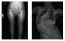
Skeletal dysplasia. Mesomelia is present at birth and appears to become more prominent over time. Slender long bones are noted at birth with an epiphyseal ossification delay and progressive widening of the metaphyses. Radial head can dislocate or sublux in childhood, leading to limited range of motion at the elbow.

Ivory and cone-shaped phalangeal epiphyses have been described; fifth finger clinodactyly and brachymesophalangy are common.

Iliac wings are small with flat acetabular angles.

Scoliosis may occur and can rapidly progress in late childhood / puberty, leading to the need for spinal fusion [Hall et al 2004, Bober & Jackson 2017].

Associated hip pathology includes coxa vara, coxa valga, developmental dysplasia, dislocation/subluxation, proximal femoral epiphysiolysis, and avascular necrosis. Eight of 12 individuals had hip pathology, some unilaterally (equaling 50% of the total hips). Developmental coxa vara was the most common finding, often beginning with a slipped capital femoral epiphysis and progressing to severe coxa vara, typically identified between ages two and five years [Karatas et al 2014] (see Table 5 and Figure 2).

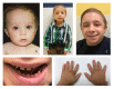
Dental abnormalities. Primary teeth are small for age but may appear proportionate to the mouth. Primary teeth can have deficient enamel and can be abnormally shaped. Hypodontia can occur.

Secondary teeth tend to be more affected. They are disproportionately small, dysplastic, and can have enamel hypoplasia. They are typically poorly rooted, and chewing can be affected. In many individuals, secondary teeth either prematurely shed or are extracted, and are replaced with dentures and/or implants [Hall et al 2004, Kantaputra et al 2011, Bober & Jackson 2017] (see Figure 1).

Hematologic abnormalities. Asymptomatic thrombocytosis, leukocytosis, and anemia are common [Unal et al 2014, Bober & Jackson 2017, Duker et al 2021].

Cerebrovascular. From birth onward, 25 of 47 individuals were diagnosed with intracranial aneurysms (with 30 having more than one aneurysm), 22 were diagnosed with moyamoya vasculopathy, 17 had both moyamoya vasculopathy and intracranial aneurysms, and 17 had neither. Approximately half of those diagnosed with moyamoya vasculopathy and/or aneurysms ultimately had a stroke (ischemic or hemorrhagic, respectively). Aneurysm risk appeared to be relatively steady through childhood, whereas moyamoya vasculopathy risk was higher prior to age five years [Duker et al 2021].

Cardiovascular. Elevated blood pressure was observed in almost 50% of teens and young adults. The many intertwined comorbidities that contribute to hypertension include moyamoya vasculopathy, renal disease, coronary artery disease, dyslipidemia, and insulin resistance.

In a cohort of 47 individuals, eight had myocardial infarctions as young adults (range 17-33 years; mean age 24±5.2 years; median age 24 years); multiple individuals had more than one acute coronary event.

Approximately 25% of individuals had cardiac malformations, including atrial septal defect, ventricular septal defect, and persistent patent foramen ovale. One individual had multiple cardiac rhabdomyomas [Duker et al 2021].

Other vasculopathies. MOPDII is associated with global vascular disease. Besides involvement of cerebral vasculature as well as renal and coronary vasculature, the external carotid artery has been stenotic as well as the site of an aneurysm. At least six individuals have had difficulties with femoral artery occlusion after catheterization procedures [Duker et al 2021].

Renal. Among 47 individuals, one third had chronic kidney disease (cause not specified). Mean age of the diagnosis of Stage III chronic kidney disease was 22 years. Additionally, two individuals had a kidney transplant, at ages 21 years and 23 years, one of the two had a renal artery aneurysm.

Another individual who had renal artery stenosis in addition to a renal artery aneurysm ultimately had a partial infarct of a kidney.

Six individuals had nephrolithiasis in early adulthood; all were on anti-hypertensive medications at the time of the diagnosis of nephrolithiasis.

Congenital anomalies included accessory/duplicated renal arteries in seven males and no females [Duker et al 2021].

Genitourinary. Two of 25 males had hypospadias and 11 had cryptorchidism or retractile testes [Duker et al 2021].

Insulin resistance and/or diabetes mellitus. Eighteen of 21 individuals older than age four years had insulin resistance or diabetes [Huang-Doran et al 2011].

Seventeen of 46 individuals (in a different but overlapping cohort) had insulin resistance and/or diabetes, typically detected in adolescence [Duker et al 2021].

These issues can also lead to dyslipidemia and fatty liver [Huang-Doran et al 2011].

Cognitive ability. Despite the significant microcephaly, intellectual development is generally in the typical to borderline range, and social skills are excellent. Many individuals have been described as hyperactive with easy distractibility in childhood [Hall et al 2004; Authors, personal observation]. However, those who have had strokes due to vascular disease have had more impairment [Bober & Jackson 2017]. Attending typical schools is expected, and some have attended college. However, it can be challenging for adults to live independently [Hall et al 2004].

Additional features

  • Skin:
    • Acanthosis nigricans, repeatedly documented, is likely related to concomitant insulin resistance [Hall et al 2004, Bober & Jackson 2017].
    • Café au lait patches have been described, with areas of hypopigmentation later in life.
    • "Wizened hands" with multiple creases developing in childhood have been noted (see Figure 1).
  • Neuroimaging findings can include simplified gyral patterns and hypoplasia of the corpus callosum [Hall et al 2004, Abdel-Salam et al 2020].
  • Subglottic stenosis, described in some individuals, required tracheostomy in at least two affected individuals [Hall et al 2004; Authors, unpublished data].
  • Craniosynostosis has been infrequently described, at times requiring surgical intervention. One child had bilateral coronal synostosis recognized in the neonatal period which required repair [Hall et al 2004; Abdel-Salam et al 2020; Authors, unpublished data].

Genotype-Phenotype Correlations

No genotype-phenotype correlations have been identified.

Nomenclature

In the 2023 revision of the Nosology of Genetic Skeletal Disorders [Unger et al 2023], MOPDII is referred to as PCNT-related microcephalic osteodysplastic primordial dwarfism and is included in the primoridal dwarfism and slender bones group.

Prevalence

Microcephalic osteodysplastic primordial dwarfism type II (MOPDII) is rare. More than 150 individuals with molecularly confirmed MOPDII have been identified across many populations worldwide [Bober & Jackson 2017].

Many different loss-of-function variants have been identified. Founder variants reported in specific populations include the following:

Differential Diagnosis

Table 3.

Selected Genes of Interest in the Differential Diagnosis of Microcephalic Osteodysplastic Primordial Dwarfism Type II

Gene(s) /
Genetic
Mechanism
DiffDx DisorderMOIKey Features of DiffDx Disorder
Shared w/MOPDIIDistinguishing from MOPDII
CDC6
CDC45
CDT1
GMNN
ORC1
ORC4
ORC6
Meier-Gorlin syndrome (OMIM PS224690)AR
AD 1
IUGR, extreme short stature w/microcephalyMicrotia, aplasia/hypoplasia of patellae; congenital emphysema; broncho-laryngomalacia; mammary hypoplasia; CDC45 Meier-Gorlin syndrome is assoc w/craniosynostosis.
CDKN1C IMAGe syndrome AD (imprinted)IUGR, extreme short stature; may have microcephalyCongenital adrenal insufficiency, cryptorchidism, small penis
LIG4 Ligase IV deficiency (LIG4 syndrome; OMIM 606593)ARIUGR, extreme short stature w/microcephalyBone marrow failure, sensitivity to ionizing radiation, immunodeficiency
POLE IMAGe-I syndrome (OMIM 618336)ARIUGR, extreme short stature; may have microcephalyImmunodeficiency; congenital adrenal insufficiency, cryptorchidism, small penis
RNU4ATAC RNU4atac-opathy (incl MOPDI/III, Roifman syndrome, & Lowry-Wood syndrome)ARIUGR, microcephaly, skeletal dysplasiaImmunodeficiency, brain malformations, retinal anomalies
XRCC4 XRCC4 deficiency (OMIM 616541)ARIUGR, extreme short stature w/microcephalyEndocrine dysfunction: hypergonadotropic hypogonadism, insulin resistance, thyroid dysfunction; rarely ataxia
4p 2
ASPM
ATR
BLM
CDK5RAP2
CENPJ
CEP152
DNA2
DNMT3A
DONSON
ERCC6
IGF1R
NBN
NCAPD3
PHGDH
PLK4
PRIM1
RTTN
SMARCAL1
SRCAP
TOP3A
TRAIP
VPS13B
See footnote 3.AR
AD
Extreme short stature w/microcephalyEither no distinguishing features, or gene-specific features that are recognized retrospectively, assoc w/syndromic diagnoses such as Nijmegen breakage syndrome, Bloom syndrome, & Seckel syndrome (see Note following table). Microcephalic dwarfism can also be an infrequent presentation of other disorders (e.g., Floating-Harbor syndrome).

AD = autosomal dominant; AR = autosomal recessive; DiffDx = differential diagnosis; IUGR = intrauterine growth restriction; MOI = mode of inheritance; MOPD = microcephalic osteodysplastic primordial dwarfism

1.

Meier-Gorlin syndrome is typically inherited in an autosomal recessive manner. GMNN-related Meier-Gorlin is inherited in an autosomal dominant manner.

2.

Or other microdeletion

3.

Listed genes are those in which pathogenic variants were identified in at least two persons with microcephalic dwarfism (defined as height & head circumference both greater than 4 SD below the mean at the time of exam) referred to the author's research laboratory for investigation [AJ, personal communication]. Other genes are also associated with extreme microcephalic dwarfism in some persons. (Note: Primary microcephaly genes are sometimes associated with short stature and therefore overlap with this definition of microcephalic dwarfism [Shaheen et al 2019].

Note: Although Seckel syndrome has been considered in the differential of MOPDII, as a diagnosis it is problematic given its variable definition. The broad original definition of Seckel syndrome includes any form of microcephalic dwarfism with intrauterine growth restriction, whereas a narrower definition includes: microcephaly that is more severe than short stature, presence of significant intellectual disability, and characteristic facial features including a sloping forehead. OMIM lists the following genes as associated with Seckel syndrome: ATR, CENPJ, CEP152, CEP63, DNA2, NSMCE2, RBBP8 (CTIP), and TRAIP (OMIM PS210600).

Management

Clinical practice guidelines for microcephalic osteodysplastic primordial dwarfism type II (MOPDII) have been proposed [Bober & Jackson 2017].

Evaluations Following Initial Diagnosis

To establish the extent of disease and needs in an individual diagnosed with MOPDII, the evaluations summarized in Table 4 (if not performed as part of the evaluation that led to the diagnosis) are recommended.

Table 4.

Recommended Evaluations Following Initial Diagnosis in Individuals with Microcephalic Osteodysplastic Primordial Dwarfism Type II

System/ConcernEvaluationComment
Growth restriction Measure height, weight, head circumference.Use MOPDII-specific growth curves. 1
Skeletal dysplasia AP/lateral x-ray of thoracolumbar spine & AP x-ray of hipsX-rays in early childhood to evaluate for slipped capital femoral epiphysis, coxa vara, & scoliosis
Cerebrovascular Brain MRI & MRAIncl at birth, as both in utero strokes & moyamoya vasculopathy have been identified in neonatal period.
Cardiac Echocardiogram, EKG
Renal
  • Renal ultrasound exam
  • Measure blood pressure.
  • If age ≥5 yrs, lab studies to assess renal function
Insulin resistance /
Diabetes mellitus
If age ≥5 yrs, perform lab studies to assess glucose homeostasis, lipids, & hepatic function.
Cognitive abilities Developmental assessment
  • Incl motor, adaptive, cognitive, & speech-language eval
  • Eval for early intervention / special education
Genetic counseling By genetics professionals 2To inform affected persons & their families re nature, MOI, & implications of MOPDII to facilitate medical & personal decision making
Family support
& resources
Assess need for:

Based on clinical practice guidelines proposed by Bober & Jackson [2017]

1.
2.

Medical geneticist, certified genetic counselor, certified advanced genetic nurse

Treatment of Manifestations

Table 5.

Treatment of Manifestations in Individuals with Microcephalic Osteodysplastic Primordial Dwarfism Type II

Manifestation/ConcernTreatmentConsiderations/Other
Growth Expect ~2 g/day weight gain throughout infancy & childhood. Avoid gastrostomy tube placement unless less-than-expected gain apparent for this disorder, after typical feeding regimens employed.
  • Use MOPDII-specific growth curves 1 for appropriate mgmt & expectations.
  • G-tube overfeeding can result in iatrogenic oral aversion.
Skeletal dysplasia
  • Hip pathology has been treated with in situ pinning or osteotomy. 2
  • Scoliosis has been treated by spinal fusion.
Dental abnormalities Ultimately treated w/dentures or implantsTypically in late teens or early adulthood
Hematologic abnormalities Anemia & thrombocytosis are common but have generally not required treatment.
Cerebrovascular disease Moyamoya vasculopathy in MOPDII has been treated w/encephaloduroarteriosynangiosis or pial synangiosis.Moyamoya vasculopathy treatment predominately required in childhood
Aneurysm treatment has been by clipping, coiling, or stenting. 3
  • Aneurysm treatment can be needed in childhood as well as throughout adulthood.
  • Aneurysm development is likely exacerbated by hypertension.
Coronary artery disease
  • Cardiovascular disease has been treated w/stents & bypass.
  • Hypercholesterolemia & hypertension have been treated w/medication. 4
  • The definition of hypertension is not well understood; standard adult ranges are likely too high for adults w/MOPDII.
  • Consider 110/70 as cutoff for hypertension in adulthood. 4
Renal disease Chronic kidney disease has been treated w/medication, dialysis, &/or kidney transplant. 4
Insulin resistance /
Diabetes mellitus
Diabetes has been treated w/medication. 4Majority treated w/metformin &/or insulin
Cognitive ability See Developmental Delay / Intellectual Disability Management Issues.Unless there have been complications from neurovascular disease, intellectual development is generally in typical-to-borderline range, & social skills are excellent.

Developmental Delay / Intellectual Disability Management Issues

The following information represents typical management recommendations for individuals with developmental delay / intellectual disability in the United States; standard recommendations may vary from country to country.

Ages 0-3 years. Referral to an early intervention program is recommended for access to occupational, physical, speech, and feeding therapy as well as infant mental health services, special educators, and sensory impairment specialists. In the US, early intervention is a federally funded program available in all states that provides in-home services to target individual therapy needs.

Ages 3-5 years. In the US, developmental preschool through the local public school district is recommended. Before placement, an evaluation is made to determine needed services and therapies and an individualized education plan (IEP) is developed for those who qualify based on established motor, language, social, or cognitive delay. The early intervention program typically assists with this transition. Developmental preschool is center based; for children too medically unstable to attend, home-based services are provided.

All ages. Consultation with a developmental pediatrician is recommended to ensure the involvement of appropriate community, state, and educational agencies (US) and to support parents in maximizing quality of life. Some issues to consider:

  • IEP services:
    • An IEP provides specially designed instruction and related services to children who qualify.
    • IEP services will be reviewed annually to determine whether any changes are needed.
    • Special education law requires that children participating in an IEP be in the least restrictive environment feasible at school and included in general education as much as possible, when and where appropriate.
    • Physical accommodations for short stature and small hands associated with MOPDII should be a part of the plan.
    • PT, OT, and speech services will be provided in the IEP to the extent that the need affects the child's access to academic material. Beyond that, private supportive therapies based on the affected individual's needs may be considered. Specific recommendations regarding type of therapy can be made by a developmental pediatrician.
    • As a child enters the teen years, a transition plan should be discussed and incorporated in the IEP. For those receiving IEP services, the public school district is required to provide services until age 21.
  • A 504 plan (Section 504: a US federal statute that prohibits discrimination based on disability) can be considered for those who require accommodations or modifications such as front-of-class seating, assistive technology devices, classroom scribes, extra time between classes, modified assignments, and modified classroom equipment/furniture.
  • Developmental Disabilities Administration (DDA) is a US public agency that provides services and support to qualified individuals. Eligibility differs by state but is typically determined by diagnosis and/or associated cognitive/adaptive disabilities.
  • Families with limited income and resources may also qualify for supplemental security income (SSI) for their child with a disability.

Surveillance

Table 6.

Recommended Surveillance for Individuals with Microcephalic Osteodysplastic Primordial Dwarfism Type II

System/ConcernEvaluationFrequency
Growth
restriction
Measurement of growth parameters 1 for appropriate mgmt & expectationsAt each visit
Skeletal
dysplasia
  • Serial x-rays of hips in early childhood
  • Evaluate for scoliosis through skeletal maturity. 2, 3
Annually or more often as needed
Dental
abnormalities
  • Routine dental care 3
  • Prosthodontist may be required for small implants in early adulthood.
Every 6 mos
Hematologic
abnormalities
Complete blood count 3Annually
Cerebrovascular
disease
Brain MRA/MRI 3, 4Childhood: Every 12-18 mos
Adulthood: Every 12-24 mos
Coronary artery
disease
  • Monitor blood pressure w/appropriately sized cuff. 3, 4
  • Consider 110/70 as cutoff for hypertension in adulthood. 4
  • Consider echocardiogram & EKG as well.
Annually
Renal Starting at age 5 yrs: Monitor renal function 3, 4
Insulin resistance
&/or
diabetes mellitus
Starting at age 5 yrs, lab assessment of glucose homeostasis, lipids, & hepatic function 3, 4, 5
Cognitive ability Monitor developmental progress & educational needs.At each visit
Family Assess family need for social work support (e.g., respite care, other local resources) or follow-up genetic counseling when new questions arise.

Agents/Circumstances to Avoid

Growth hormone supplementation in the absence of growth hormone deficiency has not been helpful, and in fact can contribute to morbidity [Hall et al 2004, Huang-Doran et al 2011, Bober et al 2012].

Evaluation of Relatives at Risk

See Genetic Counseling for issues related to testing of at-risk relatives for genetic counseling purposes.

Therapies Under Investigation

Search ClinicalTrials.gov in the US and EU Clinical Trials Register in Europe for access to information on clinical studies for a wide range of diseases and conditions. Note: There may not be clinical trials for this disorder.

Genetic Counseling

Genetic counseling is the process of providing individuals and families with information on the nature, mode(s) of inheritance, and implications of genetic disorders to help them make informed medical and personal decisions. The following section deals with genetic risk assessment and the use of family history and genetic testing to clarify genetic status for family members; it is not meant to address all personal, cultural, or ethical issues that may arise or to substitute for consultation with a genetics professional. —ED.

Mode of Inheritance

Microcephalic osteodysplastic primordial dwarfism type II (MOPDII) is inherited in an autosomal recessive manner.

Parents of a proband

  • The parents of an affected child are usually heterozygotes (i.e., presumed to be carriers of one PCNT pathogenic variant based on family history).
  • Molecular genetic testing for the parents of a proband can be performed to confirm that both parents are heterozygous for a PCNT pathogenic variant and to allow reliable recurrence risk assessment. If a pathogenic variant is detected in only one parent and parental identity testing has confirmed biological maternity and paternity, the following possibilities should be considered:
  • Heterozygotes (carriers) are asymptomatic.

Sibs of a proband

  • If both parents are known to be heterozygous for a PCNT pathogenic variant, each sib of an affected individual has at conception a 25% chance of being affected, a 50% chance of being an asymptomatic carrier, and a 25% chance of being unaffected and not a carrier.
  • Heterozygotes (carriers) are asymptomatic.

Offspring of a proband. The offspring of an individual with MOPDII are obligate heterozygotes (carriers) for a pathogenic variant in PCNT.

Other family members. Each sib of the proband's parents is at a 50% risk of being a carrier of a PCNT pathogenic variant.

Carrier Detection

Carrier testing for at-risk relatives requires prior identification of the PCNT pathogenic variants in the family.

Related Genetic Counseling Issues

Family planning

  • The optimal time for determination of genetic risk and discussion of the availability of prenatal/preimplantation genetic testing is before pregnancy.
  • It is appropriate to offer genetic counseling (including discussion of potential risks to offspring and reproductive options) to young adults who are affected, are carriers, or are at risk of being carriers.

Prenatal Testing and Preimplantation Genetic Testing

Once the PCNT pathogenic variants have been identified in an affected family member, prenatal and preimplantation genetic testing are possible.

Differences in perspective may exist among medical professionals and within families regarding the use of prenatal testing. While most centers would consider use of prenatal testing to be a personal decision, discussion of these issues may be helpful.

Resources

GeneReviews staff has selected the following disease-specific and/or umbrella support organizations and/or registries for the benefit of individuals with this disorder and their families. GeneReviews is not responsible for the information provided by other organizations. For information on selection criteria, click here.

Molecular Genetics

Information in the Molecular Genetics and OMIM tables may differ from that elsewhere in the GeneReview: tables may contain more recent information. —ED.

Table A.

Microcephalic Osteodysplastic Primordial Dwarfism Type II: Genes and Databases

GeneChromosome LocusProteinLocus-Specific DatabasesHGMDClinVar
PCNT 21q22​.3 Pericentrin PCNT @ LOVD PCNT PCNT

Data are compiled from the following standard references: gene from HGNC; chromosome locus from OMIM; protein from UniProt. For a description of databases (Locus Specific, HGMD, ClinVar) to which links are provided, click here.

Table B.

OMIM Entries for Microcephalic Osteodysplastic Primordial Dwarfism Type II (View All in OMIM)

210720MICROCEPHALIC OSTEODYSPLASTIC PRIMORDIAL DWARFISM, TYPE II; MOPD2
605925PERICENTRIN; PCNT

Molecular Pathogenesis

Biallelic loss-of-function PCNT pathogenic variants result in loss of cellular pericentrin protein [Griffith et al 2008, Rauch et al 2008]. Pericentrin is a large 360-kd coiled protein with a C-terminal PACT domain that targets it to the centrosome. It is a major component of the pericentriolar material that acts as a microtubular nucleation center to organize the mitotic spindle. Loss of pericentrin therefore perturbs mitosis in the cells of individuals with MOPDII [Rauch et al 2008], likely reducing cell number during development, resulting in extreme growth failure and microcephaly [Klingseisen & Jackson 2011]. Pericentrin also acts as a scaffold for signaling proteins, and may also reduce growth through acting in downstream ataxia-telangiectasia and Rad3-related (ATR) pathway signaling [Griffith et al 2008, Tibelius et al 2009].

Mechanism of disease causation. Loss of function

PCNT-specific laboratory technical considerations. While single amino acid substitutions could cause loss of function, none has been reported to date in individuals with MOPDII.

Table 7.

Notable PCNT Pathogenic Variants

Reference SequencesDNA Nucleotide
Change
Predicted
Protein Change
Comment
NM_006031​.6
NP_006022​.3
c.1468C>Tp.Gln490TerColombian founder variant 1
c.3465-1G>Ap.Ala1157ProfsTer36Founder variant in Israeli Druze population 2

Variants listed in the table have been provided by the authors. GeneReviews staff have not independently verified the classification of variants.

GeneReviews follows the standard naming conventions of the Human Genome Variation Society (varnomen​.hgvs.org). See Quick Reference for an explanation of nomenclature.

1.

Pachajoa et al [2014]; Authors, unpublished data

2.

Chapter Notes

Acknowledgments

The authors wish to wholeheartedly thank the Potentials Foundation and the Walking with Giants Foundation for their support of families worldwide with MOPDII, as well as for their support of research into this condition. Work in the Jackson lab has been supported by the European Union's Horizon 2020 research and innovation program ERC Advanced Grant (grant agreement 788093), and by a UK Medical Research Council (MRC) Human Genetics Unit core grant (MRC, U127580972).

Revision History

  • 30 March 2023 (sw) Revision: Nosology of Genetic Skeletal Disorders: 2023 Revision [Unger et al 2023] added to Nomenclature
  • 30 December 2021 (bp) Review posted live
  • 12 October 2021 (mb) Original submission

References

Literature Cited

  • Abdel-Salam GMH, Sayed ISM, Afifi HH, Abdel-Ghafar SF, Abouzaid MR, Ismail SI, Aglan MS, Issa MY, El-Bassyouni HT, El-Kamah G, Effat LK, Eid M, Zaki MS, Temtamy SA, Abdel-Hamid MS. Microcephalic osteodysplastic primordial dwarfism type II: additional nine patients with implications on phenotype and genotype correlation. Am J Med Genet A. 2020;182:1407–20. [PubMed: 32267100]
  • Bober MB, Jackson AP. Microcephalic osteodysplastic primordial dwarfism, type II: a clinical review. Curr Osteoporos Rep. 2017;15:61–9. [PMC free article: PMC5561166] [PubMed: 28409412]
  • Bober MB, Niiler T, Duker AL, Murray JE, Ketterer T, Harley ME, Alvi S, Flora C, Rustad C, Bongers EM, Bicknell LS, Wise C, Jackson AP. Growth in individuals with Majewski osteodysplastic primordial dwarfism type II caused by pericentrin mutations. Am J Med Genet A. 2012;158A:2719–25. [PubMed: 22821869]
  • Duker AL, Kinderman D, Jordan C, Niiler T, Baker-Smith CM, Thompson L, Parry DA, Carroll RS, Bober MB. Microcephalic osteodysplastic primordial dwarfism type II is associated with global vascular disease. Orphanet J Rare Dis. 2021;16:231. [PMC free article: PMC8139163] [PubMed: 34016138]
  • Duker AL, Niiler T, Bober MB. Expected weight gain for children with microcephalic osteodysplastic primordial dwarfsm type II. Am J Med Genet A. 2017;173:3067–9. [PubMed: 28940990]
  • Griffith E, Walker S, Martin CA, Vagnarelli P, Stiff T, Vernay B, Al Sanna N, Saggar A, Hamel B, Earnshaw WC, Jeggo PA, Jackson AP, O'Driscoll M. Mutations in pericentrin cause Seckel syndrome with defective ATR-dependent DNA damage signaling. Nat Genet. 2008;40:232–6. [PMC free article: PMC2397541] [PubMed: 18157127]
  • Hall JG, Flora C, Scott CI Jr, Pauli RM, Tanaka KI. Majewski osteodysplastic primordial dwarfism type II (MOPD II): natural history and clinical findings. Am J Med Genet A. 2004;130A:55–72. [PubMed: 15368497]
  • Huang-Doran I, Bicknell LS, Finucane FM, Rocha N, Porter KM, Tung YC, Szekeres F, Krook A, Nolan JJ, O'Driscoll M, Bober M, O'Rahilly S, Jackson AP, Semple RK, et al. Genetic defects in human pericentrin are associated with severe insulin resistance and diabetes. Diabetes. 2011;60:925–35. [PMC free article: PMC3046854] [PubMed: 21270239]
  • Jónsson H, Sulem P, Kehr B, Kristmundsdottir S, Zink F, Hjartarson E, Hardarson MT, Hjorleifsson KE, Eggertsson HP, Gudjonsson SA, Ward LD, Arnadottir GA, Helgason EA, Helgason H, Gylfason A, Jonasdottir A, Jonasdottir A, Rafnar T, Frigge M, Stacey SN, Th Magnusson O, Thorsteinsdottir U, Masson G, Kong A, Halldorsson BV, Helgason A, Gudbjartsson DF, Stefansson K. Parental influence on human germline de novo mutations in 1,548 trios from Iceland. Nature. 2017;549:519–22. [PubMed: 28959963]
  • Kantaputra P, Tanpaiboon P, Porntaveetus T, Ohazama A, Sharpe P, Rauch A, Hussadaloy A, Thiel CT. The smallest teeth in the world are caused by mutations in the PCNT gene. Am J Med Genet Part A. 2011;155:1398–403. [PubMed: 21567919]
  • Karatas AF, Bober MB, Rogers K, Duker AL, Ditro CP, Mackenzie WG. Hip pathology in Majewski osteodysplastic primordial dwarfism type II. J Pediatr Orthop. 2014;34:585–90. [PubMed: 24705347]
  • Klingseisen A, Jackson AP. Mechanisms and pathways of growth failure in primordial dwarfism. Genes Dev. 2011;25:2011–24. [PMC free article: PMC3197200] [PubMed: 21979914]
  • Lorenzo-Betancor O, Blackburn PR, Edwards E, Vázquez-do-Campo R, Klee EW, Labbé C, Hodges K, Glover P, Sigafoos AN, Soto AI, Walton RL, Doxsey S, Bober MB, Jennings S, Clark KJ, Asmann Y, Miller D, Freeman WD, Meschia J, Ross OA. PCNT point mutations and familial intracranial aneurysms. Neurology. 2018;91:e2170–e2181. [PMC free article: PMC6282233] [PubMed: 30413633]
  • Pachajoa H, Ruiz-Botero F, Isaza C. A new mutation of the PCNT gene in a Colombian patient with microcephalic osteodysplastic primordial dwarfism type II: a case report. J Med Case Rep. 2014;8:191. [PMC free article: PMC4086705] [PubMed: 24928221]
  • Rauch A, Thiel CT, Schindler D, Wick U, Crow YJ, Ekici AB, van Essen AJ, Goecke TO, Al-Gazali L, Chrzanowska KH, Zweier C, Brunner HG, Becker K, Curry CJ, Dallapiccola B, Devriendt K, Dörfler A, Kinning E, Megarbane A, Meinecke P, Semple RK, Spranger S, Toutain A, Trembath RC, Voss E, Wilson L, Hennekam R, de Zegher F, Dörr HG, Reis A. Mutations in the pericentrin (PCNT) gene cause primordial dwarfism. Science. 2008;319:816–9. [PubMed: 18174396]
  • Richards S, Aziz N, Bale S, Bick D, Das S, Gastier-Foster J, Grody WW, Hegde M, Lyon E, Spector E, Voelkerding K, Rehm HL, et al. Standards and guidelines for the interpretation of sequence variants: a joint consensus recommendation of the American College of Medical Genetics and Genomics and the Association for Molecular Pathology. Genet Med. 2015;17:405–24. [PMC free article: PMC4544753] [PubMed: 25741868]
  • Sauvigny T, Alawi M, Krause L, Renner S, Spohn M, Busch A, Kolbe V, Altmüller J, Löscher BS, Franke A, Brockmann C, Lieb W, Westphal M, Schmidt NO, Regelsberger J, Rosenberger G. Exome sequencing in 38 patients with intracranial aneurysms and subarachnoid hemorrhage. J Neurol. 2020;267:2533–45. [PMC free article: PMC7419486] [PubMed: 32367296]
  • Shaheen R, Maddirevula S, Ewida N, Alsahli S, Abdel-Salam GMH, Zaki MS, Tala SA, Alhashem A, Softah A, Al-Owain M, Alazami AM, Abadel B, Patel N, Al-Sheddi T, Alomar R, Alobeid E, Ibrahim N, Hashem M, Abdulwahab F, Hamad M, Tabarki B, Alwadei AH, Alhazzani F, Bashiri FA, Kentab A, Şahintürk S, Sherr E, Fregeau B, Sogati S, Alshahwan SAM, Alkhalifi S, Alhumaidi Z, Temtamy S, Aglan M, Otaify G, Girisha KM, Tulbah M, Seidahmed MZ, Salih MA, Abouelhoda M, Momin AA, Saffar MA, Partlow JN, Arold ST, Faqeih E, Walsh C, Alkuraya FS. Genomic and phenotypic delineation of congenital microcephaly. Genet Med. 2019;21:545–52. [PMC free article: PMC6986385] [PubMed: 30214071]
  • Stenson PD, Mort M, Ball EV, Chapman M, Evans K, Azevedo L, Hayden M, Heywood S, Millar DS, Phillips AD, Cooper DN. The Human Gene Mutation Database (HGMD®): optimizing its use in a clinical diagnostic or research setting. Hum Genet. 2020;139:1197–207. [PMC free article: PMC7497289] [PubMed: 32596782]
  • Teo M, Johnson JN, Bell-Stephens TE, Marks MP, Do HM, Dodd RL, Bober MB, Steinberg GK. Surgical outcomes of Majewski osteodysplastic primordial dwarfism Type II with intracranial vascular anomalies. J Neurosurg Pediatr. 2016;25:717–23. [PubMed: 27611897]
  • Tibelius A, Marhold J, Zentgraf H, Heilig CE, Neitzel H, Ducommun B, Rauch A, Ho AD, Bartek J, Krämer A. Microcephalin and pericentrin regulate mitotic entry via centrosome-associated Chk1. J Cell Biol. 2009;185:1149–57. [PMC free article: PMC2712957] [PubMed: 19546241]
  • Unal S, Alanay Y, Cetin M, Boduroglu K, Utine E, Cormier-Daire V, Huber C, Ozsurekci Y, Kilic E, Simsek Kiper OP, Gumruk F. Striking hematological abnormalities in patients with microcephalic osteodysplastic primordial dwarfism type II (MOPD II): a potential role of pericentrin in hematopoiesis. Pediatr Blood Cancer. 2014;61:302–5. [PubMed: 24106199]
  • Unger S, Ferreira CR, Mortier GR, Ali H, Bertola DR, Calder A, Cohn DH, Cormier-Daire V, Girisha KM, Hall C, Krakow D, Makitie O, Mundlos S, Nishimura G, Robertson SP, Savarirayan R, Sillence D, Simon M, Sutton VR, Warman ML, Superti-Furga A. Nosology of genetic skeletal disorders: 2023 revision. Am J Med Genet A. 2023. Epub ahead of print. [PMC free article: PMC10081954] [PubMed: 36779427]
  • Weiss K, Ekhilevitch N, Cohen L, Bratman-Morag S, Bello R, Martinez AF, Hadid Y, Shlush LI, Kurolap A, Paperna T, Mory A, Baris HN, Muenke M. Identification of a novel PCNT founder pathogenic variant in the Israeli Druze population. Eur J Med Genet. 2020;63:103643. [PubMed: 30922925]
Copyright © 1993-2024, University of Washington, Seattle. GeneReviews is a registered trademark of the University of Washington, Seattle. All rights reserved.

GeneReviews® chapters are owned by the University of Washington. Permission is hereby granted to reproduce, distribute, and translate copies of content materials for noncommercial research purposes only, provided that (i) credit for source (http://www.genereviews.org/) and copyright (© 1993-2024 University of Washington) are included with each copy; (ii) a link to the original material is provided whenever the material is published elsewhere on the Web; and (iii) reproducers, distributors, and/or translators comply with the GeneReviews® Copyright Notice and Usage Disclaimer. No further modifications are allowed. For clarity, excerpts of GeneReviews chapters for use in lab reports and clinic notes are a permitted use.

For more information, see the GeneReviews® Copyright Notice and Usage Disclaimer.

For questions regarding permissions or whether a specified use is allowed, contact: ude.wu@tssamda.

Bookshelf ID: NBK575926PMID: 34978779

Views

Tests in GTR by Gene

Related information

  • OMIM
    Related OMIM records
  • PMC
    PubMed Central citations
  • PubMed
    Links to PubMed
  • Gene
    Locus Links

Similar articles in PubMed

See reviews...See all...

Recent Activity

Your browsing activity is empty.

Activity recording is turned off.

Turn recording back on

See more...