U.S. flag

An official website of the United States government

NCBI Bookshelf. A service of the National Library of Medicine, National Institutes of Health.

Adam MP, Feldman J, Mirzaa GM, et al., editors. GeneReviews® [Internet]. Seattle (WA): University of Washington, Seattle; 1993-2024.

Cover of GeneReviews®

GeneReviews® [Internet].

Show details

MN1 C-Terminal Truncation Syndrome

, MBChB, PhD, , BBiomedSc, , MSc, , MD, , MD, PhD, , MD, PhD, , PhD, and , MD.

Author Information and Affiliations

Initial Posting: .

Estimated reading time: 20 minutes

Summary

Clinical characteristics.

Individuals with MN1 C-terminal truncation (MCTT) syndrome have mild-to-moderate intellectual disability, severe expressive language delay, dysmorphic facial features (midface hypoplasia, downslanting palpebral fissures, hypertelorism, exophthalmia, short upturned nose, and small low-set ears), and distinctive findings on brain imaging (including perisylvian polymicrogyria and atypical rhombencephalosynapsis). Mild-to-moderate prelingual hearing loss (usually bilateral, conductive, and/or sensorineural) is common. Generalized seizures (observed in the minority of individuals) are responsive to anti-seizure medication. There is an increased risk for craniosynostosis and, thus, increased intracranial pressure. To date, 25 individuals with MCTT syndrome have been identified.

Diagnosis/testing.

No consensus clinical diagnostic criteria for MCTT syndrome have been published. The diagnosis is established in a proband with suggestive findings and a heterozygous pathogenic variant in MN1 identified by molecular genetic testing.

Management.

Treatment of manifestations: Multidisciplinary specialists to help manage developmental delay / intellectual disability, feeding issues, seizures, hearing loss, and speech and language needs, especially alternative communication.

Surveillance: Routine follow up by multidisciplinary specialists per their recommendations.

Genetic counseling.

MCTT syndrome is an autosomal dominant disorder typically caused by a de novo MN1 pathogenic variant. The risk to the sibs of a proband depends on the genetic status of the proband's parents: if the MN1 pathogenic variant found in the proband cannot be detected in the leukocyte DNA of either parent, the recurrence risk to sibs is estimated to be slightly greater than that of the general population because of the possibility of parental somatic/germline mosaicism. Prenatal testing for a pregnancy at increased risk and preimplantation genetic testing are possible once the MN1 pathogenic variant has been identified in an affected family member.

Diagnosis

No consensus clinical diagnostic criteria for MN1 C-terminal truncation (MCTT) syndrome have been published.

Suggestive Findings

MN1 C-terminal truncation (MCTT) syndrome should be suspected in individuals with the following clinical findings and brain MRI findings.

Clinical findings

  • Intellectual disability (ID) with severe expressive language delay
  • Hypotonia
  • Delays in motor development
  • Hearing loss (conductive or sensorineural)
  • Distinctive craniofacial features (See Figure 1.)
Figure 1. . Facial features of individuals with C-terminal truncating variants in MN1, illustrating how features evolve from before to after age five years.

Figure 1.

Facial features of individuals with C-terminal truncating variants in MN1, illustrating how features evolve from before to after age five years. Frontal bossing may be more prominent at infancy, with a tall forehead at later stages. Skull shape anomalies (more...)

Brain MRI findings. In MCTT syndrome, an atypical distinctive form of rhombencephalosynapsis is observed in which there is partial or complete loss of the cerebellar vermis landmarks with fusion of the cerebellar hemispheres characterized by abnormal midline crossing of cerebellar folia and white matter tracts. Other distinctive brain MRI findings are summarized in Table 1 (see Figure 2 and Figure 3).

Figure 2. . Partial rhombencephalosynapsis in patients with C-terminal truncating MN1 variants.

Figure 2.

Partial rhombencephalosynapsis in patients with C-terminal truncating MN1 variants. Changes of the cerebellum include foliar dysplasia, abnormal folia crossing the midline, and small or almost absent vermis. For details, refer to Figure 4 of Mak et al (more...)

Figure 3.

Figure 3.

Persistent trigeminal artery and prominent posterior clinoid process in patients with C-terminal truncating MN1 variants A. Carotid and basilar arteries (axial view): persistent trigeminal artery flow-voids (dark signal) connecting the carotid (C) and (more...)

Table 1.

Brain Imaging Features in Individuals with MCTT Syndrome

FeaturePrevalence
Perisylvian polymicrogyria 19/10
Rhombencephalosynapsis8/10
Persistent trigeminal artery7/10
Prominent posterior clinoid process7/10
Thick rostral corpus callosum6/11
Hypoplastic olfactory bulbs4/8
Ventriculomegaly3/11
Subcortical heterotopia2/11

Based on 11 MRIs reviewed at one center [Mak et al 2020]

1.

Observed in insula; can sometimes extend more broadly in perisylvian region.

Establishing the Diagnosis

The diagnosis of MN1 C-terminal truncation (MCTT) syndrome is established in a proband with suggestive findings and a heterozygous pathogenic (or likely pathogenic) variant in MN1 identified by molecular genetic testing (see Table 2).

(1) Per ACMG/AMP variant interpretation guidelines, the terms "pathogenic variants" and "likely pathogenic variants" are synonymous in a clinical setting, meaning that both are considered diagnostic and both can be used for clinical decision making [Richards et al 2015]. Reference to "pathogenic variants" in this section is understood to include any likely pathogenic variants. (2) Identification of a heterozygous MN1 variant of uncertain significance does not establish or rule out the diagnosis of this disorder.

Molecular genetic testing approaches can include a combination of gene-targeted testing (single-gene testing and multigene panel) and comprehensive genomic testing (exome sequencing, genome sequencing) depending on the phenotype.

Gene-targeted testing requires that the clinician determine which gene(s) are likely involved, whereas genomic testing does not. Individuals with the distinctive craniofacial or brain MRI findings described in Suggestive Findings are likely to be diagnosed using gene-targeted testing (see Option 1), whereas those in whom the diagnosis of MCTT syndrome has not been considered are more likely to be diagnosed using genomic testing (see Option 2).

Option 1

Single-gene testing. Sequence analysis of MN1 is performed first to detect small intragenic deletions/insertions and missense, nonsense, and splice site variants. Note: Depending on the sequencing method used, single-exon, multiexon, or whole-gene deletions/duplications may not be detected. Typically, if no variant is detected by the sequencing method used, the next step is to perform gene-targeted deletion/duplication analysis to detect exon and whole-gene deletions or duplications; however, since MCTT occurs through a gain-of-function mechanism and large intragenic deletion or duplication has not been reported, testing for intragenic deletions or duplication is much less likely to identify a disease-causing variant given the current understanding.

Note: To date all described variants have been C-terminal truncating variants at the 3' end of exon 1 or in exon 2.

An intellectual disability multigene panel that includes MN1 and other genes of interest (see Differential Diagnosis) is most likely to identify the genetic cause of the condition while limiting identification of variants of uncertain significance and pathogenic variants in genes that do not explain the underlying phenotype. Note: (1) The genes included in the panel and the diagnostic sensitivity of the testing used for each gene vary by laboratory and are likely to change over time. (2) Some multigene panels may include genes not associated with the condition discussed in this GeneReview. Given that MCTT syndrome is quite rare and only recently identified, some intellectual disability panels may not include MN1. (3) In some laboratories, panel options may include a custom laboratory-designed panel and/or custom phenotype-focused exome analysis that includes genes specified by the clinician. (4) Methods used in a panel may include sequence analysis, deletion/duplication analysis, and/or other non-sequencing-based tests.

For an introduction to multigene panels click here. More detailed information for clinicians ordering genetic tests can be found here.

Option 2

Comprehensive genomic testing does not require the clinician to determine which gene(s) are likely involved. Exome sequencing is most commonly used; genome sequencing is also possible.

For an introduction to comprehensive genomic testing click here. More detailed information for clinicians ordering genomic testing can be found here.

Table 2.

Molecular Genetic Testing Used in MN1 C-Terminal Truncation Syndrome

Gene 1MethodProportion of Probands with a Pathogenic Variant 2 Detectable by Method
MN1 Sequence analysis 324/24 4
Gene-targeted deletion/duplication analysis 5None reported 4
1.
2.

See Molecular Genetics for information on variants detected in this gene.

3.

Sequence analysis detects variants that are benign, likely benign, of uncertain significance, likely pathogenic, or pathogenic. Variants may include small intragenic deletions/insertions and missense, nonsense, and splice site variants; typically, exon or whole-gene deletions/duplications are not detected. For issues to consider in interpretation of sequence analysis results, click here.

4.
5.

Gene-targeted deletion/duplication analysis detects intragenic deletions or duplications. Methods used may include a range of techniques such as quantitative PCR, long-range PCR, multiplex ligation-dependent probe amplification (MLPA), and a gene-targeted microarray designed to detect single-exon deletions or duplications.

Clinical Characteristics

Clinical Description

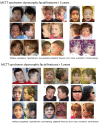
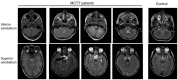
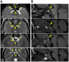
To date, 25 individuals have been identified with a germline (de novo or inherited from a mosaic parent) pathogenic C-terminal variant in MN1 [Mak et al 2020, Miyake et al 2020]. Individuals with MN1 C-terminal truncation (MCTT) syndrome have intellectual disability, severe expressive language delay, dysmorphic facial features (see Figure 1), and distinctive findings on brain imaging (including perisylvian polymicrogyria and atypical rhombencephalosynapsis) (see Table 1). The summary of the phenotypic features associated with MCTT syndrome in Table 3 is based on these reports.

Table 3.

Clinical Features of MN1 C-Terminal Truncation Syndrome

FeaturePersons w/Feature
Intellectual disability19/20 (95%)
Motor delay19/20 (95%)
Speech delay21/23 (91%)
Hypotonia19/21 (90%)
Hearing loss16/20 (80%)
Feeding difficulties14/21 (67%)
Spinal anomalies8/12 (67%)
Dental anomalies9/15 (60%)
Ophthalmologic anomalies10/17 (59%)
Cardiovascular anomalies6/20 (30%)
Seizures6/22 (27%)
Craniosynostosis3/22 (14%)

Developmental delay (DD) / intellectual disability (ID). Individuals with MCTT syndrome have mild-to-moderate intellectual disability and severe expressive language delay. The majority have nonverbal communication with notable exceptions. For example, one child was able to speak at age two years, and at age 14 years was able to function at the level of a seven-year-old. For others, single-word speech began between ages three and six years. Some can communicate in sign language with up to 50 signs [Mak et al 2020].

Delay in gross motor development included hypotonia; at least four of 22 children walked independently by age two to three years. Others required orthotics or a wheelchair for mobilization. As for fine motor and self-help skills, most individuals require help with writing, feeding, or dressing [Mak et al 2020].

Hearing loss, when present, is mild to moderate and prelingual. It is usually bilateral, conductive, and/or sensorineural. Dysplasia of the cochlea, semicircular canals, and bony structures of the middle ear (e.g., incus) has been reported.

Feeding difficulties are more prominent early in infancy and may resolve after the first year of life. Hyperphagia has also been reported in three children [Miyake et al 2020].

Spinal anomalies include lordosis, scoliosis, or kyphosis, which may be detected clinically or by imaging.

Dental anomalies can include conical teeth, crowded teeth, and serrated teeth. Malocclusion has also been reported.

Ophthalmologic anomalies can include oculomotor defects, strabismus, and/or shallow orbits, giving the appearance of exorbitism.

Cardiovascular anomalies include atrial septal defect or ventricular septal defects.

Seizures are generalized and may be myoclonic in nature. Among the six individuals with seizures, the majority were isolated events and were responsive to anti-seizure medication. While polymicrogyria may be associated with increased risk of seizures, more information is needed.

Craniosynostosis. Individuals with MCTT syndrome are at increased risk for craniosynostosis with no specific pattern to the sutures affected. Although head circumference is normal in individuals with craniosynostosis, head shape is consistently affected. Skull shape anomalies that may be observed with or without underlying craniosynostosis include dolichocephaly, turricephaly, and/or bitemporal narrowing, plagiocephaly, and macrocephaly. Individuals with craniosynostosis are at increased risk for elevated intracranial pressure.

Behavioral problems. While some affected individuals may experience frustration due to poor verbal communication, no consistent behavior problems have been reported. The natural history into adulthood has not yet been delineated.

Growth. Growth parameters tend to remain within the normal range.

Prognosis. It is unknown if life span is affected in MCTT syndrome. One individual is alive at age 21 years [Mak et al 2020]. An unreported male is well at age 39 years [Angela Lin, personal commmunication], demonstrating that survival into adulthood is possible. Since many adults with disabilities have not undergone advanced genetic testing, it is likely that adults with this condition are unrecognized and underreported.

Genotype-Phenotype Correlations

No genotype-phenotype correlations have been identified.

Nomenclature

MCTT syndrome is referred to as CEBALID (craniofacial defects, dysmorphic ears, structural brain abnormalities, expressive language delay, and impaired intellectual development) syndrome in OMIM (OMIM 618774).

Prevalence

To date, 25 individuals with MCTT syndrome have been reported [Mak et al 2020, Miyake et al 2020]. This disorder is expected to be rare; the prevalence among individuals with unexplained developmental disorders is estimated at 4.2:10,000 [Kaplanis et al 2020].

Differential Diagnosis

Gomez-Lopez-Hernandez syndrome (GLHS) (OMIM 601853). Like MN1 C-terminal truncation (MCTT) syndrome, GLHS is associated with turricephaly, midface hypoplasia, craniosynostosis, and rhombencephalosynapsis. Unlike MCTT syndrome, GLHS is also known to be associated with trigeminal anesthesia, scalp alopecia, corneal opacities, short stature, and ataxia. The genetic cause of GLHS is unknown.

Management

Evaluations Following Initial Diagnosis

To establish the extent of disease and needs in an individual diagnosed with MN1 C-terminal truncation (MCTT) syndrome, the evaluations summarized in Table 4 (if not performed as part of the evaluation that led to the diagnosis) are recommended.

Table 4.

Recommended Evaluations Following Initial Diagnosis in Individuals with MN1 C-Terminal Truncation Syndrome

System/ConcernEvaluationComment
Constitutional Measure height, weight, & head circumference.
Craniofacial
  • Dental eval
  • Oral-maxillofacial eval
Incl:
  • Dental anomalies (e.g., conical teeth, crowded teeth, malocclusion)
  • Palate for submucous cleft assoc w/bifid uvula
  • Cranial exam & skull radiographs for craniosynostosis
  • CT if indicated
There is ↑ risk for craniosynostosis & ↑ intracranial pressure.
Neurologic Neurologic eval
  • Brain MRI to identify polymicrogyria (possibly assoc w/seizure risk) & persistent trigeminal artery (possible risks for neurosurgical procedures involving skull base & pituitary)
  • EEG, if seizures present
Development Developmental assessment
  • Incl motor, adaptive, cognitive, & speech/language eval
  • Eval for early intervention / special education
Speech & language Speech-language therapistAnticipate need for augmentative communication incl sign language.
Psychiatric/
Behavioral
Assessment by primary care provider
  • No consistent behavior problems reporte
  • Some may experience frustration due to poor verbal communication.
Musculoskeletal Orthopedics / physical medicine & rehab / PT & OT evalIncl assessment of:
  • Spinal involvement (lordosis, scoliosis, kyphosis)
  • Need for PT (to improve gross motor skills) &/or OT (to improve fine motor skills)
Gastrointestinal/
Feeding
Gastroenterology / nutrition / feeding team evalIncl eval of aspiration risk & nutritional status
Eyes Ophthalmology evalTo assess for ↓ vision, oculomotor defects, strabismus, or shallow orbits
Hearing Audiology eval
  • Assess for conductive &/or sensorineural hearing loss.
  • If present, refer to otolaryngologist.
Cardiovascular Cardiology evalFor congenital heart defects, most commonly ASD & VSD
Genetic counseling By genetics professionals 1To inform affected individuals & their families re nature, MOI, & implications of MCTT syndrome to facilitate medical & personal decision making
Family support
& resources
Assess need for:

ASD = atrial septal defect; MOI = mode of inheritance; OT = occupational therapy; PT = physical therapy; VSD = ventricular septal defects

1.

Medical geneticist, certified genetic counselor, or certified advanced genetic nurse

Treatment of Manifestations

Table 5.

Treatment of Manifestations in Individuals with MN1 C-Terminal Truncation Syndrome

Manifestation/ConcernTreatmentConsiderations/Other
Craniofacial Per oral-maxillofacial & neurosurgical specialist
Developmental delay /
Intellectual disability
See Developmental Delay / Intellectual Disability Management Issues.W/emphasis on alternative (nonverbal) communication
Speech & language Alternative communication as needed
Epilepsy Standardized treatment w/ASM by experienced neurologist
  • Many ASMs may be effective; none demonstrated effective specifically for this disorder.
  • Education of parents/caregivers 1
Poor weight gain /
Failure to thrive
Feeding therapyLow threshold for clinical feeding eval &/or radiographic swallowing study when showing clinical signs or symptoms of dysphagia
Musculoskeletal Orthopedics / physical medicine & rehab / PT & OT
  • Incl stretching to help avoid contractures & falls
  • Consider need for positioning & mobility devices, disability parking placard.
Abnormal vision
&/or strabismus
Standard treatment(s) per ophthalmologist
Hearing Community hearing services through early intervention or school district
Cardiovascular Per treating cardiologist
Family/Community
  • Ensure appropriate social work involvement to connect families w/local resources, respite, & support.
  • Coordinate care to manage multiple subspecialty appointments, equipment, medications, & supplies.

ASM = anti-seizure medication; OT = occupational therapy; PT = physical therapy

1.

Education of parents/caregivers regarding common seizure presentations is appropriate. For information on non-medical interventions and coping strategies for children diagnosed with epilepsy, see Epilepsy Foundation Toolbox.

Developmental Delay / Intellectual Disability Management Issues

The following information represents typical management recommendations for individuals with developmental delay / intellectual disability in the United States; standard recommendations may vary from country to country.

Ages 0-3 years. Referral to an early intervention program is recommended for access to occupational, physical, speech, and feeding therapy as well as infant mental health services, special educators, and sensory impairment specialists. In the US, early intervention is a federally funded program available in all states that provides in-home services to target individual therapy needs.

Ages 3-5 years. In the US, developmental preschool through the local public school district is recommended. Before placement, an evaluation is made to determine needed services and therapies and an individualized education plan (IEP) is developed for those who qualify based on established motor, language, social, or cognitive delay. The early intervention program typically assists with this transition. Developmental preschool is center based; for children too medically unstable to attend, home-based services are provided.

All ages. Consultation with a developmental pediatrician is recommended to ensure the involvement of appropriate community, state, and educational agencies (US) and to support parents in maximizing quality of life. Some issues to consider:

  • IEP services:
    • An IEP provides specially designed instruction and related services to children who qualify.
    • IEP services will be reviewed annually to determine whether any changes are needed.
    • Special education law requires that children participating in an IEP be in the least restrictive environment feasible at school and included in general education as much as possible, when and where appropriate.
    • Vision and hearing consultants should be a part of the child's IEP team to support access to academic material.
    • PT, OT, and speech services will be provided in the IEP to the extent that the need affects the child's access to academic material. Beyond that, private supportive therapies based on the affected individual's needs may be considered. Specific recommendations regarding type of therapy can be made by a developmental pediatrician.
    • As a child enters the teen years, a transition plan should be discussed and incorporated in the IEP. For those receiving IEP services, the public school district is required to provide services until age 21.
  • A 504 plan (Section 504: a US federal statute that prohibits discrimination based on disability) can be considered for those who require accommodations or modifications such as front-of-class seating, assistive technology devices, classroom scribes, extra time between classes, modified assignments, and enlarged text.
  • Developmental Disabilities Administration (DDA) enrollment is recommended. DDA is a US public agency that provides services and support to qualified individuals. Eligibility differs by state but is typically determined by diagnosis and/or associated cognitive/adaptive disabilities.
  • Families with limited income and resources may also qualify for supplemental security income (SSI) for their child with a disability.

Motor Dysfunction

Gross motor dysfunction

  • Physical therapy is recommended to maximize mobility and to reduce the risk for later-onset orthopedic complications (e.g., contractures, scoliosis, hip dislocation).
  • Consider use of durable medical equipment and positioning devices as needed (e.g., wheelchairs, walkers, bath chairs, orthotics, adaptive strollers).

Fine motor dysfunction. Occupational therapy is recommended for difficulty with fine motor skills that affect adaptive function such as feeding, grooming, dressing, and writing.

Oral motor dysfunction should be assessed at each visit and clinical feeding evaluations and/or radiographic swallowing studies should be obtained for choking/gagging during feeds, poor weight gain, frequent respiratory illnesses, or feeding refusal that is not otherwise explained. Assuming that the child is safe to eat by mouth, feeding therapy (typically from an occupational or speech therapist) is recommended to help improve coordination or sensory-related feeding issues. Feeds can be thickened or chilled for safety. When feeding dysfunction is severe, an NG-tube or G-tube may be necessary.

Communication issues. Consider evaluation for alternative means of communication (e.g., augmentative and alternative communication [AAC]) for individuals who have expressive language difficulties. An AAC evaluation can be completed by a speech-language pathologist who has expertise in the area. The evaluation will consider cognitive abilities and sensory impairments to determine the most appropriate form of communication. AAC devices can range from low-tech, such as picture exchange communication, to high-tech, such as voice-generating devices. Contrary to popular belief, AAC devices do not hinder verbal development of speech, but rather support optimal speech and language development.

Social/Behavioral Concerns

Children may qualify for and benefit from interventions used in treatment of autism spectrum disorder, including applied behavior analysis (ABA). ABA therapy is targeted to the individual child's behavioral, social, and adaptive strengths and weaknesses and typically performed one on one with a board-certified behavior analyst.

Consultation with a developmental pediatrician may be helpful in guiding parents through appropriate behavior management strategies or providing prescription medications, such as medication used to treat attention-deficit/hyperactivity disorder, when necessary.

Concerns about serious aggressive, self-injurious, or destructive behavior can be addressed by a pediatric psychiatrist, developmental pediatrician, or psychologist with particular interest in management of these types of behaviors.

Surveillance

Table 6.

Recommended Surveillance for Individuals with MN1 C-Terminal Truncation Syndrome

System/ConcernEvaluationFrequency
Feeding Measurement of growth parameters; eval of nutritional status & safety of oral intakeAt each visit
Neurologic Monitor those w/seizures as clinically indicated.
Assess for new manifestations incl seizures, changes in tone, & movement disorders.Per initial assessment or specialist advice
Development Monitor developmental progress & educational needs.At each visit
Psychiatric/
Behavioral
Behavior assessment for anxiety, attention, & aggressive or self-injurious behaviorPer initial assessment or specialist advice
Musculoskeletal Physical medicine, OT/PT assessment of mobility, self-help skillsAt each visit
Hearing Per initial assessment or specialist advicePer initial assessment or specialist advice
Risk for
craniosynostosis
Cardiovascular
Miscellaneous/
Other
Assess family need for social work support (e.g., palliative/respite care, home nursing, other local resources) & care coordination.At each visit

OT = occupational therapy; PT = physical therapy

Evaluation of Relatives at Risk

See Genetic Counseling for issues related to testing of at-risk relatives for genetic counseling purposes.

Therapies Under Investigation

Search ClinicalTrials.gov in the US and EU Clinical Trials Register in Europe for access to information on clinical studies for a wide range of diseases and conditions. Note: There may not be clinical trials for this disorder.

Genetic Counseling

Genetic counseling is the process of providing individuals and families with information on the nature, mode(s) of inheritance, and implications of genetic disorders to help them make informed medical and personal decisions. The following section deals with genetic risk assessment and the use of family history and genetic testing to clarify genetic status for family members; it is not meant to address all personal, cultural, or ethical issues that may arise or to substitute for consultation with a genetics professional. —ED.

Mode of Inheritance

MN1 C-terminal truncation (MCTT) syndrome is an autosomal dominant disorder typically caused by a de novo pathogenic variant.

Risk to Family Members

Parents of a proband

  • Molecular genetic testing is recommended to confirm the genetic status of the parents and to allow reliable recurrence risk assessment.
  • If the pathogenic variant identified in the proband is not identified in either parent, the following possibilities should be considered:
    • The proband has a de novo pathogenic variant. Note: A pathogenic variant is reported as "de novo" if: (1) the pathogenic variant found in the proband is not detected in parental DNA; and (2) parental identity testing has confirmed biological maternity and paternity. If parental identity testing is not performed, the variant is reported as "assumed de novo" [Richards et al 2015].
    • The proband inherited a pathogenic variant from a parent with germline (or somatic and germline) mosaicism.* Note: Testing of parental leukocyte DNA may not detect all instances of somatic mosaicism.
      * A parent with somatic and germline mosaicism for an MN1 pathogenic variant may be mildly/minimally affected. In one family, the mosaic father of affected sibs had mild/nondistinctive features of MCTT syndrome (i.e., mildly dysplastic ears and a high, narrow palate) [Mak et al 2020].

Sibs of a proband. The risk to the sibs of the proband depends on the genetic status of the proband's parents:

Offspring of a proband

  • The majority of probands reported to date with MCTT syndrome whose parents have undergone molecular genetic testing have the disorder as a result of a de novo MN1 pathogenic variant.
  • Each child of an individual with MCTT syndrome has a 50% chance of inheriting the MN1 pathogenic variant.
  • Individuals with MCTT syndrome are not known to reproduce; however, many are not yet of reproductive age.

Other family members. Given that most probands with MCTT syndrome reported to date have the disorder as a result of a de novo MN1 pathogenic variant, the risk to other family members is presumed to be low.

Related Genetic Counseling Issues

Family planning

  • The optimal time for determination of genetic risk and discussion of the availability of prenatal/preimplantation genetic testing is before pregnancy.
  • It is appropriate to offer genetic counseling (including discussion of potential risks to offspring and reproductive options) to parents of affected individuals.

Prenatal Testing and Preimplantation Genetic Testing

Once the MN1 pathogenic variant has been identified in an affected family member, prenatal/preimplantation genetic testing are possible. Risk to future pregnancies is presumed to be low as the proband most likely has a de novo MN1 pathogenic variant. However, risk to future pregnancies is slightly greater than that of the general population because of the possibility of parental germline mosaicism [Mak et al 2020].

Differences in perspective may exist among medical professionals and within families regarding the use of prenatal testing. While most centers would consider use of prenatal testing to be a personal decision, discussion of these issues may be helpful.

Resources

GeneReviews staff has selected the following disease-specific and/or umbrella support organizations and/or registries for the benefit of individuals with this disorder and their families. GeneReviews is not responsible for the information provided by other organizations. For information on selection criteria, click here.

  • Human Disease Genes Website Series - Registry
    This website was created to share and collect information about clinic, management and research projects to gather more knowledge and provide better treatment of patients with mutations in the MN1 gene.
    MN1

Molecular Genetics

Information in the Molecular Genetics and OMIM tables may differ from that elsewhere in the GeneReview: tables may contain more recent information. —ED.

Table A.

MN1 C-Terminal Truncation Syndrome: Genes and Databases

GeneChromosome LocusProteinClinVar
MN1 22q12.3-qter Transcriptional activator MN1 MN1

Data are compiled from the following standard references: gene from HGNC; chromosome locus from OMIM; protein from UniProt. For a description of databases (Locus Specific, HGMD, ClinVar) to which links are provided, click here.

Table B.

OMIM Entries for MN1 C-Terminal Truncation Syndrome (View All in OMIM)

156100MN1 PROTOONCOGENE, TRANSCRIPTIONAL REGULATOR; MN1
618774CEBALID SYNDROME; CEBALID

Molecular Pathogenesis

MN1, a two-exon gene, encodes a transcriptional coregulator that is predicted to be involved in transcriptional regulation of target genes through interaction with the transcription factors PBX1, PKNOX1, and ZBTB24.

All MN1 C-terminal truncation (MCTT) syndrome-causing variants fall in the terminal exon, exon 2, or the extreme 3' region of exon 1. These late-truncating variants are predicted to escape nonsense-mediated mRNA decay and produce a truncated protein [Mak et al 2020]. An in vitro study demonstrated that a C-terminal region deletion also led to increased protein stability, diminished cell proliferation, and enhanced MN1 aggregation in nuclei, consistent with a gain-of-function disease mechanism [Miyake et al 2020].

Truncated MN1 impairs the binding of the E3 ubiquitin ligase RING1; therefore, C-terminal deletion may interfere with the interaction of MN1 with molecules related to the ubiquitin-mediated proteasome pathway, increasing the amount of abnormal protein. This increase would lead to the dysregulation of MN1 target genes [Miyake et al 2020].

Mechanism of disease causation. Likely gain of function mediated by expression of truncated MN1 protein

Table 7.

Notable MN1 Pathogenic Variants

Reference SequencesDNA Nucleotide
Change
Predicted
Protein Change
Comment [Reference]
NM_002430​.2
NP_002421​.3
c.3817C>Tp.Gln1273TerRecurrent pathogenic variant: 3/25 persons [Mak et al 2020]
c.3883C>Tp.Arg1295TerRecurrent pathogenic variant: 9/25 persons [McRae et al 2017, Rossi et al 2017, Mak et al 2020, Miyake et al 2020]
c.3903G>Ap.Trp1301TerRecurrent pathogenic variant: 3/25 persons [Mak et al 2020]

Variants listed in the table have been provided by the authors. GeneReviews staff have not independently verified the classification of variants.

GeneReviews follows the standard naming conventions of the Human Genome Variation Society (varnomen​.hgvs.org). See Quick Reference for an explanation of nomenclature.

Cancer and Benign Tumors

Fusion of MN1 is implicated in somatic variants observed in leukemic cells [Shao et al 2018] and neuromalignancies [Burford et al 2018]. However, cancer has not been reported in any individuals with MCTT syndrome to date.

Chapter Notes

Author Notes

Author's website: paed.hku.hk/menu/staff/bhychung/bhychung.html

Dr Brian Hon-Yin Chung is Clinical Associate Professor / Honorary Consultant in the Department of Pædiatrics and Adolescent Medicine and the Department of Obstetrics & Gynæcology of the University of Hong Kong. Dr Chung takes care of patients and families with genetic disorders. A clinical geneticist and academic paediatrician by training, he focuses on accurate delineation of the clinical phenotype/natural history of genetic syndromes and how genetic and epigenetic factors contribute to disease susceptibility, using state-of-the-art genomic technologies.

Acknowledgments

We are extremely grateful to the families for their participation, and our collaborators for their valuable contribution to our knowledge about these disorders.

Revision History

  • 13 August 2020 (bp) Review posted live
  • 22 April 2020 (bc) Original submission

References

Literature Cited

  • Burford A, Mackay A, Popov S, Vinci M, Carvalho D, Clarke M, Izquierdo E, Avery A, Jacques TS, Ingram WJ, Moore AS. The ten-year evolutionary trajectory of a highly recurrent paediatric high grade neuroepithelial tumour with MN1: BEND2 fusion. Sci Rep. 2018;8:1032. [PMC free article: PMC5773598] [PubMed: 29348602]
  • Kaplanis J, Samocha KE, Wiel L, Zhang Z, Arvai KJ, Eberhardt RY, Gallone G, Lelieveld SH, Martin HC, McRae JF, Short PJ, Torene RI, de Boer E, Danecek P, Gardner EJ, Huang N, Lord J, Martincorena I, Pfundt R, Reijnders MRF, Yeung A, Yntema HG. Deciphering Developmental Disorders Study, Vissers LELM, Juusola J, Wright CF, Brunner HG, Firth HV, FitzPatrick DR, Barrett JC, Hurles ME, Gilissen C, Retterer K. Evidence for 28 genetic disorders discovered by combining healthcare and research data. Nature. 2020;586:757–62. [PMC free article: PMC7116826] [PubMed: 33057194]
  • Mak CCY, Doherty D, Lin AE, Vegas N, Cho MT, Viot G, Dimartino C, Weisfeld-Adams JD, Lessel D, Joss S, Li C, Gonzaga-Jauregui C, Zarate YA, Ehmke N, Horn D, Troyer C, Kant SG, Lee Y, Ishak GE, Leung G, Barone Pritchard A, Yang S, Bend EG, Filippini F, Roadhouse C, Lebrun N, Mehaffey MG, Martin PM, Apple B, Millan F, Puk O, Hoffer MJV, Henderson LB, McGowan R, Wentzensen IM, Pei S, Zahir FR, Yu M, Gibson WT, Seman A, Steeves M, Murrell JR, Luettgen S, Francisco E, Strom TM, Amlie-Wolf L, Kaindl AM, Wilson WG, Halbach S, Basel-Salmon L, Lev-El N, Denecke J, Vissers LELM, Radtke K, Chelly J, Zackai E, Friedman JM, Bamshad MJ, Nickerson DA, Reid RR, Devriendt K, Chae JH, Stolerman E, McDougall C, Powis Z, Bienvenu T, Tan TY, Orenstein N, Dobyns WB, Shieh JT, Choi M, Waggoner D, Gripp KW, Parker MJ, Stoler J, Lyonnet S, Cormier-Daire V, Viskochil D, Hoffman TL, Amiel J, Chung BHY, Gordon CT, et al. MN1 C-terminal truncation syndrome is a novel neurodevelopmental and craniofacial disorder with partial rhombencephalosynapsis. Brain. 2020;143:55–68. [PMC free article: PMC7962909] [PubMed: 31834374]
  • McRae JF, Clayton S, Fitzgerald TW, Kaplanis J, Prigmore E, Rajan D, Sifrim A, Aitken S, Akawi N, Alvi M, et al. Prevalence and architecture of de novo mutations in developmental disorders. Nature. 2017;542:433–8. [PMC free article: PMC6016744] [PubMed: 28135719]
  • Miyake N, Takahashi H, Nakamura K, Isidor B, Hiraki Y, Koshimizu E, Shiina M, Sasaki K, Suzuki H, Abe R, Kimura Y, Akiyama T, Tomizawa SI, Hirose T, Hamanaka K, Miyatake S, Mitsuhashi S, Mizuguchi T, Takata A, Obo K, Kato M, Ogata K, Matsumoto N. Gain-of-function MN1 truncation variants cause a recognizable syndrome with craniofacial and brain abnormalities. Am J Hum Genet. 2020;106:13–25. [PMC free article: PMC7042485] [PubMed: 31839203]
  • Richards S, Aziz N, Bale S, Bick D, Das S, Gastier-Foster J, Grody WW, Hegde M, Lyon E, Spector E, Voelkerding K, Rehm HL, et al. Standards and guidelines for the interpretation of sequence variants: a joint consensus recommendation of the American College of Medical Genetics and Genomics and the Association for Molecular Pathology. Genet Med. 2015;17:405–24. [PMC free article: PMC4544753] [PubMed: 25741868]
  • Rossi M, El-Khechen D, Black MH, Farwell Hagman KD, Tang S, Powis Z. Outcomes of diagnostic exome sequencing in patients with diagnosed or suspected autism spectrum disorders. Pediatr Neurol. 2017;70:34–43.e2. [PubMed: 28330790]
  • Shao H, Cen J, Chen S, Qiu H, Pan J. Myeloid neoplasms with t (12; 22)(p13; q12)/MN1-EVT6: a systematic review of 12 cases. Ann Hematol. 2018;97:417–24. [PubMed: 29273914]
Copyright © 1993-2024, University of Washington, Seattle. GeneReviews is a registered trademark of the University of Washington, Seattle. All rights reserved.

GeneReviews® chapters are owned by the University of Washington. Permission is hereby granted to reproduce, distribute, and translate copies of content materials for noncommercial research purposes only, provided that (i) credit for source (http://www.genereviews.org/) and copyright (© 1993-2024 University of Washington) are included with each copy; (ii) a link to the original material is provided whenever the material is published elsewhere on the Web; and (iii) reproducers, distributors, and/or translators comply with the GeneReviews® Copyright Notice and Usage Disclaimer. No further modifications are allowed. For clarity, excerpts of GeneReviews chapters for use in lab reports and clinic notes are a permitted use.

For more information, see the GeneReviews® Copyright Notice and Usage Disclaimer.

For questions regarding permissions or whether a specified use is allowed, contact: ude.wu@tssamda.

Bookshelf ID: NBK560443PMID: 32790267

Views

Tests in GTR by Gene

Related information

  • OMIM
    Related OMIM records
  • PMC
    PubMed Central citations
  • PubMed
    Links to PubMed

Similar articles in PubMed

See reviews...See all...

Recent Activity

Your browsing activity is empty.

Activity recording is turned off.

Turn recording back on

See more...