U.S. flag

An official website of the United States government

NCBI Bookshelf. A service of the National Library of Medicine, National Institutes of Health.

Adam MP, Feldman J, Mirzaa GM, et al., editors. GeneReviews® [Internet]. Seattle (WA): University of Washington, Seattle; 1993-2024.

Cover of GeneReviews®

GeneReviews® [Internet].

Show details

Lipoid Proteinosis

Synonyms: Hyalinosis Cutis et Mucosae, Urbach-Wiethe Disease

, PhD, , PhD, and , MD, PhD.

Author Information and Affiliations

Initial Posting: ; Last Update: July 22, 2021.

Estimated reading time: 19 minutes

Summary

Clinical characteristics.

Lipoid proteinosis (LP) is characterized by deposition of hyaline-like material in various tissues resulting in a hoarse voice from early infancy, vesicles and hemorrhagic crusts in the mouth and on the face and extremities, verrucous and keratotic cutaneous lesions on extensor surfaces (especially the elbows), and moniliform blepharosis (multiple beaded papules along the eyelid margins and inner canthus). Extracutaneous manifestations may include epilepsy, neuropsychiatric disorders, spontaneous CNS hemorrhage, and asymptomatic multiple yellowish nodules throughout the gastrointestinal tract. Generally, the disease course is chronic and fluctuating. Males and females are affected equally. Affected individuals have a normal life span unless they experience laryngeal obstruction.

Diagnosis/testing.

The diagnosis of lipoid proteinosis is established in a proband with characteristic clinical findings and either biallelic ECM1 pathogenic variants identified on molecular genetic testing or characteristic histologic and/or immunolabeling findings on skin biopsy.

Management.

Treatment of manifestations: There is no curative therapy for LP. Microlaryngoscopic excision of laryngeal deposits can improve airway access and voice quality. Significant airway obstruction may require tracheostomy to ensure a safe airway. Oral dimethylsulfoxide, D-penicillamine, and oral retinoid may be used for skin softening and amelioration of mucosal lesions and hoarseness. Seizures should be assessed and managed by a neurologist with anti-seizure medications.

Surveillance: Assessment of the airway and vocal cords by an otolaryngologist and dermatologic examinations every six months; yearly neurologic and neuropsychiatric evaluations for seizures and emotional and cognitive development.

Genetic counseling.

LP is inherited in an autosomal recessive manner. If both parents are known to be heterozygous for an ECM1 pathogenic variant, each sib of an affected individual has at conception a 25% chance of inheriting biallelic pathogenic variants and being affected, a 50% chance of being an asymptomatic carrier, and a 25% chance of being unaffected and not a carrier. Once the ECM1 pathogenic variants have been identified in an affected family member, carrier testing for at-risk relatives, prenatal testing for a pregnancy at increased risk, and preimplantation genetic testing are possible.

Diagnosis

Suggestive Findings

Lipoid proteinosis (LP), which is characterized by deposition of hyaline-like material in the larynx, oral cavity, skin, and internal organs, should be suspected in individuals with the following clinical manifestations, neuroimaging findings and family history.

Clinical manifestations (in order of their importance for diagnosis):

  • Hoarse voice. The first manifestation in almost all individuals is a weak cry or hoarse voice (due to infiltration and deposition of hyaline-like material in the vocal cords) appearing during the first year of life, and often in early infancy. Hoarseness usually persists lifelong [Savage et al 1988].
  • Moniliform blepharosis (the presence of multiple beaded papules along the eyelid margins and inner canthus; see Figure 1d, 1e), is a pathognomic sign [Belliveau et al 2015]. The papular infiltration can be quite subtle in some individuals.
  • Cutaneous findings (appearing in two overlapping stages):
    • First, vesicles and hemorrhagic crusts, often caused by minor trauma or friction, appear in the mouth and on the face and extremities (Figure 1a). Vesicles can be one of the earliest clinical manifestations in up to 50% of affected neonates.
    • Later, with increasing hyaline deposition in the dermis, the skin becomes diffusely thickened and appears waxy with a yellowish discoloration; papules, nodules, and plaques appear on the face and lips (Figure 1d). Verrucous and keratotic cutaneous lesions may develop on extensor surfaces, especially the elbows (Figure 1f) [Hamada 2002].
  • Central nervous system and neuropsychiatric manifestations commonly include epilepsy (predominantly temporal lobe variant) and behavioral manifestations (memory impairment, paranoia, aggressive behavior, hallucinations, and absence of fear) [Siebert et al 2003, Thornton et al 2008] in association with calcification of the temporal lobes or hippocampi (Figure 1h).
  • Ear-nose-throat. Infiltration of the mucosae of the pharynx, tongue, soft palate, tonsils, and lips may lead to upper respiratory tract infections as well as recurrent episodes of parotitis (caused by stenosis of the parotid duct), submandibular gland inflammation, and poor dental health. The tongue may be short; thickening of the sublingual frenulum may result in difficulty protruding the tongue (Figure1c) [Chan et al 2007].
  • Hair and nails. Patchy alopecia of the scalp, beard, eyelashes, and eyebrows as well as nail dystrophy may be present [Hamada 2002].
Figure 1.

Figure 1.

Clinical manifestations of lipoid proteinosis a. Vesicles and hemorrhagic crusts and scars on the face

Neuroimaging findings

Family history is consistent with autosomal recessive inheritance (e.g., affected sibs and/or parental consanguinity). Absence of a known family history does not preclude the diagnosis.

Establishing the Diagnosis

The diagnosis of lipoid proteinosis is established in a proband with suggestive clinical findings and EITHER of the following:

Note: Identification of biallelic ECM1 variants of uncertain significance (or identification of one known ECM1 pathogenic variant and one ECM1 variant of uncertain significance) does not establish or rule out the diagnosis of this disorder.

Molecular genetic testing approaches can include a combination of gene-targeted testing (single-gene testing, multigene panel) and comprehensive genomic testing (exome sequencing, genome sequencing) depending on the phenotype.

Gene-targeted testing requires that the clinician determine which gene(s) are likely involved, whereas genomic testing does not. Individuals with the distinctive findings described in Suggestive Findings are likely to be diagnosed using gene-targeted testing (see Option 1), whereas those in whom the diagnosis of lipoid proteinosis has not been considered are more likely to be diagnosed using genomic testing (see Option 2).

Option 1

Single-gene testing. Sequence analysis of ECM1 is performed first to detect small intragenic deletions/insertions and missense, nonsense, and splice site variants. Note: Depending on the sequencing method used, single-exon, multiexon, or whole-gene deletions/duplications may not be detected. If only one or no variant is detected by the sequencing method used, the next step is to perform gene-targeted deletion/duplication analysis to detect exon and whole-gene deletions or duplications.

A multigene panel that includes ECM1 and other genes of interest (see Differential Diagnosis) is most likely to identify the genetic cause of the condition at the most reasonable cost while limiting identification of variants of uncertain significance and pathogenic variants in genes that do not explain the underlying phenotype. Note: (1) The genes included in the panel and the diagnostic sensitivity of the testing used for each gene vary by laboratory and are likely to change over time. (2) Some multigene panels may include genes not associated with the condition discussed in this GeneReview. (3) In some laboratories, panel options may include a custom laboratory-designed panel and/or custom phenotype-focused exome analysis that includes genes specified by the clinician. (4) Methods used in a panel may include sequence analysis, deletion/duplication analysis, and/or other non-sequencing-based tests.

For an introduction to multigene panels click here. More detailed information for clinicians ordering genetic tests can be found here.

Option 2

Comprehensive genomic testing does not require the clinician to determine which gene is likely involved. Exome sequencing is most commonly used; genome sequencing is also possible.

For an introduction to comprehensive genomic testing click here. More detailed information for clinicians ordering genomic testing can be found here.

Table 1.

Molecular Genetic Testing Used in Lipoid Proteinosis

Gene 1MethodProportion of Pathogenic Variants 2 Detectable by Method
ECM1 Sequence analysis 3>99% 4
Gene-targeted deletion/duplication analysis 5Rare 4, 6
1.
2.

See Molecular Genetics for information on variants detected in this gene.

3.

Sequence analysis detects variants that are benign, likely benign, of uncertain significance, likely pathogenic, or pathogenic. Variants may include small intragenic deletions/insertions and missense, nonsense, and splice site variants; typically, exon or whole-gene deletions/duplications are not detected. For issues to consider in interpretation of sequence analysis results, click here.

4.

Data derived from the subscription-based professional view of Human Gene Mutation Database [Stenson et al 2017]

5.

Gene-targeted deletion/duplication analysis detects intragenic deletions or duplications. Methods used may include quantitative PCR, long-range PCR, multiplex ligation-dependent probe amplification (MLPA), and a gene-targeted microarray designed to detect single-exon deletions or duplications.

6.

Gross deletions and duplications have been reported in several individuals [Hamada et al 2002, Hameed et al 2009, Nasir et al 2009, Lee et al 2015a].

Skin Biopsy

Hematoxylin and eosin (H&E) staining shows hyperkeratosis and accumulation of hyaline-like material in the dermis.

Periodic acid-Schiff (PAS)-diastase staining shows PAS-positive, diastase-resistant basement membrane thickening and reduplication at the dermal-epidermal junction around blood vessels [An et al 2019].

Immunolabeling using polyclonal anti-ECM1 antibody shows reduced levels of ECM1 protein, providing a means of rapid diagnosis especially in the early stages of the disease [Chan et al 2004].

Clinical Characteristics

Clinical Description

Lipoid proteinosis (LP) is characterized by deposition of hyaline-like material that results in a hoarse voice from early infancy and characteristic skin lesions.

To date, more than 400 individuals have been identified with biallelic pathogenic variants in ECM1 [LeWitt et al 2021]. The following description of the phenotypic features associated with this condition is based on these reports.

Table 2.

Lipoid Proteinosis: Frequency of Select Features

FeatureFrequency
In nearly allCommonInfrequent
Hoarse voice
Severe dysphonia &/or complete aphonia
Breathing difficulties
Recurrent parotitis
Poor dental health
Thickened sublingual frenulum
Vesicles & hemorrhagic crusts
Moniliform blepharosis
Hyperkeratotic & verrucous lesions
Patchy & diffuse alopecia
Epilepsy
Neuropsychiatric disorders
Spontaneous CNS hemorrhage
Gastrointestinal bleeding

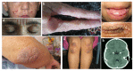
Oral and upper-airway manifestations. A hoarse voice, often evident at birth or in early infancy as a weak cry, is present in essentially all individuals with LP, usually persists lifelong, and can progress to severe dysphonia and/or complete aphonia [Savage et al 1988]. In addition, involvement of the mucosae of the pharynx, soft palate, tonsils, and lips may lead to pulmonary manifestations, especially upper-respiratory tract infection. In some individuals, infiltration of the laryngeal mucosa may lead to breathing difficulties.

Recurrent episodes of parotitis caused by stenosis of the parotid duct and submandibular gland inflammation are reported. Infiltration of the tongue may destroy the dorsal papillae, causing the tongue to have a smooth surface.

Dental health is often poor [Chan et al 2007, Ravi Prakash et al 2013].

Skin lesions consist of vesicles and hemorrhagic crusts in the mouth and on the face and extremities induced by minor trauma, verrucous and keratotic cutaneous lesions on extensor surfaces (especially the elbows), and moniliform blepharosis (multiple beaded papules along the eyelid margins and inner canthus). Skin lesions usually progress during the first few years of life in two overlapping stages:

  • During childhood the skin may be easily damaged by minor trauma or friction, resulting in blisters and scar formation. Pock-like or acneiform scars, vesicles, and hemorrhagic crust are particularly evident on the face and extremities (Figure1a).
  • At later stages with increasing hyaline-like deposition within the dermis, skin becomes diffusely thickened and appears waxy with yellowish discoloration; papules, nodules, and plaques appear on the face and the lips (Figure1d).

Hyperkeratotic and verrucous lesions may appear in regions exposed to mechanical trauma, such as the hands, elbows, knees, buttocks, and axillae (Figure 1b, 1f, 1g) [Hamada 2002].

Patchy and diffuse alopecia of the scalp, beard, eyelashes, and eyebrows may be present; however, alopecia is not a significant finding in most. Nail dystrophy has been reported [Hamada 2002].

Extracutaneous manifestations may include the following:

  • Epilepsy. Seizure onset may be in childhood or adulthood. Temporal lobe epilepsy is the most common type, with both partial and (less frequently) secondarily generalized seizures observed. Seizures may be resistant to therapy [Claeys et al 2007].
  • Neuropsychiatric disorders can include impairment of day-to-day memory (but not distant memory), paranoia, aggressive behavior and rage, hallucinations, absence of fear, and lack of normal sense of distrust or danger. Neuropsychiatric disorders can appear in childhood and are progressive. Individuals with lipoid proteinosis perform poorly on facial recognition of positive and negative emotions. These neuropsychiatric manifestations sometimes occur in association with calcification in the temporal lobes in the region of the amygdalae (Figure1h) [Siebert et al 2003, Thornton et al 2008].
  • Spontaneous CNS hemorrhage (including small deep brain hemorrhages and large brain hematomata) has been reported and can lead to hemiparesis and hemiplegia [Siebert et al 2003, Messina et al 2012, Teive et al 2013].
  • Gastrointestinal manifestations. Multiple yellowish nodules can be found throughout the esophagus, stomach, duodenum, and colon, and are usually asymptomatic [Custódio Lima et al 2014]; however, small intestinal bleeding has been observed in one individual [Caccamo et al 1994].

Clinical variability. A wide range of clinical manifestations and disease progression, including presence of neurologic abnormalities in the absence of skin manifestations, occurs in individuals with the same ECM1 pathogenic variants even within the same family or population isolate [Youssefian et al 2015].

Course and prognosis. Generally, LP follows a chronic and fluctuating course. Males and females are affected equally. Affected individuals have a normal life span unless they experience laryngeal obstruction.

Genotype-Phenotype Correlations

No ECM1 genotype-phenotype correlations have been identified [Hamada et al 2003].

Prevalence

More than 400 individuals with lipoid proteinosis (LP) have been reported worldwide [LeWitt et al 2021].

LP tends to be more common in countries with extensive consanguinity and/or in areas (e.g., South Africa) in which a founder variant has been postulated [Van Hougenhouck-Tulleken et al 2004, Chan et al 2007].

Differential Diagnosis

Table 3.

Genes and Disorders of Interest in the Differential Diagnosis of Lipoid Proteinosis

GeneDisorderMOIOverlapping FeaturesDistinguishing Features
ABCC6 Pseudoxanthoma elasticum (PXE)ARSome skin changes in LP (e.g., yellowish papules seen on the neck) are reminiscent of those in PXE.Ocular manifestations are different:
  • LP is characterized by moniliform blepharosis.
  • PXE is characterized by subretinal neovascularization w/hemorrhage that can cause significant visual impairment.
FECH Autosomal recessive erythropoietic protoporphyria (EPP)ARThe early vesicular lesions in LP can resemble those in EPP.
  • Hepatic dysfunction is rare in LP but may occur in 20%-30% of those w/EPP.
  • Hoarseness & moniliform blepharosis (features characteristic of LP) are not found in those w/EPP.

AR = autosomal recessive; LP = lipoid proteinosis; MOI = mode of inheritance

Management

No clinical practice guidelines for lipoid proteinosis (LP) have been published.

Evaluations Following Initial Diagnosis

To establish the extent of disease and needs in an individual diagnosed with LP, the evaluations summarized in Table 4 (if not performed as part of the evaluation that led to the diagnosis) are recommended.

Table 4.

Recommended Evaluations Following Initial Diagnosis in Individuals with Lipoid Proteinosis (LP)

System/ConcernEvaluationComment
Skin lesions DermatologistAssess for hemorrhagic or crusting lesions.
Upper airway OtolaryngologistAssess for airway obstruction assoc w/vocal cord infiltration.
Epilepsy NeurologistIf clinical findings suggest seizures
Neuropsychiatric
disorders
Neurobehavioral testingAssess for memory impairment, hallucinations, paranoia, & aggressive behavior.
Brain MRIAssess for calcification in temporal lobes in the region of the amygdalae.
Genetic
counseling
By genetics professionals 1To inform patients & families re nature, MOI, & implications of LP in order to facilitate medical & personal decision making
Family support
& resources
Assess:
1.

Medical geneticist, certified genetic counselor, certified advanced genetic nurse

Treatment of Manifestations

There is no curative therapy for LP.

Table 5.

Treatment of Manifestations in Individuals with Lipoid Proteinosis (LP)

Manifestation/ConcernTreatmentConsiderations/Other
Airway obstruction Microlaryngoscopic excision of laryngeal depositsImproves airway access & voice quality
TracheostomyFor significant airway obstruction
Cutaneous papules
& plaques
Oral dimethylsulfoxide, D-penicillamine, & oral retinoid (e.g., acitretin)For skin softening & concomitant amelioration of mucosal lesions & hoarseness
Seizures ASM as determined by neurologist
  • Some persons are resistant to ASMs.
  • Successful treatment w/ASMs incl Tegretol® & Keppra® has been reported [Omrani et al 2012].

ASM = anti-seizure medication

Surveillance

Table 6.

Recommended Surveillance for Individuals with Lipoid Proteinosis (LP)

System/ConcernEvaluationFrequency
Airway obstruction Otolaryngologist assessment of airway & vocal cordsEvery 6 mos
Cutaneous papules
& plaques
Dermatologic exams
Seizures Neurologic evalAnnually
Neuropsychiatric
disorders
Monitor for emotional & cognitive development.

Evaluation of Relatives at Risk

See Genetic Counseling for issues related to testing of at-risk relatives for genetic counseling purposes.

Therapies Under Investigation

Search ClinicalTrials.gov in the US and EU Clinical Trials Register in Europe for access to information on clinical studies for a wide range of diseases and conditions. Note: There may not be clinical trials for this disorder.

Genetic Counseling

Genetic counseling is the process of providing individuals and families with information on the nature, mode(s) of inheritance, and implications of genetic disorders to help them make informed medical and personal decisions. The following section deals with genetic risk assessment and the use of family history and genetic testing to clarify genetic status for family members; it is not meant to address all personal, cultural, or ethical issues that may arise or to substitute for consultation with a genetics professional. —ED.

Mode of Inheritance

Lipoid proteinosis (LP) is inherited in an autosomal recessive manner.

Risk to Family Members

Parents of a proband

  • The parents of an affected individual are obligate heterozygotes (i.e., presumed to be carriers of one ECM1 pathogenic variant based on family history).
  • If a molecular diagnosis has been established in the proband, molecular genetic testing is recommended for the parents of a proband to confirm that both parents are heterozygous for an ECM1 pathogenic variant and to allow reliable recurrence risk assessment. If a pathogenic variant is detected in only one parent and parental identity testing has confirmed biological maternity and paternity, the following possibilities should be considered:
  • Heterozygotes are typically asymptomatic but may have variable findings. Youssefian et al [2015] reported voice hoarseness in cold seasons and thickening of the frenulum and a firm tongue in heterozygous family members.

Sibs of a proband

  • If both parents are known to be heterozygous for an ECM1 pathogenic variant, each sib of an affected individual has at conception a 25% chance of inheriting biallelic pathogenic variants and being affected, a 50% chance of being an asymptomatic carrier, and a 25% chance of being unaffected and not a carrier.
  • A wide range of clinical manifestations and disease progression may be observed in sibs with the same ECM1 pathogenic variants [Youssefian et al 2015].
  • Heterozygotes are typically asymptomatic but may have variable findings. Youssefian et al [2015] reported voice hoarseness in cold seasons and thickening of the frenulum and a firm tongue in family members heterozygous for the c.507delT pathogenic variant in ECM1.

Offspring of a proband. Unless an individual with LP has children with an affected individual or a carrier, his/her offspring will be obligate heterozygotes (carriers) for a pathogenic variant in ECM1.

Other family members. Each sib of the proband's parents is at a 50% risk of being a carrier of an ECM1 pathogenic variant.

Carrier (Heterozygote) Detection

Carrier testing for at-risk relatives requires prior identification of the ECM1 pathogenic variants in the family.

Related Genetic Counseling Issues

Family planning

  • The optimal time for determination of genetic risk and discussion of the availability of prenatal/preimplantation genetic testing is before pregnancy.
  • It is appropriate to offer genetic counseling (including discussion of potential risks to offspring and reproductive options) to young adults who are affected, are carriers, or are at risk of being carriers.

DNA banking. Because it is likely that testing methodology and our understanding of genes, allelic variants, and diseases will improve in the future, consideration should be given to banking DNA from probands in whom a molecular diagnosis has not been confirmed (i.e., the causative genetic alteration/s are unknown).

Prenatal Testing and Preimplantation Genetic Testing

Once the ECM1 pathogenic variants have been identified in an affected family member, prenatal testing for a pregnancy at increased risk and preimplantation genetic testing for LP are possible.

Differences in perspective may exist among medical professionals and within families regarding the use of prenatal testing. While most centers would consider use of prenatal testing to be a personal decision, discussion of these issues may be helpful.

Resources

GeneReviews staff has selected the following disease-specific and/or umbrella support organizations and/or registries for the benefit of individuals with this disorder and their families. GeneReviews is not responsible for the information provided by other organizations. For information on selection criteria, click here.

Molecular Genetics

Information in the Molecular Genetics and OMIM tables may differ from that elsewhere in the GeneReview: tables may contain more recent information. —ED.

Table A.

Lipoid Proteinosis: Genes and Databases

GeneChromosome LocusProteinLocus-Specific DatabasesHGMDClinVar
ECM1 1q21​.2 Extracellular matrix protein 1 ECM1 database ECM1 ECM1

Data are compiled from the following standard references: gene from HGNC; chromosome locus from OMIM; protein from UniProt. For a description of databases (Locus Specific, HGMD, ClinVar) to which links are provided, click here.

Table B.

OMIM Entries for Lipoid Proteinosis (View All in OMIM)

247100LIPOID PROTEINOSIS OF URBACH AND WIETHE
602201EXTRACELLULAR MATRIX PROTEIN 1; ECM1

Molecular Pathogenesis

Lipoid proteinosis (LP) is characterized by deposition of hyaline-like material in the larynx, oral cavity, skin, and internal organs and caused by pathogenic variants in ECM1, which codes for extracellular matrix protein 1 (ECM1).

Three transcripts of ECM1 (ECM1a, ECM1b and ECM1c) in human have been discovered. This gene encodes an 85-kd soluble and widely expressed glycoprotein that is involved in endochondral bone formation, mineralization, angiogenesis, and epidermal differentiation. ECM1 is found within the epidermis and also as a secreted protein in the dermis. It interacts with a variety of extracellular and structural proteins, contributing to the maintenance of skin integrity and homeostasis. It has a physiologic role in the proliferation of endothelial cells and maintains skin integrity and homeostasis [Hamada et al 2003].

The ECM1 protein structure has four functional domains including a cysteine-free N-terminal segment, two tandem repeats, and a C-terminal segment. The tandem domains contain highly conserved sequence and numerous cysteine residues that are involved in protein-protein interactions and allow the ECM1 protein to function as a transport protein or to be involved in binding of growth or differentiation factors [Chan 2004]. It was shown by a yeast two-hybrid genetic system and confirmed by coimmunoprecipitations that ECM1 interacts with the C-terminal segments of fibulins 1C and 1D splice variants through its tandem repeat domain. It was proposed that alteration of this strong protein-protein interaction of ECM1/fibulin-1 is involved in LP disease pathogenesis [Fujimoto et al 2005]. Also, it has been shown that ECM1 inhibits the activity of MMP-9 through protein-protein interaction, and increased MMP-9 activity in individuals with LP may explain the hyaline changes of the dermis in these individuals [Fujimoto et al 2006].

Mechanism of disease causation. Lipoid proteinosis is caused by ECM1 loss-of-function variants, which may lead to absence of ECM1 protein or production of nonfunctional protein [Chan et al 2007].

Table 7.

Notable ECM1 Pathogenic Variants

Reference SequencesDNA Nucleotide ChangePredicted Protein ChangeComment [Reference]
NM_004425​.4
NP_004416​.2
c.826C>Tp.Gln276TerSouth African founder variant [Hamada 2002, Van Hougenhouck-Tulleken et al 2004]
c.742G>Tp.Glu248TerIn Pakistani families [Nasir et al 2014]
c.658T>Gp.Cys220GlyIn Chinese families [Zhang et al 2014]

Variants listed in the table have been provided by the authors. GeneReviews staff have not independently verified the classification of variants.

GeneReviews follows the standard naming conventions of the Human Genome Variation Society (varnomen​.hgvs.org). See Quick Reference for an explanation of nomenclature.

Cancer and Benign Tumors

No correlation between increased cancer incidence and lipoid proteinosis has been reported; however, evidence linking expression of ECM1 with aspects of malignancy has emerged as ECM1 is dysregulated in some carcinomas [Wang et al 2003, Gómez-Contreras et al 2017]. ECM1 has been shown to have an important role in growth, angiogenesis, metastasis, and epithelial-stromal interactions [Lee et al 2015b, Steinhaeuser et al 2020].

Chapter Notes

Author Notes

Jouni Uitto, MD, PhD, has been Professor of Dermatology and Cutaneous Biology, and Biochemistry and Molecular Biology, and Chair of the Department of Dermatology and Cutaneous Biology at The Sidney Kimmel Medical College at Thomas Jefferson University, in Philadelphia, Pennsylvania, since 1986. He is also Director of the Jefferson Institute of Molecular Medicine at Thomas Jefferson University. He received his MD and PhD degrees from the University of Helsinki, Finland, and completed his residency training in dermatology at Washington University School of Medicine, St Louis, Missouri. Dr Uitto is internationally recognized for his research on connective tissue biology and molecular genetics in relation to cutaneous diseases. Dr Uitto's publications include 745 original articles in peer-reviewed journals, 346 textbook chapters and review articles, and 1,043 abstracts on presentations in national and international meetings. Dr Uitto has been the recipient of numerous national and international awards, including honorary doctorate degrees from the University of Kuopio, University of Oulu, and University of Turku (all in Finland) as well as the University of Buenos Aires. He also holds honorary professorships at China Medical University, Shenyang, Hebei United University, Tangshan, and The Fourth Military Medical University, Xi'an, all in China. Dr Uitto has held office in several scientific and professional societies, including as President of the Society for Investigative Dermatology and President and Chairman of the Board of Trustees of Dermatology Foundation. Dr Uitto is also Section Editor of the Journal of Investigative Dermatology and Associate Editor of the American Journal of Pathology; he is on the editorial boards of numerous peer-reviewed journals. In 2021, he was bestowed the honor of The Knight of White Rose, First Order, Republic of Finland, in recognition of his contributions to science.

Hassan Vahidnezhad, PhD in medical genetics, has been a principal investigator and faculty member of the Departments of Dermatology and Cutaneous Biology, and Biochemistry and Molecular Biology of The Sidney Kimmel Medical College at Thomas Jefferson University, Philadelphia, Pennsylvania, since 2019.

Leila Youssefian, PhD in genetics, genomics and cancer biology, is a postdoctoral fellow working on genodermatoses in the department of Dermatology and Cutaneous Biology, Sidney Kimmel Medical College of Thomas Jefferson University, Philadelphia, Pennsylvania.

Leila Youssefian and Hassan Vahidnezhad contributed equally to this work.

Acknowledgments

Carol Kelly assisted in manuscript preparation.

Revision History

  • 22 July 2021 (ha) Comprehensive update posted live
  • 21 January 2016 (bp) Review posted live
  • 28 July 2015 (ju) Original submission

References

Literature Cited

  • Agredano PM, Del Barrio CM, Martinez MC, Cabrera CA. Intracranial calcifications associated with epilepsy: a case report of lipoid proteinosis. Seizure. 2020;83:172–4. [PubMed: 33161246]
  • An I, Güldür ME, Aksoy M, Yeşilova Y, Ozturk M. Histopathological findings in patients with lipoid proteinosis. Turk J Dermatol. 2019;13:99.
  • Belliveau MJ, Alkhotani A, Ali A. Moniliform blepharosis of lipoid proteinosis. JAMA Ophthalmol. 2015;133:e150688. [PubMed: 26158437]
  • Caccamo D, Jaen A, Telenta M, Varela E, Tiscornia O. Lipoid proteinosis of the small bowel. Arch Pathol Lab Med. 1994;118:572–4. [PubMed: 7514865]
  • Chan I. The role of extracellular matrix protein 1 in human skin. Clin Exp Dermatol. 2004;29:52–6. [PubMed: 14723723]
  • Chan I, Liu L, Hamada T, Sethuraman G, McGrath JA. The molecular basis of lipoid proteinosis: mutations in extracellular matrix protein 1. Exp Dermatol. 2007;16:881–90. [PubMed: 17927570]
  • Chan I, South AP, McGrath JA, Oyama N, Bhogal BS, Black MM, Hamada T. Rapid diagnosis of lipoid proteinosis using an anti-extracellular matrix protein 1 (ECM1) antibody. J Dermatol Sci. 2004;35:151–3. [PubMed: 15265527]
  • Claeys KG, Claes LR, Van Goethem JW, Sercu S, Merregaert J, Lambert J, Van Marck EA, Parizel PM, De Jonghe P. Epilepsy and migraine in a patient with Urbach-Wiethe disease. Seizure. 2007;16:465–8. [PubMed: 17403608]
  • Custódio Lima J, Nagasako CK, Montes CG, Barcelos IH, de Carvalho RB, Mesquita MA. Gastrointestinal involvement in lipoid proteinosis: a ten-year follow-up of a Brazilian female patient. Case Rep Med. 2014;2014:952038. [PMC free article: PMC4089944] [PubMed: 25045357]
  • Fujimoto N, Terlizzi J, Aho S, Brittingham R, Fertala A, Oyama N, McGrath JA, Uitto J. Extracellular matrix protein 1 inhibits the activity of matrix metalloproteinase 9 through high-affinity protein/protein interactions. Exp Dermatol. 2006;15:300–7. [PubMed: 16512877]
  • Fujimoto N, Terlizzi J, Brittingham R, Fertala A, McGrath JA, Uitto J. Extracellular matrix protein 1 interacts with the domain III of fibulin-1C and 1D variants through its central tandem repeat 2. Biochem Biophys Res Commun. 2005;333:1327–33. [PubMed: 15990087]
  • Gómez-Contreras P, Ramiro-Díaz JM, Sierra A, Stipp C, Domann FE, Weigel RJ, Lal G. Extracellular matrix 1 (ECM1) regulates the actin cytoskeletal architecture of aggressive breast cancer cells in part via S100A4 and Rho-family GTPases. Clin Exp Metastasis. 2017;34:37–49. [PMC free article: PMC5288287] [PubMed: 27770373]
  • Gonçalves FG, de Melo MB, del Matos V, Barra FR, Figueroa RE. Amygdalae and striatum calcification in lipoid proteinosis. AJNR Am J Neuroradiol. 2010;31:88–90. [PMC free article: PMC7964074] [PubMed: 19696137]
  • Hamada T, McLean WH, Ramsay M, Ashton GH, Nanda A, Jenkins T, Edelstein I, South AP, Bleck O, Wessagowit V, Mallipeddi R, Orchard GE, Wan H, Dopping-Hepenstal PJ, Mellerio JE, Whittock NV, Munro CS, van Steensel MA, Steijlen PM, Ni J, Zhang L, Hashimoto T, Eady RA, McGrath JA. Lipoid proteinosis maps to 1q21 and is caused by mutations in the extracellular matrix protein 1 gene (ECM1). Hum Mol Genet. 2002;11:833–40. [PubMed: 11929856]
  • Hamada T, Wessagowit V, South AP, Ashton GH, Chan I, Oyama N, Siriwattana A, Jewhasuchin P, Charuwichitratana S, Thappa DM, Jeevankumar B, Lenane P, Krafchik B, Kulthanan K, Shimizu H, Kaya TI, Erdal ME, Paradisi M, Paller AS, Seishima M, Hashimoto T, McGrath JA. Extracellular matrix protein 1 gene (ECM1) mutations in lipoid proteinosis and genotype-phenotype correlation. J Invest Dermatol. 2003;120:345–50. [PubMed: 12603844]
  • Hamada T. Lipoid proteinosis. Clin Exp Dermatol. 2002;27:624–9. [PubMed: 12472532]
  • Hameed A, Nasir M, Ajmal M, Latif L. Novel human pathological mutations. Gene symbol: ECM1. Disease: Lipoid Proteinosis. Hum Genet. 2009;126:336. [PubMed: 19694009]
  • Jónsson H, Sulem P, Kehr B, Kristmundsdottir S, Zink F, Hjartarson E, Hardarson MT, Hjorleifsson KE, Eggertsson HP, Gudjonsson SA, Ward LD, Arnadottir GA, Helgason EA, Helgason H, Gylfason A, Jonasdottir A, Jonasdottir A, Rafnar T, Frigge M, Stacey SN, Th Magnusson O, Thorsteinsdottir U, Masson G, Kong A, Halldorsson BV, Helgason A, Gudbjartsson DF, Stefansson K. Parental influence on human germline de novo mutations in 1,548 trios from Iceland. Nature. 2017;549:519–22. [PubMed: 28959963]
  • Lee KM, Nam K, Oh S, Lim J, Kim RK, Shim D, Choi JH, Lee SJ, Yu JH, Lee JW, Ahn SH, Shin I. ECM1 regulates tumor metastasis and CSC-like property through stabilization of β-catenin. Oncogene. 2015b;34:6055–65. [PubMed: 25746001]
  • Lee MY, Wang HJ, Han Y, Zhou Y, Zhao JH, Duo LN, Feng C, Hua H, Liu HW, Lin ZM, Yang Y. Lipoid proteinosis resulting from a large homozygous deletion affecting part of the ECM1 gene and adjacent long non-coding RNA. Acta Derm Venereol. 2015a;95:608–10. [PubMed: 25518807]
  • LeWitt TM, Paller AS, Bell A, Zhou X. Lipoid proteinosis. In: StatPearls. Treasure Island, FL: StatPearls Publishing. Available online. 2021. Accessed 7-15-21.
  • Messina MJ, Nuzzaco G, Barbieri A, Scarlato M, Gerevini S, Martinelli V, Comi G, Sessa M. Spontaneous intracerebral hemorrhage in Urbach-Wiethe disease. Neurology. 2012;79:1740–1. [PubMed: 23035073]
  • Nasir M, Latif A, Ajmal M, Ismail M, Hameed A. A novel homozygous 62-bp insertion in ECM1 causes lipoid proteinosis in a multigeneration Pakistani family. Br J Dermatol. 2009;161:688–90. [PubMed: 19519837]
  • Nasir M, Rahman SB, Sieber CM, Mir A, Latif A, Ahmad N, Malik SA, Hameed A. Identification of recurrent c. 742G> T nonsense mutation in ECM1 in Pakistani families suffering from lipoid proteinosis. Mol Biol Rep. 2014;41:2085–92. [PubMed: 24413997]
  • Omrani HG, Tajdini M, Ghelichnia B, Hosseini SM, Tafakhori A, Rahimian E, Aghamollaii V. Should we think of Urbach–Wiethe disease in refractory epilepsy? Case report and review of the literature. J Neurol Sci. 2012;320:149–52. [PubMed: 22795383]
  • Ravi Prakash S, Verma S, Sumalatha MN, Chattopadhyay S. Oral manifestations of lipoid proteinosis: a case report and literature review. Saudi Dent J. 2013;25:91–4. [PMC free article: PMC3723256] [PubMed: 23960561]
  • Savage MM, Crockett DM, McCabe BF. Lipoid proteinosis of the larynx: a cause of voice change in the infant and young child. Int J Pediatr Otorhinolaryngol. 1988;15:33–8. [PubMed: 3372140]
  • Siebert M, Markowitsch HJ, Bartel P. Amygdala, affect and cognition: evidence from 10 patients with Urbach-Wiethe disease. Brain. 2003;126:2627–37. [PubMed: 12937075]
  • Steinhaeuser SS, Morera E, Budkova Z, Schepsky A, Wang Q, Rolfsson O, Riedel A, Krueger A, Hilmarsdottir B, Maelandsmo GM, Valdimarsdottir B. ECM1 secreted by HER2-overexpressing breast cancer cells promotes formation of a vascular niche accelerating cancer cell migration and invasion. Lab Invest. 2020;100:928–44. [PubMed: 32203150]
  • Stenson PD, Mort M, Ball EV, Evans K, Hayden M, Heywood S, Hussain M, Phillips AD, Cooper DN. The Human Gene Mutation Database: towards a comprehensive repository of inherited mutation data for medical research, genetic diagnosis and next-generation sequencing studies. Hum Genet. 2017;136:665–77. [PMC free article: PMC5429360] [PubMed: 28349240]
  • Teive HA, Ruschel E, Munhoz RP. Spontaneous intracerebral hemorrhage in Urbach-Wiethe disease. Neurology. 2013;80:1720–1. [PubMed: 23628933]
  • Thornton HB, Nel D, Thornton D, van Honk J, Baker GA, Stein DJ. The neuropsychiatry and neuropsychology of lipoid proteinosis. J Neuropsychiatry Clin Neurosci. 2008;20:86–92. [PubMed: 18305289]
  • Vahidnezhad H, Youssefian L, Tafakhori A, Li Q, Uitto J, Rajabpour FV, Pishnamazi M, Modabbernia A, Tabrizi M. Lipoid proteinosis due to homozygous deletion mutation (c. 735delTG) in the ECM1 gene presents with seizures and hoarseness but no skin involvement. Int J Dermatol Venereol. 2020;3:43–5.
  • Van Hougenhouck-Tulleken W, Chan I, Hamada T, Thornton H, Jenkins T, McLean WH, McGrath JA, Ramsay M. Clinical and molecular characterization of lipoid proteinosis in Namaqualand, South Africa. Br J Dermatol. 2004;151:413–23. [PubMed: 15327549]
  • Wang L, Yu J, Ni J, Xu XM, Wang J, Ning H, Pei XF, Chen J, Yang S, Underhill CB, Liu L, Liekens J, Merregaert J, Zhang L. Extracellular matrix protein 1 (ECM1) is over-expressed in malignant epithelial tumors. Cancer Lett. 2003;200:57–67. [PubMed: 14550953]
  • Youssefian L, Vahidnezhad H, Daneshpazhooh M, Abdollahzadeh S, Talari H, Khoshnevisan A, Chams-Davatchi C, Mobasher R, Li Q, Uitto J, Akhondzadeh S, Tabrizi M. Lipoid proteinosis: phenotypic heterogeneity in Iranian families with c.507delT mutation in ECM1. Exp Dermatol. 2015;24:220–2. [PubMed: 25529926]
  • Zhang R, Liu Y, Xue Y, Wang Y, Wang X, Shi S. Cai T1, Wang Q. Treatment of lipoid proteinosis due to the p.C220G mutation in ECM1, a major allele in Chinese patients. J Transl Med. 2014;12:85. [PMC free article: PMC4021827] [PubMed: 24708644]
Copyright © 1993-2024, University of Washington, Seattle. GeneReviews is a registered trademark of the University of Washington, Seattle. All rights reserved.

GeneReviews® chapters are owned by the University of Washington. Permission is hereby granted to reproduce, distribute, and translate copies of content materials for noncommercial research purposes only, provided that (i) credit for source (http://www.genereviews.org/) and copyright (© 1993-2024 University of Washington) are included with each copy; (ii) a link to the original material is provided whenever the material is published elsewhere on the Web; and (iii) reproducers, distributors, and/or translators comply with the GeneReviews® Copyright Notice and Usage Disclaimer. No further modifications are allowed. For clarity, excerpts of GeneReviews chapters for use in lab reports and clinic notes are a permitted use.

For more information, see the GeneReviews® Copyright Notice and Usage Disclaimer.

For questions regarding permissions or whether a specified use is allowed, contact: ude.wu@tssamda.

Bookshelf ID: NBK338540PMID: 26803878

Views

Tests in GTR by Gene

Related information

  • OMIM
    Related OMIM records
  • PMC
    PubMed Central citations
  • PubMed
    Links to PubMed
  • Gene
    Locus Links

Similar articles in PubMed

See reviews...See all...

Recent Activity

Your browsing activity is empty.

Activity recording is turned off.

Turn recording back on

See more...