U.S. flag

An official website of the United States government

NCBI Bookshelf. A service of the National Library of Medicine, National Institutes of Health.

Adam MP, Feldman J, Mirzaa GM, et al., editors. GeneReviews® [Internet]. Seattle (WA): University of Washington, Seattle; 1993-2024.

Cover of GeneReviews®

GeneReviews® [Internet].

Show details

IMAGe Syndrome

Synonym: Intrauterine Growth Restriction, Metaphyseal Dysplasia, Adrenal Hypoplasia Congenita, and Genital Anomalies

, MD and , MD, PhD.

Author Information and Affiliations

Initial Posting: ; Last Update: August 5, 2021.

Estimated reading time: 24 minutes

Summary

Clinical characteristics.

IMAGe syndrome is an acronym for the major findings of intrauterine growth restriction (IUGR), metaphyseal dysplasia, adrenal hypoplasia congenita, and genitourinary abnormalities (in males). Findings reported in individuals with a clinical and/or molecular diagnosis include:

  • IUGR;
  • Some type of skeletal abnormality (most commonly delayed bone age and short stature, and occasionally, metaphyseal and epiphyseal dysplasia of varying severity);
  • Adrenal insufficiency often presenting in the first month of life as an adrenal crisis or (rarely) later in childhood with failure to thrive and recurrent vomiting;
  • Genital abnormalities in males (cryptorchidism, micropenis, and hypospadias) but not in females.

Hypotonia and developmental delay are reported in some individuals; cognitive outcome appears to be normal in the majority of individuals.

Diagnosis/testing.

The diagnosis of IMAGe syndrome is established in a proband with suggestive findings and/or a heterozygous CDKN1C pathogenic variant in the PCNA (proliferating cell nuclear antigen)-binding domain of the maternally expressed allele identified by molecular genetic testing.

Management.

Treatment of manifestations: Management of adrenal insufficiency in IMAGe syndrome is similar to management of adrenal insufficiency from other causes and should be under the supervision of an endocrinologist. Chronic treatment includes replacement doses of glucocorticoids and mineralocorticoids and oral sodium chloride supplements. Steroid doses should be optimized to allow for linear growth without risking an adrenal crisis. Consider assessment for growth hormone deficiency to determine if growth hormone should be considered. Routine management of cryptorchidism and hypospadias by a urologist, and routine hormone replacement by an endocrinologist for hypogonadotropic hypogonadism. Management by an orthopedist as needed for skeletal complications such as scoliosis and hip dysplasia. Occupational, speech, and/or physical therapy as needed, particularly in those with hypotonia.

Surveillance: Growth assessment at each visit; annual evaluations by an endocrinologist to monitor adrenal function and for development of hypercalciuria and nephrocalcinosis; evaluation as needed by an orthopedist to monitor for skeletal complications as needed; assessment of hypotonia, developmental progress, and educational needs at each visit.

Evaluation of relatives at risk: To allow early diagnosis and management of adrenal insufficiency in at-risk newborns, molecular genetic testing should be pursued if the CDKN1C pathogenic variant in the family is known; if the familial pathogenic variant is not known, screen for serum electrolyte abnormalities, elevated serum ACTH level, and skeletal features of IMAGe syndrome.

Pregnancy management: Risks to a mother with IMAGe syndrome during pregnancy include possible adrenal insufficiency; risks during delivery include cephalopelvic disproportion.

Genetic counseling.

Typically, a CDKN1C pathogenic variant causing IMAGe syndrome is inherited in an autosomal dominant manner; however, only maternal transmission of the pathogenic variant results in IMAGe syndrome. Each child of a woman with a heterozygous pathogenic CDKN1C variant has a 50% chance of inheriting the variant and being affected. Each child of a man with a heterozygous pathogenic CDKN1C variant has a 50% chance of inheriting the variant but is expected to be unaffected. If the pathogenic variant has been identified in an affected family member, prenatal testing is possible for a pregnancy at increased risk (i.e., when the mother has the pathogenic variant).

Diagnosis

IMAGe syndrome is an acronym for the major findings in this disorder: intrauterine growth restriction, metaphyseal dysplasia, adrenal hypoplasia congenita, and genitourinary abnormalities (in males) [Vilain et al 1999].

No formal clinical diagnostic criteria for IMAGe syndrome have been defined.

Suggestive Findings

IMAGe syndrome should be suspected in individuals with the following clinical, imaging, and suggestive laboratory findings.

Clinical findings

  • Intrauterine growth restriction (IUGR) *
  • Postnatal growth deficiency, with variable growth hormone deficiency
  • Adrenal hypoplasia congenita (AHC)*, often presenting as spontaneous adrenal crisis in the first week to month of life, with hypotension, hyponatremia, and hyperkalemia, which can be life-threatening
    • Some individuals with AHC may present with adrenal insufficiency during childhood or early adulthood.
    • Later-onset adrenal insufficiency can be precipitated by stress, such as that associated with illness or surgery.
  • Genital abnormalities in males including unilateral or bilateral cryptorchidism, hypospadias, micropenis, and chordee

* Note: The clinical features of IUGR and AHC, with or without a family history of IMAGe syndrome, are highly suggestive of the diagnosis.

Imaging findings

  • Metaphyseal and/or epiphyseal dysplasia, mesomelia, osteopenia, gracile long bones, and delayed bone age on radiographs
    Note: Skeletal abnormalities, which are age dependent, can be absent or subtle.
  • Adrenal imaging that suggests small or normal-sized adrenal glands, in contrast to enlarged adrenal glands seen in individuals with congenital adrenal hyperplasia (CAH)

Suggestive laboratory findings

  • Evidence of adrenal insufficiency during a crisis including hyponatremia, hyperkalemia, and elevated ACTH levels (frequently >1000 pg/mL [normal: 10-60 pg/mL])
  • Lack of findings consistent with other causes of adrenal insufficiency

Establishing the Diagnosis

The diagnosis of IMAGe syndrome is established in a proband with suggestive findings and/or a heterozygous CDKN1C pathogenic variant in the PCNA (proliferating cell nuclear antigen)-binding domain of the maternally expressed allele identified by molecular genetic testing (see Table 1). Identification of a heterozygous CDKN1C variant of uncertain significance does not establish or rule out the diagnosis of this disorder.

Molecular genetic testing approaches can include a combination of gene-targeted testing (single-gene testing, multigene panel) and comprehensive genomic testing (exome sequencing, genome sequencing) depending on the phenotype.

When the clinical, radiographic, and laboratory findings suggest the diagnosis of IMAGe syndrome, molecular genetic testing approaches can include single-gene testing or use of a multigene panel (see Option 1).

Option 1

Single-gene testing. Sequence analysis of CDKN1C is performed first to detect small intragenic deletions/insertions and missense, nonsense, and splice site variants. To date, deletions and duplications in CDKN1C have not been reported to cause IMAGe syndrome.

A multigene panel that includes CDKN1C and other genes of interest (see Differential Diagnosis) may also be considered. Note: (1) The genes included in the panel and the diagnostic sensitivity of the testing used for each gene vary by laboratory and are likely to change over time. (2) Some multigene panels may include genes not associated with the condition discussed in this GeneReview; thus, clinicians need to determine which multigene panel is most likely to identify the genetic cause of the condition while limiting identification of variants of uncertain significance and pathogenic variants in genes that do not explain the underlying phenotype. (3) In some laboratories, panel options may include a custom laboratory-designed panel and/or custom phenotype-focused exome analysis that includes genes specified by the clinician. (4) Methods used in a panel may include sequence analysis, deletion/duplication analysis, and/or other non-sequencing-based tests.

For an introduction to multigene panels click here. More detailed information for clinicians ordering genetic tests can be found here.

Option 2

When the phenotype is indistinguishable from many other inherited disorders characterized by growth deficiency and adrenal hypoplasia, comprehensive genomic testing, which does not require the clinician to determine which gene is likely involved, is the best option. Exome sequencing is most commonly used; genome sequencing is also possible.

For an introduction to comprehensive genomic testing click here. More detailed information for clinicians ordering genomic testing can be found here.

Table 1.

Molecular Genetic Testing Used in IMAGe Syndrome

Gene 1MethodProportion of Probands with a Pathogenic Variant 2 Detectable by Method
CDKN1C Sequence analysis 311/11 families 4
Gene-targeted deletion/duplication analysis 5See footnote 6.
1.
2.

See Molecular Genetics for information on variants detected in this gene.

3.

Sequence analysis detects variants that are benign, likely benign, of uncertain significance, likely pathogenic, or pathogenic. Variants may include small intragenic deletions/insertions and missense, nonsense, and splice site variants; typically, exon or whole-gene deletions/duplications are not detected. For issues to consider in interpretation of sequence analysis results, click here.

4.

At present all six reported pathogenic variants in CDKN1C in persons with IMAGe syndrome have been located within an eight-amino-acid region of the PCNA-binding domain (nucleotides c.814 to c.836, based on NM_000076​.2) [Arboleda et al 2012, Hamajima et al 2013, Bolomiti et al 2021].

5.

Gene-targeted deletion/duplication analysis detects intragenic deletions or duplications. Methods used may include a range of techniques such as quantitative PCR, long-range PCR, multiplex ligation-dependent probe amplification (MLPA), and a gene-targeted microarray designed to detect single-exon deletions or duplications.

6.

No deletions or duplications of CDKN1C have been reported or are anticipated in persons with IMAGe syndrome due to the gain-of-function disease mechanism.

Clinical Characteristics

Clinical Description

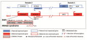
Thirty-one individuals reported from 19 families have features consistent with the clinical diagnosis of IMAGe syndrome [Vilain et al 1999, Lienhardt et al 2002, Pedreira et al 2004, Bergadá et al 2005, Hutz et al 2006, Tan et al 2006, Ko et al 2007, Amano et al 2008, Balasubramanian et al 2010, Arboleda et al 2012, Hamajima et al 2013, Bodian et al 2014, Kato et al 2014, Bolomiti et al 2021, Çamtosun et al 2021]. Of these 31 individuals, 19 from eleven unrelated families have had the diagnosis confirmed molecularly [Arboleda et al 2012, Hamajima et al 2013, Bolomiti et al 2021]. Of the twelve individuals who have not had confirmatory genetic testing, nearly all have clinical findings that significantly overlap those of the individuals with a molecularly confirmed diagnosis.

A diagnosis of IMAGe syndrome has been considered in other published cases; however, the clinical information was either significantly different from the 31 typical cases or insufficient to determine the diagnosis with certainty, and pathogenic CDKN1C variants were not reported [Blethen et al 1990, Hall & Stelling 1991, Le & Kutteh 1996, Coman et al 2007, McDonald et al 2010, Lindemeyer et al 2014]. Several of these cases are further discussed in Differential Diagnosis.

It is likely that the spectrum and natural history of IMAGe syndrome will be refined as more affected individuals are identified.

Onset. Although intrauterine growth restriction (IUGR) may be detected prenatally, IMAGe syndrome is typically evident at birth with IUGR, mild dysmorphic features, adrenal insufficiency, and (in males) genitourinary abnormalities. Of 23 individuals with information about age and findings at presentation, one was identified prenatally, 13 at birth, four in the first month of life, three from age one month to one year, one at age five years, and one at age 15 years.

Growth. All neonates have had IUGR with birth weights from -2 to -4 SD and birth lengths from -1.8 to -4.5 SD. Of the nine for whom information was available at birth, five had a normal occipitofrontal circumference (OFC) and four had an OFC from -2 to -3 SD.

Individuals with IMAGe syndrome consistently demonstrate continued short stature (height -2.7 to -6.5 SD) and postnatal failure to thrive (weight -2 to -7 SD). Of the nine on whom longitudinal information was available, postnatal OFC was normal in seven and below -2 SD in two individuals.

Skeletal. All affected individuals have had some skeletal abnormality with the most common manifestations being delayed bone age and short stature. Metaphyseal and epiphyseal dysplasia of the long bones are common. The metaphyses are frequently described as striated and the diaphyses as gracile.

A significant degree of age-dependent variation is observed: in some, the metaphyseal dysplasia may be late, mild, and/or easily missed. Most children have radiologic evidence of skeletal abnormality by age five years.

Less common skeletal features include progressive and severe scoliosis with onset before age five years, ovoid-shaped vertebral bodies, short first metatarsals, hallux valgus, and hip dysplasia.

In one individual, fractures of the humerus and tibia were present at birth [Lienhardt et al 2002].

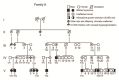
Adrenal insufficiency appears to be universal. Although this may be due to an ascertainment bias for probands, in one family it appeared fully penetrant, with adrenal insufficiency present in 7/7 individuals who possessed a maternally inherited CDKN1C pathogenic variant [Arboleda et al 2012].

Adrenal crisis, presenting with hyponatremia, hyperkalemia, and life-threatening hypotension, can occur within the first month of life, typically within the first week; extremely elevated ACTH levels, frequently above 1000 pg/mL (normal 10-60 pg/mL), can cause severe hyperpigmentation in these infants [Vilain et al 1999].

A few individuals do not have adrenal crisis, but rather milder adrenal insufficiency, presenting with failure to thrive and recurrent vomiting. One child, who experienced recurrent vomiting associated with mild infections from birth, was diagnosed with adrenal insufficiency at age five years following the diagnosis of IMAGe syndrome in her younger brother [Lienhardt et al 2002]. Another affected individual was diagnosed with hypoaldosteronism without glucocorticoid deficiency [Bodian et al 2014]. Hypercalciuria, soft tissue calcification, and renal calculi were noted in one individual [Authors, unpublished observation].

Genitourinary abnormalities. Genital abnormalities, which are nearly universal in males with IMAGe syndrome, have not been reported in females. Reported abnormalities include cryptorchidism (usually bilateral), micropenis, and hypospadias.

Of the 31 individuals reported with IMAGe syndrome 22 are male, which may represent ascertainment bias due to the presence of genital abnormalities in males only. Two females with IMAGe syndrome have had children [Authors, unpublished observations]. No males with IMAGe syndrome are known to have reproduced.

Hydronephrosis has been reported; however, the majority of affected individuals are reported to have normal renal ultrasound examinations.

Neurologic. Developmental outcome is believed to be normal, as 15 of the 16 individuals in whom cognitive outcome was mentioned were reported as normal; the oldest reported was age 26 years.

Hypotonia was reported in six individuals and noted to be absent in four others; some of those reported with developmental delay likely had motor delays secondary to hypotonia.

In one affected individual who had wasting of facial and distal muscles, muscle biopsy showed nonspecific myopathic changes [Lienhardt et al 2002].

Of 31 individuals with IMAGe syndrome, head imaging was reported for seven (3 via cranial ultrasound examination, 1 via head CT, and 3 via brain MRI), all were normal.

Other

  • Characteristic facial features that have been reported in the vast majority of individuals include frontal bossing, depressed or wide nasal bridge, and small/low-set ears; features are typically appreciated by age one year. The facial profile can be similar to the "triangular" facies seen in Silver-Russell syndrome. The facial profile may change with time. Micrognathia or retrognathia are also frequently reported. Less common features include cleft palate or cleft uvula, craniosynostosis, short palpebral fissures, smooth philtrum, and microglossia.
  • One individual has been reported with rhabdomyosarcoma [Bolomiti et al 2021].
  • Variable hypercalcemia of unclear etiology, occasionally with evidence of soft tissue calcifications, was reported in eight of 16 affected individuals on whom information was available. Several individuals have had nephrocalcinosis-associated hypercalciuria. While hypercalcemia may be a feature of IMAGe syndrome, it may also be secondary to sodium chloride supplementation, which is part of the treatment of the mineralocorticoid deficiency associated with adrenal insufficiency [Bergadá et al 2005].

Genotype-Phenotype Correlations

Currently, no genotype-phenotype correlations are known.

Penetrance

Although few large pedigrees with IMAGe syndrome have been reported to date, it is clear that the mode of inheritance is autosomal dominant in which only maternal transmission of the imprinted pathogenic variant results in IMAGe syndrome [Arboleda et al 2012].

In one large family of 24 individuals, all seven individuals with IMAGe syndrome inherited the CDKN1C pathogenic variant from their mother. Consistent with the imprinted expression of CDKN1C, unaffected individuals either inherited the pathogenic variant from their father (n=9) or did not have the pathogenic variant (n=8) [Bergadá et al 2005, Arboleda et al 2012].

Nomenclature

Prior to the identification of its molecular basis [Arboleda et al 2012], IMAGe syndrome was referred to as IMAGe association [Vilain et al 1999].

Historically the term "intrauterine growth retardation" was used. More recently, the term "intrauterine growth restriction" has come into favor.

Prevalence

The prevalence of IMAGe syndrome is currently unknown. A total of 31 affected individuals from 19 families have been reported to date.

Differential Diagnosis

Adrenal Insufficiency

Table 2.

Disorders with Adrenal Insufficiency in the Differential Diagnosis of IMAGe Syndrome

Gene(s) / Genetic MechanismDiffDx DisorderMOIClinical Features of DiffDx Disorder
Overlapping w/
IMAGe Syndrome
Distinguishing from IMAGe Syndrome
CYP11B1
CYP17A1
CYP21A2
HSD3B2
Congenital adrenal hyperplasia resulting from: 11-beta hydroxylase deficiency (OMIM 202010); 17-alpha hydroxylase deficiency (OMIM 202110); 21-hydroxylase-deficiency; or 3-beta hydroxysteroid dehydrogenase deficiency (OMIM 201810)ARAmbiguous genitalia at birth, adrenal crisis in infancy or childhood
  • Infants w/CAH rarely have IUGR.
  • Genital abnormalities in CAH: typically virilization of females (vs undervirilization of males incl cryptorchidism & hypospadias in IMAGe syndrome)
  • Adrenal glands in CAH: hypertrophic (vs hypoplastic in IMAGe syndrome).
  • Note: CAH is far more common than IMAGe syndrome.
NR0B1

Xp21 deletion 1

NR0B1-related adrenal hypoplasia congenita (incl XL AHC & Xp21 deletion)XLIn newborn period, phenotypic features in males w/XL AHC can overlap w/males w/IMAGe syndrome, as both conditions incl adrenal insufficiency, adrenal hypoplasia, & cryptorchidism.Males w/XL AHC typically do not have growth restriction, metaphyseal dysplasia, or facial features of IMAGe syndrome.
POLE IMAGe-I (OMIM 618336)ARIUGR, metaphyseal dysplasia, adrenal hypoplasia congenita, genital anomaliesImmunodeficiency, microcephaly, distinct facial features (long thin nose, small low-set posteriorly rotated ears, crowded dentition, micrognathia), & short wide neck
POR Antley-Bixler syndrome w/genital anomalies & disordered steroidogenesis (See Cytochrome P450 Oxidoreductase Deficiency.)ARGrowth deficiency, skeletal anomalies, genital abnormalities, & adrenal crisesPersons w/ABS w/genital anomalies & disordered steroidogenesis typically present w/craniosynostosis & often have radiohumeral synostosis, midface retrusion, choanal stenosis or atresia, & multiple joint contractures.
SAMD9 MIRAGE syndrome ADAdrenal hypoplasia, growth restriction, genital phenotypesMyelodysplasia, enteropathy; often fatal w/in 1st decade of life due to infection

ABS = Antley-Bixler syndrome; AD = autosomal dominant; AHC = adrenal hypoplasia congenita; AR = autosomal recessive; CAH = congenital adrenal hyperplasia; DiffDx = differential diagnosis; IUGR = intrauterine growth restriction; MOI = mode of inheritance; XL = X-linked

1.

Non-recurrent Xp21 deletion that includes NR0B1

Disorders of Growth

Frequently children with IMAGe syndrome have short stature with a normal head size and normal cognitive development. Other disorders of growth with these features should be considered (see Table 3).

Table 3.

Disorders of Growth in the Differential Diagnosis of IMAGe Syndrome

Gene(s) / Genetic MechanismDiffDx DisorderMOIClinical Features of DiffDx Disorder
Overlapping w/
IMAGe syndrome
Distinguishing from IMAGe syndrome
Genetically heterogeneous 1Silver-Russell syndrome (SRS)See footnote 2.IUGR, genital abnormalities, frontal bossing, & nl head sizePersons w/SRS typically display 5th digit clinodactyly, limb-length asymmetry, & café au lait macules. Growth velocity is nl in children w/SRS.
CCDC8
CUL7
OBSL1
3-M syndrome ARSevere pre- & postnatal growth deficiency (final height -5-6 SD) & nl intelligence. Males w/3-M have hypogonadism & occasionally hypospadias.Dolichocephaly, prominent heels. Facial features of 3M: typically full brows, downturned corners of mouth, hypotonic appearance

AR = autosomal recessive; DiffDx = differential diagnosis; IUGR = intrauterine growth restriction; MOI = mode of inheritance; nl = normal

1.

Genetic testing confirms clinical diagnosis in ~60% of affected individuals. Hypomethylation of the imprinted control region 1 at 11p15.5 causes SRS in 35%-50% of individuals, and maternal uniparental disomy causes SRS in 7%-10% of individuals. There are a small number of individuals with SRS who have duplications, deletions or translocations involving the imprinting centers at 11p15.5 or duplications, deletions, or translocations involving chromosome 7. Rarely, affected individuals with pathogenic variants in CDKN1C (see Genetically Related Disorders), IGF2, PLAG1, and HMGA2 have been described.

2.

SRS has multiple etiologies and typically has a low recurrence risk. In most families, a proband with SRS represents a simplex case (a single affected family member) and has SRS as the result of an apparent de novo epigenetic or genetic alteration.

Management

Evaluations Following Initial Diagnosis

To establish the extent of disease and needs in an individual diagnosed with IMAGe syndrome, the evaluations summarized in Table 4 (if not performed as part of the evaluation that led to the diagnosis) are recommended.

Table 4.

Recommended Evaluations Following Initial Diagnosis in Individuals with IMAGe Syndrome

System/ConcernEvaluationComment
Endocrine

Consultation w/endocrinologist for adrenal insufficiency, typically:

  • Serum & urine concentration of electrolytes, incl calcium
  • Serum concentration of glucose & ACTH
  • Assessment of arterial blood gases
  • Growth assessment
  • Assessment for hypogonadotropic hypogonadism in males
Of note, patients who are in shock often have hyponatremia, hyperkalemia, hypoglycemia, acidosis, markedly ↑ serum ACTH, & ↑ urinary excretion of sodium.
Genitourinary Urogenital eval & consultation w/urologistTo determine mgmt of undescended testicles &/or genital surgery
Musculoskeletal Clinical orthopedic assessment for hip dysplasia &/or scoliosis
Developmental
assessment
Assess for delays assoc w/hypotonia &/or other developmental concerns
Genetic
counseling
By genetics professionals 1To inform patients & their families re nature, MOI, & implications of IMAGe syndrome in order to facilitate medical & personal decision making
Family support
& resources
Assess:
1.

Medical geneticist, certified genetic counselor, certified advanced genetic nurse

Treatment of Manifestations

Adrenal insufficiency. Management of adrenal insufficiency in IMAGe syndrome is similar to management of adrenal insufficiency from other causes and should be under the supervision of an endocrinologist.

Episodes of acute adrenal insufficiency require close monitoring of blood pressure, hydration, clinical status, and serum concentration of glucose and electrolytes. Treatment with IV saline, glucose, and cortisol are utilized. If the serum electrolytes do not improve, a mineralocorticoid (fludrocortisone) is added or the dose of cortisol is increased.

Once the acute episode has been managed, replacement doses of glucocorticoids and mineralocorticoids and oral supplements of sodium chloride are given. Steroid dosages must be increased with stress, such as that associated with intercurrent illness, surgery, or trauma. Steroid doses need to be managed to enable optimal linear growth without risking an adrenal crisis.

Vigilance for proactive medical management and early recognition of adrenal insufficiency is required during illnesses and surgeries to prevent adrenal crisis.

The wearing of a Medic Alert® bracelet is strongly recommended.

Growth hormone (GH) therapy. Although information regarding anticipated adult height in IMAGe syndrome is limited, assessment for GH deficiency should be considered, as one child showed poor GH response to glucagon stimulation [Pedreira et al 2004] and other children with normal GH secretion have demonstrated a response in growth with growth hormone therapy [Lienhardt et al 2002, Kato et al 2014].

Genitourinary abnormalities. Routine surgical management of cryptorchidism and hypospadias by a urologist is indicated.

Males with hypogonadotropic hypogonadism are likely to need increasing doses of testosterone to induce puberty. Long-term adrenal steroid replacement and testosterone replacement should be managed by an experienced endocrinologist.

Orthopedic intervention as necessary for skeletal complications including scoliosis and hip dysplasia is appropriate.

Occupational, speech, or physical therapy as needed in those with hypotonia and developmental delays is appropriate.

Surveillance

Table 5.

Recommended Surveillance for Individuals with IMAGe Syndrome

System/ConcernEvaluationFrequency
Growth Measurement of growth parametersAt each visit
Endocrine Endocrinologist evals for adrenal insufficiency & potentially for hypercalciuria & nephrocalcinosisAnnually & more often as needed
Skeletal
complications
Orthopedist eval to monitor scoliosis, tibial/femoral bowing, or pain assoc w/skeletal dysplasiaAs needed
Development
  • Neurologic assessment for hypotonia
  • Monitor developmental progress & educational needs.
At each visit

Evaluation of Relatives at Risk

If prenatal testing for IMAGe syndrome has not been performed, it is appropriate to evaluate newborn sibs of a proband to enable prompt diagnosis and management of adrenal insufficiency.

  • Molecular genetic testing is indicated if the CDKN1C pathogenic variant in the family is known.
  • If the CDKN1C pathogenic variant in the family is not known, family members can be screened for serum electrolyte abnormalities, elevated serum ACTH level, and skeletal features of IMAGe syndrome including short stature and delayed bone age.

See Genetic Counseling for issues related to testing of at-risk relatives for genetic counseling purposes.

Pregnancy Management

Pregnant women with IMAGe syndrome are at increased risk for adrenal insufficiency and should be followed by an endocrinologist. Risks during delivery include cephalopelvic disproportion.

Therapies Under Investigation

Search ClinicalTrials.gov in the US and EU Clinical Trials Register in Europe for access to information on clinical studies for a wide range of diseases and conditions. Note: There may not be clinical trials for this disorder.

Genetic Counseling

Genetic counseling is the process of providing individuals and families with information on the nature, mode(s) of inheritance, and implications of genetic disorders to help them make informed medical and personal decisions. The following section deals with genetic risk assessment and the use of family history and genetic testing to clarify genetic status for family members; it is not meant to address all personal, cultural, or ethical issues that may arise or to substitute for consultation with a genetics professional. —ED.

Mode of Inheritance

The CDKN1C pathogenic variant is inherited in an autosomal dominant maternally imprinted manner. In any given affected individual, a pathogenic variant that causes IMAGe syndrome can either be inherited from the mother or arise de novo on the maternally derived CDKN1C allele [Arboleda et al 2012]. See Figure 2.

Figure 2. . Pedigree of a family demonstrating autosomal dominant maternally imprinted inheritance of IMAGe syndrome.

Figure 2.

Pedigree of a family demonstrating autosomal dominant maternally imprinted inheritance of IMAGe syndrome. Individuals labeled T/G are heterozygous for the normal "T" allele and the "G" pathogenic variant (CDKN1C variant c.826T>G); individuals (more...)

Risk to Family Members

Parents of a proband

Sibs of a proband. The risk to the sibs of the proband depends on the genetic status of the proband’s mother:

  • If the mother of the proband has the CDKN1C pathogenic variant, the risk to the sibs of inheriting the CDKN1C pathogenic variant and being affected with IMAGe syndrome is 50%.
  • If the CDKN1C pathogenic variant found in the proband cannot be detected in maternal leukocyte DNA, the risk to sibs is slightly greater than that of the general population (though still <1%) because of the theoretic possibility of maternal germline mosaicism.

Offspring of a proband

  • Each child of a woman with IMAGe syndrome has a 50% chance of inheriting the CDKN1C pathogenic variant and being affected.
  • Each child of a man with IMAGe syndrome has a 50% chance of inheriting the CDKN1C pathogenic variant but is expected to be unaffected.

Other family members. The risk to other family members depends on the status of the proband's mother: if the proband’s mother has the CDKN1C pathogenic variant, her family members may be at risk.

Related Genetic Counseling Issues

See Management, Evaluation of Relatives at Risk for information on evaluating at-risk relatives for the purpose of early diagnosis and treatment.

Family planning

  • The optimal time for determination of genetic risk and discussion of the availability of prenatal/preimplantation genetic testing is before pregnancy.
  • It is appropriate to offer genetic counseling (including discussion of potential risks to offspring and reproductive options) to young adults who are affected or at risk of having a child with IMAGe syndrome.

DNA banking. Because it is likely that testing methodology and our understanding of genes, pathogenic mechanisms, and diseases will improve in the future, consideration should be given to banking DNA from probands in whom a molecular diagnosis has not been confirmed (i.e., the causative pathogenic mechanism is unknown).

Prenatal Testing and Preimplantation Genetic Testing

Once the CDKN1C pathogenic variant has been identified in an affected family member, prenatal testing for a pregnancy at increased risk (i.e., when the mother has the pathogenic variant) and preimplantation genetic testing are possible.

Differences in perspective may exist among medical professionals and within families regarding the use of prenatal testing. While most centers would consider use of prenatal testing to be a personal decision, discussion of these issues may be helpful.

Resources

GeneReviews staff has selected the following disease-specific and/or umbrella support organizations and/or registries for the benefit of individuals with this disorder and their families. GeneReviews is not responsible for the information provided by other organizations. For information on selection criteria, click here.

Molecular Genetics

Information in the Molecular Genetics and OMIM tables may differ from that elsewhere in the GeneReview: tables may contain more recent information. —ED.

Table A.

IMAGe Syndrome: Genes and Databases

GeneChromosome LocusProteinLocus-Specific DatabasesHGMDClinVar
CDKN1C 11p15​.4 Cyclin-dependent kinase inhibitor 1C CDKN1C database CDKN1C CDKN1C

Data are compiled from the following standard references: gene from HGNC; chromosome locus from OMIM; protein from UniProt. For a description of databases (Locus Specific, HGMD, ClinVar) to which links are provided, click here.

Table B.

OMIM Entries for IMAGe Syndrome (View All in OMIM)

600856CYCLIN-DEPENDENT KINASE INHIBITOR 1C; CDKN1C
614732INTRAUTERINE GROWTH RETARDATION, METAPHYSEAL DYSPLASIA, ADRENAL HYPOPLASIA CONGENITA, AND GENITAL ANOMALIES; IMAGE

Molecular Pathogenesis

CDKN1C is located within an imprinted locus at 11p15 (Figure 1). Imprinting is the epigenetic modification of one of the two alleles of a gene which leads to differential expression depending on the parental origin of the allele. In the case of CDKN1C, the maternal allele is preferentially expressed.

See OMIM 130650 and 180860 for other phenotypes resulting from dysregulation of the genes shown in Figure 1.

The longest isoform of CDKN1C, NP_000067.1, is cyclin-dependent kinase inhibitor 1C, also known as p57KIP2, a 316-residue protein that binds to and inhibits G1 phase cyclin-dependent kinases. It functions as a tumor suppressor and negative regulator of cellular proliferation. CDKN1C possesses an amino-terminal cyclin-dependent kinase-binding domain, a central PAPA domain consisting of highly polymorphic proline-alanine repeats, and a carboxy-terminal PCNA domain.

Coimmunoprecipitation studies in HEK293T cells suggest that pathogenic variants associated with IMAGe syndrome disrupt binding to PCNA, which likely is required for ubiquitin-mediated degradation of p57KIP2/CDKN1C [Arboleda et al 2012]. Transient transfection experiments in HEK293T cells showed that IMAGe syndrome-associated CDKN1C pathogenic variants were more stable than wild type CDKN1C protein, likely because the mutated CDKN1C protein is more resistant to proteasome-mediated degradation [Hamajima et al 2013].

Mechanism of disease causation. Gain of function. Pathogenic variants in the PCNA-binding site of CDKN1C significantly increase CDKN1C protein stability and prevent cell-cycle progression into the S phase [Borges et al 2015]. Interestingly, p.Arg281Ile, which was associated with IUGR, short stature, and/or early-adulthood-onset diabetes but not adrenal insufficiency, did not abrogate PCNA binding [Kerns et al 2014].

This pathogenic mechanism causing IMAGe syndrome and related conditions is in contrast to loss-of-function pathogenic variants in CDKN1C that cause Beckwith-Wiedemann syndrome (BWS).

CDKN1C-specific laboratory technical considerations. All reported CDKN1C pathogenic variants in individuals with IMAGe syndrome are missense variants on the maternally inherited allele that fall within an eight amino-acid region of the PCNA (proliferating cell nuclear antigen)-binding domain (residues 274-279) [Arboleda et al 2012].

Table 6.

Notable CDKN1C Pathogenic Variants

Reference SequencesDNA Nucleotide ChangePredicted Protein ChangeComment [Reference]
NM_000076​.2
NP_000067​.1
c.826T>Gp.Phe276ValAssoc w/IMAGe syndrome; see Figure 2 [Arboleda et al 2012].
c.836G>Cp.Arg279ProAssoc w/IMAGe syndrome [Arboleda et al 2012]
c.836G>Tp.Arg279LeuAssoc w/Silver-Russell syndrome [Binder et al 2020]
c.835C>Ap.Arg279Ser

Variants listed in the table have been provided by the authors. GeneReviews staff have not independently verified the classification of variants.

GeneReviews follows the standard naming conventions of the Human Genome Variation Society (varnomen​.hgvs.org). See Quick Reference for an explanation of nomenclature.

Chapter Notes

Author History

James Bennett, MD, PhD; Seattle Children’s Hospital (2014-2021)
Matthew A Deardorff, MD, PhD (2014-present)
Samantha A Schrier Vergano, MD (2014-present)

Revision History

  • 5 August 2021 (sw) Comprehensive update posted live
  • 8 September 2016 (ma) Comprehensive update posted live
  • 13 March 2014 (me) Review posted live
  • 11 September 2013 (md) Original submission

References

Literature Cited

  • Amano N, Naoaki H, Ishii T, Narumi S, Hachiya R, Nishimura G, Hasegawa T. Radiological evolution in IMAGe association: a case report. Am J Med Genet A. 2008;146A:2130–3. [PubMed: 18627061]
  • Arboleda VA, Lee H, Parnaik R, Fleming A, Banerjee A, Ferraz-de-Souza B, Délot EC, Rodriguez-Fernandez IA, Braslavsky D, Bergadá I, Dell'Angelica EC, Nelson SF, Martinez-Agosto JA, Achermann JC, Vilain E. Mutations in the PCNA-binding domain of CDKN1C cause IMAGe syndrome. Nat Genet. 2012;44:788–92. [PMC free article: PMC3386373] [PubMed: 22634751]
  • Balasubramanian M, Sprigg A, Johnson DS. IMAGe syndrome: Case report with a previously unreported feature and review of published literature. Am J Med Genet A. 2010;152A:3138–42. [PubMed: 21108398]
  • Bergadá I, Del Rey G, Lapunzina P, Bergadá C, Fellous M, Copelli S. Familial occurrence of the IMAGe association: additional clinical variants and a proposed mode of inheritance. J Clin Endocrinol Metab. 2005;90:3186–90. [PubMed: 15769992]
  • Berland S, Haukanes BI, Juliusson PB, Houge G. Deep exploration of a CDKN1C mutation causing a mixture of Beckwith-Wiedemann and IMAGe syndromes revealed a novel transcript associated with developmental delay. J Med Genet. 2022;59:155–64. [PMC free article: PMC8788247] [PubMed: 33443097]
  • Binder G, Ziegler J, Schweizer R, Habhab W, Haack TB, Heinrich T, Eggermann T. Novel mutation points to a hot spot in CDKN1C causing Silver-Russell syndrome. Clin Epigenetics. 2020;12:152. [PMC free article: PMC7574352] [PubMed: 33076988]
  • Blethen SL, Wenick GB, Hawkins LA. Congenital adrenal hypoplasia in association with growth hormone deficiency, developmental delay, partial androgen resistance, unusual facies, and skeletal abnormalities. Clin Genet. 1990;4:110–6.
  • Bodian DL, Solomon BD, Khromykh A, Thach DC, Iyer RK, Link K, Baker RL, Baveja R, Vockley JG, Niederhuber JE. Diagnosis of an imprinted-gene syndrome by a novel bioinformatics analysis of whole-genome sequences from a family trio. Mol Genet Genomic Med. 2014;2:530–8. [PMC free article: PMC4303223] [PubMed: 25614875]
  • Bolomiti M, Batnes-Pedersen E, Telman G, Januszkiewicz-Lewandowska D. A Case report: Co-occurrence of IMAGe syndrome and Rhabdomyosarcoma. Cancer Genet. 2021;256-257:100–5. [PubMed: 34098225]
  • Borges KS, Arboleda VA, Vilain E. Mutations in the PCNA-binding site of CDKN1C inhibit cell proliferation by impairing the entry into S phase. Cell Div. 2015;10:2. [PMC free article: PMC4389716] [PubMed: 25861374]
  • Brioude F, Oliver-Petit I, Blaise A, Praz F, Rossignol S, Le Jule M, Thibaud N, Faussat AM, Tauber M, Le Bouc Y, Netchine I. CDKN1C mutation affecting the PCNA-binding domain as a cause of familial Russell Silver syndrome. J Med Genet. 2013;50:823–30. [PubMed: 24065356]
  • Çamtosun E, Dündar İ, Akıncı A, Kayaş L, Çiftci N. Pediatric primary adrenal insufficiency: a 21-year single center experience. J Clin Res Pediatr Endocrinol. 2021;13:88–99. [PMC free article: PMC7947721] [PubMed: 32938577]
  • Coman DJ, White SM, Amor DJ. Two siblings with 46,XY DSD, congenital adrenal hypoplasia, aniridia, craniofacial, and skeletal abnormalities and intrauterine growth retardation: a new syndrome? Am J Med Genet A. 2007;143A:2085–8. [PubMed: 17702017]
  • Hall BD, Stelling MW. Adrenal hypoplasia associated with severe growth deficiency, specific pattern of malformations and psychomotor retardation. Clin Res. 1991;39:63A.
  • Hamajima N, Johmura Y, Suzuki S, Nakanishi M, Saitoh S. Increased protein stability of CDKN1C causes a gain-of-function phenotype in patients with IMAGe syndrome. PLoS One. 2013;8:e75137. [PMC free article: PMC3787065] [PubMed: 24098681]
  • Hutz JE, Krause AS, Achermann JC, Vilain E, Tauber M, Lecointre C, McCabe ER, Hammer GD, Keegan CE. IMAGe association and congenital adrenal hypoplasia: no disease-causing mutations found in the ACD gene. Mol Genet Metab. 2006;88:66–70. [PubMed: 16504561]
  • Kato F, Hamajima T, Hasegawa T, Amano N, Horikawa R, Nishimura G, Nakashima S, Fuke T, Sano S, Fukami M, Ogata T. IMAGe syndrome: clinical and genetic implications based on investigations in three Japanese patients. Clin Endocrinol (Oxf). 2014;80:706–13. [PubMed: 24313804]
  • Kerns SL, Guevara-Aguirre J, Andrew S, Geng J, Guevara C, Guevara-Aguirre M, Guo M, Oddoux C, Shen Y, Zurita A, Rosenfeld RG, Ostrer H, Hwa V, Dauber A. A novel variant in CDKN1C is associated with intrauterine growth restriction, short stature, and early-adulthood-onset diabetes. J Clin Endocrinol Metab. 2014;99:E2117–22. [PMC free article: PMC4184067] [PubMed: 25057881]
  • Ko JM, Lee JH, Kim GH, Kim AR, Yoo HW. A case of a Korean newborn with IMAGe association presenting with hyperpigmented skin at birth. Eur J Pediatr. 2007;166:879–80. [PubMed: 17120039]
  • Le SQ, Kutteh WH. Monosomy 7 syndrome associated with congenital adrenal hypoplasia and male pseudohermaphroditism. Obstet Gynecol. 1996;87:854–6. [PubMed: 8677114]
  • Lienhardt A, Mas JC, Kalifa G, Chaussain JL, Tauber M. IMAGe association: additional clinical features and evidence for recessive autosomal inheritance. Horm Res. 2002;57 Suppl 2:71–8. [PubMed: 12065932]
  • Lindemeyer RG, Rashewsky SE, Louie PJ, Schleelein L. Anesthetic and dental management of a child with IMAGe syndrome. Anesth Prog. 2014;61:165–8. [PMC free article: PMC4269357] [PubMed: 25517553]
  • McDonald S, Wilson DB, Pumbo E, Kulkarni S, Mason PJ, Else T, Bessler M, Ferkol T, Shenoy S. Acquired monosomy 7 myelodysplastic syndrome in a child with clinical features suggestive of dyskeratosis congenita and IMAGe association. Pediatr Blood Cancer. 2010;54:154–7. [PubMed: 19760774]
  • Pedreira CC, Savarirayan R, Zacharin MR. IMAGe syndrome: a complex disorder affecting growth, adrenal and gonadal function, and skeletal development. J Pediatr. 2004;144:274–7. [PubMed: 14760276]
  • Sabir AH, Ryan G, Mohammed Z, Kirk J, Kiely N, Thyagarajan M, Cole T. Familial Russell-Silver syndrome like phenotype in the PCNA domain of the CDKN1C gene, a further case. Case Rep Genet. 2019;2019:1398250. [PMC free article: PMC6959155] [PubMed: 31976094]
  • Soejima H, Higashimoto K. Epigenetic and genetic alterations of the imprinting disorder Beckwith-Wiedemann syndrome and related disorders. J Hum Genet. 2013;58:402–9. [PubMed: 23719190]
  • Tan TY, Jameson JL, Campbell PE, Ekert PG, Zacharin M, Savarirayan R. Two sisters with IMAGe syndrome: cytomegalic adrenal histopathology, support for autosomal recessive inheritance and literature review. Am J Med Genet A. 2006;140:1778–84. [PubMed: 16835919]
  • Vilain E, Le Merrer M, Lecointre C, Desangles F, Kay MA, Maroteaux P, McCabe ER. IMAGe, a new clinical association of intrauterine growth retardation, metaphyseal dysplasia, adrenal hypoplasia congenita, and genital anomalies. J Clin Endocrinol Metab. 1999;84:4335–40. [PubMed: 10599684]
Copyright © 1993-2024, University of Washington, Seattle. GeneReviews is a registered trademark of the University of Washington, Seattle. All rights reserved.

GeneReviews® chapters are owned by the University of Washington. Permission is hereby granted to reproduce, distribute, and translate copies of content materials for noncommercial research purposes only, provided that (i) credit for source (http://www.genereviews.org/) and copyright (© 1993-2024 University of Washington) are included with each copy; (ii) a link to the original material is provided whenever the material is published elsewhere on the Web; and (iii) reproducers, distributors, and/or translators comply with the GeneReviews® Copyright Notice and Usage Disclaimer. No further modifications are allowed. For clarity, excerpts of GeneReviews chapters for use in lab reports and clinic notes are a permitted use.

For more information, see the GeneReviews® Copyright Notice and Usage Disclaimer.

For questions regarding permissions or whether a specified use is allowed, contact: ude.wu@tssamda.

Bookshelf ID: NBK190103PMID: 24624461

Views

Tests in GTR by Gene

Related information

  • OMIM
    Related OMIM records
  • PMC
    PubMed Central citations
  • PubMed
    Links to PubMed
  • Gene
    Locus Links

Similar articles in PubMed

See reviews...See all...

Recent Activity

Your browsing activity is empty.

Activity recording is turned off.

Turn recording back on

See more...