U.S. flag

An official website of the United States government

NCBI Bookshelf. A service of the National Library of Medicine, National Institutes of Health.

Adam MP, Feldman J, Mirzaa GM, et al., editors. GeneReviews® [Internet]. Seattle (WA): University of Washington, Seattle; 1993-2024.

Cover of GeneReviews®

GeneReviews® [Internet].

Show details

Inclusion Body Myopathy with Paget Disease of Bone and/or Frontotemporal Dementia

Synonyms: IBMPFD, Inclusion Body Myopathy with Early-Onset Paget Disease of Bone and/or Frontotemporal Dementia, Multisystem Proteinopathy

, MD.

Author Information and Affiliations

Initial Posting: ; Last Update: September 12, 2019.

Estimated reading time: 27 minutes

Summary

Clinical characteristics.

Inclusion body myopathy associated with Paget disease of bone (PDB) and/or frontotemporal dementia (IBMPFD) is characterized by adult-onset proximal and distal muscle weakness (clinically resembling a limb-girdle muscular dystrophy syndrome), early-onset PDB, and premature frontotemporal dementia (FTD). Muscle weakness progresses to involve other limb and respiratory muscles. PDB involves focal areas of increased bone turnover that typically lead to spine and/or hip pain and localized enlargement and deformity of the long bones; pathologic fractures occur on occasion. Early stages of FTD are characterized by dysnomia, dyscalculia, comprehension deficits, and paraphasic errors, with minimal impairment of episodic memory; later stages are characterized by inability to speak, auditory comprehension deficits for even one-step commands, alexia, and agraphia. Mean age at diagnosis for muscle disease and PDB is 42 years; for FTD, 56 years. Dilated cardiomyopathy, amyotrophic lateral sclerosis, and Parkinson disease are now known to be part of the spectrum of findings associated with IBMPFD.

Diagnosis/testing.

The diagnosis of IBMPFD is established in a proband with typical clinical findings and a heterozygous pathogenic variant in HNRNPA1, HNRNPA2B1, or VCP identified by molecular genetic testing.

Management.

Treatment of manifestations: Weight control to avoid obesity; physical therapy and stretching exercises to promote mobility and prevent contractures; mechanical aids (canes, walkers, orthotics, wheelchairs) for ambulation/mobility; surgical intervention for foot deformity and scoliosis; respiratory aids when indicated; social and emotional support; assisted living arrangements for muscle weakness and/or dementia; bisphosphonates to relieve pain and disability from PDB.

Surveillance: At periodic intervals: echocardiogram and EKG to monitor for evidence of cardiomyopathy; pulmonary function studies; sleep study; alkaline phosphatase, skeletal x-rays and bone scans to monitor for PDB onset and effectiveness of therapy; assessment of behavior and mental status.

Genetic counseling.

IBMPFD is inherited in an autosomal dominant manner. An estimated 80% of affected individuals have an affected parent; approximately 20% have the disorder as a result of a de novo pathogenic variant. Each child of an individual with IBMPFD has a 50% chance of inheriting the pathogenic variant. Once the IBMPFD-causing pathogenic variant has been identified in an affected family member, prenatal testing for a pregnancy at increased risk and preimplantation genetic testing are possible.

Diagnosis

Suggestive Findings

Inclusion body or nonspecific myopathy associated with Paget disease of bone with or without frontotemporal dementia (IBMPFD) should be suspected in individuals with a combination of the following findings.

Myopathy that is usually proximal, progressive, and adult-onset:

  • Serum CK concentration is normal to mildly elevated (mean: 195 U/L; range: 40-1145 U/L; normal range: 20-222 U/L).
  • EMG (electromyogram) shows myopathic changes, and neuropathic changes including acute and chronic denervation.

Skeletal muscle pathology is typically nonspecific (both light microscopy and electron microscopy). On light microscopy, findings characteristic of inclusion body myopathy consisting of rimmed vacuoles and cytoplasmic TAR DNA-binding protein 43 (TDP-43) and ubiquitin-positive inclusions may be visible in some fibers; the inclusions appear with time and can be observed at a later stage of the disease in some individuals.

Paget disease of bone (PDB), suspected in individuals with spine or hip pain, bony tenderness, reduced height, pathologic fractures, long-bone or cranial-bone deformity, or hearing loss resulting from eighth-nerve compression by calvarial bony overgrowth. The diagnosis of PDB can be established with the following findings:

  • Elevated serum alkaline phosphatase concentration (mean: 359 U/L; range: 58-1724 U/L; normal range: 30-130 U/L)
  • Elevated urine concentrations of pyridinoline (PYD) and deoxypyridinoline (DPD):
    • Mean PYD: 153 IU/L (normal: 31.1 IU/L)
    • Mean DPD: 40 IU/L (normal: 6.8 IU/L)
    Note: The DPD/PYD ratio is not significantly different between affected persons (0.291) and normal controls (0.214).
  • Bone findings – either of the following:
    • Skeletal radiographs reveal diagnostic changes of coarse trabeculation; cortical thickening; and spotty sclerosis in the skull, pelvis, spine, and scapula that later becomes widespread. Radiographic findings of PDB are typically present ten to 15 years before the diagnosis of PDB can be made based on clinical findings.
    • Radionuclide scan shows focally increased bony uptake (a more sensitive indicator of PDB than skeletal radiographs).

Frontotemporal dementia (FTD), diagnosed by comprehensive neuropsychological assessment that reveals behavioral alteration (e.g., lack of personal/social awareness, perseveration, disinhibition), early expressive or receptive language dysfunction, and relative preservation of memory, orientation, and praxis [Miller et al 1997]. Brain MRI studies reveal atrophy of anterior temporal and frontal lobes.

Establishing the Diagnosis

The diagnosis of IBMPFD is established in a proband with typical clinical findings and a heterozygous pathogenic (or likely pathogenic) variant in HNRNPA1, HNRNPA2B1, or VCP identified by molecular genetic testing (see Table 1).

Note: (1) Per ACMG/AMP variant interpretation guidelines, the terms "pathogenic variants" and "likely pathogenic variants" are synonymous in a clinical setting, meaning that both are considered diagnostic and both can be used for clinical decision making [Richards et al 2015]. Reference to "pathogenic variants" in this section is understood to include any likely pathogenic variants. (2) Identification of a heterozygous HNRNPA1, HNRNPA2B1, or VCP variant of uncertain significance does not establish or rule out the diagnosis.

Molecular genetic testing approaches can include a combination of gene-targeted testing (multigene panel, single-gene testing) and comprehensive genomic testing (exome sequencing, genome sequencing) depending on the phenotype.

Gene-targeted testing requires that the clinician determine which gene(s) are likely involved, whereas genomic testing does not. Because the phenotype of IBMPFD is broad, individuals with the distinctive findings described in Suggestive Findings are likely to be diagnosed using gene-targeted testing (see Option 1), whereas those in whom the diagnosis of IBMPFD has not been considered are more likely to be diagnosed using genomic testing (see Option 2).

Option 1

When the phenotypic and laboratory findings suggest the diagnosis of IBMPFD molecular genetic testing approaches can include use of a multigene panel or single-gene testing:

  • A multigene panel that includes HNRNPA1, HNRNPA2B1, VCP and other genes of interest (see Differential Diagnosis) is most likely to identify the genetic cause of the condition while limiting identification of variants of uncertain significance and pathogenic variants in genes that do not explain the underlying phenotype. Note: (1) The genes included in the panel and the diagnostic sensitivity of the testing used for each gene vary by laboratory and are likely to change over time. (2) Some multigene panels may include genes not associated with the condition discussed in this GeneReview. (3) In some laboratories, panel options may include a custom laboratory-designed panel and/or custom phenotype-focused exome analysis that includes genes specified by the clinician. (4) Methods used in a panel may include sequence analysis, deletion/duplication analysis, and/or other non-sequencing-based tests.
    For an introduction to multigene panels click here. More detailed information for clinicians ordering genetic tests can be found here.
  • Single-gene testing. If a multigene panel is not available, single-gene testing could be performed starting with VCP, pathogenic variants in which cause the vast majority of IBMPFD. Sequence analysis of VCP detects small intragenic deletions/insertions and missense, nonsense, and splice site variants; typically, exon or whole-gene deletions/duplications are not detected. Perform sequence analysis first. If no pathogenic variant is found, gene-targeted deletion/duplication analysis can be considered; however, to date no large deletions or complex rearrangements involving VCP have been reported.
    If no pathogenic variant is found in VCP, sequence analysis of HNRNPA1 and HNRNPA2B1 can be performed.

Option 2

When the diagnosis of IBMPFD is not considered because an individual has atypical phenotypic features, comprehensive genomic testing (which does not require the clinician to determine which gene[s] are likely involved) is the best option. Exome sequencing is the most commonly used genomic testing method; genome sequencing is also possible.

For an introduction to comprehensive genomic testing click here. More detailed information for clinicians ordering genomic testing can be found here.

Table 1.

Molecular Genetic Testing Used in Inclusion Body Myopathy with Paget Disease of Bone and/or Frontotemporal Dementia (IBMPFD)

Gene 1, 2Proportion of IBMPFD Attributed to Pathogenic Variants in GeneProportion of Pathogenic Variants 3 Detectable by Method
Sequence analysis 4Gene-targeted deletion/duplication analysis 5
HNRNPA1 <1%1 family 6Unknown 7
HNRNPA2B1 <1%1 family 6Unknown 7
VCP >99%~100% 8Unknown 7
1.

Genes are listed in alphabetic order.

2.
3.

See Molecular Genetics for information on allelic variants detected in this gene.

4.

Sequence analysis detects variants that are benign, likely benign, of uncertain significance, likely pathogenic, or pathogenic. Variants may include small intragenic deletions/insertions and missense, nonsense, and splice site variants; typically, exon or whole-gene deletions/duplications are not detected. For issues to consider in interpretation of sequence analysis results, click here.

5.

Gene-targeted deletion/duplication analysis detects intragenic deletions or duplications. Methods used may include a range of techniques such as quantitative PCR, long-range PCR, multiplex ligation-dependent probe amplification (MLPA), and a gene-targeted microarray designed to detect single-exon deletions or duplications. Gene-targeted deletion/duplication analysis detects intragenic deletions or duplications. Methods that may be used include: quantitative PCR, long-range PCR, multiplex ligation-dependent probe amplification (MLPA), and a gene-targeted microarray designed to detect single-exon deletions or duplications.

6.
7.

No data on detection rate of gene-targeted deletion/duplication analysis are available.

8.

Clinical Characteristics

Clinical Description

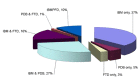
Inclusion body myopathy associated with Paget disease of bone and/or frontotemporal dementia (IBMPFD) is characterized by adult-onset proximal and distal muscle weakness (clinically resembling a limb-girdle muscular dystrophy syndrome), early-onset Paget disease of bone (PDB), and premature frontotemporal dementia (FTD).

Death typically occurs in the sixth or seventh decade from progressive respiratory failure.

Recently Al-Obeidi et al [2018] studied 231 individuals (118 males and 113 females) from 36 families and found that myopathy, PDB, and FTD were present in 90%, 42%, and 30% of the individuals, respectively, beginning at an average age of 43, 41, and 56 years, respectively. Intra- and interfamilial variability is observed in this disorder.

Myopathy. In families studied thus far, 90% of affected individuals had proximal limb-girdle weakness.

  • Diagnosis was at a mean age of 43 years (range: 3-61 years; typically 20s-40s).
  • Muscle weakness is usually proximal, involving the hip and shoulder girdle muscles; however, several individuals have had initial weakness of the muscles of the hands and feet.
  • Affected individuals experience difficulty walking upstairs and raising the arms above the shoulders.
  • The gait is typically waddling and the stance lordotic.
  • Weakness progresses and other limb and respiratory muscle groups become involved over time. Many affected individuals become wheelchair bound.
  • Muscle biopsy findings:
    • Light microscopy of muscle biopsy reveals nonspecific changes: variability in fiber size, type I fiber predominance, and atrophic and hypertrophic fibers. Fibers may contain single or multiple vacuoles. Rimmed vacuoles and cytoplasmic ubiquitin and TAR DNA-binding protein 43 (TDP-43) positive inclusions visible in some fibers are characteristic of inclusion body myopathy [Weihl et al 2008]. The inclusions appear with time and can be observed at a later stage of the disease in some individuals. In advanced cases, severe degenerative muscle changes and fatty replacement of muscle fibers may be noted. Inflammatory cells are absent.
    • Electron microscopy may show nonspecific cytoplasmic changes. The characteristic inclusions, composed of randomly oriented tubulofilaments roughly 15-21 nm in diameter, are seen in muscle nuclei and in cytoplasm. In one family, atrophic and vacuolated muscle fibers containing abundant cytoplasmic-paired helical filaments with epitopes of phosphorylated tau, congophilia, abnormal accumulation of β-amyloid precursor protein (βAPP) epitopes, and accumulation of apolipoprotein E (ApoE) were observed [Alvarez et al 1998].

Paget disease of bone (PDB). In families studied by Al-Obeidi et al [2018], 42% of affected individuals had PDB. The mean age at diagnosis was 41 years (range: 31-61 years). PDB was often asymptomatic, but was diagnosed based on the serum concentration of alkaline phosphatase and bone scans; therefore, it may be underdiagnosed.

PDB involves focal areas of increased bone turnover that lead to complications such as bone pain, localized painful enlargement and deformity of the long bones, pathologic fractures (rare), and deafness. PDB typically manifests as spine and/or hip pain.

Frontotemporal dementia (FTD). FTD is a degenerative condition of the frontal and anterior temporal lobes that differs from the dementia seen in disorders such as Alzheimer disease (see Alzheimer Disease Overview), Pick disease, and Creutzfeldt-Jakob disease (see Genetic Prion Disease). The areas of the brain affected by FTD control reasoning, personality, movement, speech, social graces, and language; memory is preserved.

Among those studied, features were consistent with frontotemporal dementia. In the early stages, dysnomia, dyscalculia, comprehension deficits, and paraphasic errors were evident. Adjusting for aphasia, episodic memory is minimally impaired in the early stages. Progressive aphasia with inability to speak, auditory comprehension deficits for even one-step commands, alexia, and agraphia are noted.

In families studied by Al-Obeidi et al [2018], approximately 30% of affected individuals had dementia. Mean age at diagnosis of dementia was 56 years (range: 30-86 years). This was a cross-sectional study and several individuals were not old enough to have developed FTD. Several individuals were in advanced stages of dementia when diagnosed with IBMPFD and detailed evaluation of the FTD was not possible in them.

Dilated cardiomyopathy. In several individuals in the first family originally reported by Kimonis et al [2000] with limb-girdle myopathy and Paget disease of bone, cardiac failure and cardiomyopathy were noted in the later stages of the disease. Hübbers et al [2007] reported dilated cardiomyopathy characterized by ubiquitin-positive cytoplasmic aggregates and nuclear inclusions in an affected woman. This relatively uncommon finding was most recently reported in four of 18 affected individuals in a large family [Miller et al 2009]. See Dilated Cardiomyopathy Overview.

Amyotrophic lateral sclerosis (ALS). Al-Obeidi et al [2018] reported that approximately 10% of individuals with IBMPFD had a previous diagnosis of ALS. Benatar et al [2013] conducted a systematic EMG characterization of 17 individuals with a diagnosis of IBMPFD from eight families and found that the EMG was abnormal in all individuals. The abnormality was purely neurogenic in four and mixed neurogenic/myopathic in seven individuals; thus, motor neuron involvement as characteristic of ALS was identified in 11/17 (65%) of the participants.

An earlier study by Johnson et al [2010] identified a pathogenic variant in VCP in five of 289 (1%-2%) cases of familial ALS. The parent of one proband died at age 58 years with dementia, parkinsonism, Paget disease, and upper-limb muscle weakness, findings that strongly suggested IBMPFD. In another individual with a pathogenic variant in VCP and diagnosis of ALS, neuropsychological testing performed within one year of symptom onset suggested mild frontal lobe dysfunction. The study findings widened the spectrum of clinical findings associated with IBMPFD to include ALS. See Amyotrophic Lateral Sclerosis Overview.

Parkinson disease (PD). PD is now known to be a feature of IBMPFD. Spina et al [2013] reported affected individuals with PD, but complete details were lacking. Al-Obeidi et al [2018] reported an incidence of 3.8% of Parkinson disease in a cohort of 231 individuals. Individuals with PD in IBMPFD tend to have classic symptoms and respond well to standard treatment [Chan et al 2012]. More recently, Regensburger et al [2017] reported an individual with VCP-related multisystem proteinopathy presenting as early-onset PD.

Other phenotypic features including hepatic steatosis, cataracts, sensorimotor axonal neuropathy, pyramidal tract dysfunction, sphincter disturbance, and sensorineural hearing loss have been reported [Haubenberger et al 2005, Guyant-Maréchal et al 2006, Hübbers et al 2007, Djamshidian et al 2009, Miller et al 2009, Kumar et al 2010].

Neuropathology. VCP-related IBMPFD represents a novel class of neurodegenerative diseases called TDP-43 proteinopathies. Neuropathologic findings associated with IBMPFD include ubiquitin-positive neuronal intranuclear inclusions, dystrophic neuritis, and rare intracytoplasmic inclusions. These findings are abundant in the neocortex, less robust in limbic and subcortical nuclei, and absent in the dentate gyrus [Forman et al 2006, Neumann et al 2007, van der Zee et al 2009].

IBMPFD associated with pathogenic variants in either HNRNPA2B1 or HNRNPA1 has similar neuropathologic findings.

Genotype-Phenotype Correlations

Al-Obeidi et al [2018] analyzed clinical, radiologic, biochemical, and pathogenic variant data in 231 individuals from 36 families with 15 different pathogenic variants in VCP. Inter- and intrafamilial variability made establishing correlations difficult. No significant genotype-phenotype correlations were identified.

No major differences are noted in the IBMPFD phenotype associated with pathogenic variants in either HNRNPA or HNRNPA2B1 except that the Paget disease of the bone seen with a pathogenic variant in HNRNPA2B1 is much more severe and involves the extremities – unlike the distribution in VCP-related disease, in which the sites of predilection are the spine, hip, pelvis, skull, and scapulae with relative sparing of the extremities [Waggoner et al 2002, Kim et al 2013]

Penetrance

Penetrance is almost complete; however, it is age related.

Penetrance by phenotype (see Figure 1).There is marked intra- and interfamilial variability in severity, age of onset, distribution of weakness, and presence or absence of Paget disease, myopathy, and cognitive impairment [Al-Obeidi et al 2018]:

Figure 1.

Figure 1.

IBMPFD phenotypes FTD = frontotemporal dementia; IBM = inclusion body myopathy; IBMPFD = inclusion body myopathy with Paget disease of bone and/or frontotemporal dementia; PDB = Paget disease of bone

  • Presence of all three major manifestations: 10% of affected individuals
  • Presence of only two major manifestations in any combination: 50% of affected individuals
  • Each of the three major manifestations as an apparently isolated finding:
    • Inclusion body myopathy: 37%
    • Paget disease of bone: 5%
    • Frontotemporal dementia: 3%

Prevalence

IBMPFD is rare; the true prevalence is unknown. A study from the UK estimated a prevalence of approximately 1:300,000, although this was not a population ascertainment and the true incidence may be higher. Because previous studies have shown that individuals receive a diagnosis after a diagnostic odyssey of several years and are typically seen by numerous specialists in a number of disciplines (neurology, rheumatology, endocrinology, pain management, genetics), this disorder is considered to be significantly underdiagnosed. As the spectrum of disorders associated with pathogenic variants in VCP expands (as indicated by the number of worldwide publications) it is anticipated that the disorder will be increasingly recognized.

Very few families have been reported with IBMPFD associated with a pathogenic variant in either HNRNPA1 or HNRNPA2B1; thus no estimates of prevalence are available.

Differential Diagnosis

The differential diagnosis of inclusion body myopathy with Paget disease and frontotemporal dementia (IBMPFD) includes the following disorders.

Limb-girdle muscular dystrophy (LGMD). Because the muscle biopsy is nonspecific in the majority of individuals with IBMPFD, the disorder has been labeled as an LGMD.

GNE-related myopathy is characterized by adult-onset, slowly progressive distal muscle weakness that begins with gait disturbance and foot drop secondary to anterior tibialis muscle weakness. Weakness eventually includes the hand and thigh muscles, but commonly spares the quadriceps muscles, even in advanced disease. Affected individuals are usually wheelchair bound approximately 20 years after onset. If quadriceps sparing is incomplete, loss of ambulation tends to occur earlier. Muscle histopathology typically shows rimmed vacuoles and characteristic filamentous inclusions. GNE-related myopathy is inherited in an autosomal recessive manner.

Sporadic inclusion-body myositis (sIBM) (OMIM 147421) is the most common acquired muscle disease in individuals of European heritage older than age 50 years. Pathologically it is characterized by inflammatory, degenerative, and mitochondrial changes that interact in an as-yet-unknown manner to cause progressive muscle degeneration and weakness. The cause is unknown, but it is thought to involve a complex interplay between environmental factors, genetic susceptibility, and aging [Askanas & Engel 2002].

Facioscapulohumeral muscular dystrophy (FSHD). FSHD typically presents before age 20 years with weakness of the facial muscles and the stabilizers of the scapula or the dorsiflexors of the foot. Severity is variable. Weakness is slowly progressive and approximately 20% of affected individuals eventually require a wheelchair. Life expectancy is not shortened. Although some controversy remains, FSHD is likely caused by inappropriate expression of the double homeobox-containing gene DUX4 in muscle cells. Inheritance of FSHD1 is autosomal dominant; inheritance of FSHD2 is digenic.

Scapuloperoneal myopathy (SPM). Scapuloperoneal syndromes are heterogeneous (see OMIM 616852,181430, and 300695). They are characterized by weakness in the distribution of the shoulder girdle and peroneal muscles. Scapuloperoneal myopathy can resemble FSHD clinically.

Amyotrophic lateral sclerosis (ALS). Because of asymmetric involvement and association of both distal and proximal muscle groups, individuals with IBMPFD have been misdiagnosed as having ALS. Published data indicate that up to 10% of individuals with VCP-confirmed IBMPFD had a previous diagnosis of ALS [Kimonis et al 2008]. Furthermore, studies indicate that pathogenic variants in VCP cause ALS, broadening the phenotype of IBMPFD to include motor neuron degeneration [Johnson et al 2010]. More than 30 genes are known to be associated with ALS.

Paget disease of bone (PDB) (OMIM PS167250). Pathogenic variants in SQSTM1, ZNF687, TNFRSF11A, and TNFRSF11A have been associated with PDB. The SQSTM1 p.Pro392Leu pathogenic variant accounts for 16% of simplex cases (i.e., a single occurrence in a family) and 46% of familial cases in the French Canadian population.

Frontotemporal dementia (FTD) causes a substantial proportion of primary degenerative dementia occurring before age 65 years. (See CHMP2B Frontotemporal Dementia, GRN Frontotemporal Dementia.)

Frontotemporal dementia with parkinsonism-17 (FTDP-17) is a presenile dementia affecting the frontal and temporal cortex and some subcortical nuclei. Clinical presentation is variable. Individuals may present with slowly progressive behavioral changes, language disturbances, and/or extrapyramidal signs. Some present with rigidity, bradykinesia, supranuclear palsy, and saccadic eye movement disorders. Symptoms usually start between ages 40 and 60 years, but may occur earlier or later. Disease duration is usually between five and ten years, but occasionally may be up to 20 to 30 years. The disease progresses over a few years into a profound dementia with mutism. FTDP-17 is caused by pathogenic variants in MAPT and inherited in an autosomal dominant manner. (See MAPT-Related Frontotemporal Dementia.)

Alzheimer disease. Imaging studies in IBMPFD reveal atrophy of anterior temporal and frontal lobes. By contrast, more widespread atrophy or perfusion deficits – for example, involving parietal lobes – are more compatible with Alzheimer disease.

Other disorders

  • Limb-girdle myopathy with bone fragility (also referred to as diaphyseal medullary stenosis with malignant fibrous histiocytoma [DMSMFH]) (OMIM 112250), associated with progressive myopathy of a limb-girdle distribution, bone fragility, poor healing of long bones, premature graying with thin hair, thin skin, hernias, and clotting disorders that may resemble IBMPFD. Skeletal radiographs demonstrate coarse trabeculation, patchy sclerosis, cortical thickening, and narrowing of medullary cavities. DMSMFH is caused by pathogenic variants in MTAP and inherited in an autosomal dominant manner.
  • Nasu Hakola disease (also known as polycystic lipomembranous osteodysplasia with sclerosing leukoencephalopathy, or PLOSL) is a presenile dementia associated with loss of myelin, basal ganglia calcification, and bone cysts caused by pathogenic variants in TYROBP or TREM2 and inherited in an autosomal recessive manner.

Management

Evaluations Following Initial Diagnosis

To establish the extent of disease in an individual diagnosed with inclusion body myopathy with Paget disease and frontotemporal dementia (IBMPFD), the evaluations summarized in Table 2 (if not performed as part of the evaluation that led to the diagnosis) are recommended.

Table 2.

Recommended Evaluations Following Initial Diagnosis in Individuals with IBMPFD

System/ConcernEvaluation
Muscle Assessment of muscle strength, muscle wasting, & tendon reflexes. EMG &/or muscle biopsy may be necessary.
Cardiac Baseline echocardiogram & EKG
Lungs Baseline pulmonary function studies
Bone Blood alkaline phosphatase, urine pyridinoline studies, & bone scan studies followed by skeletal x-ray to evaluate distribution & severity of Paget disease of bone
Neurologic Baseline neuropsychological studies of behavior & mental status
Other Consultation w/clinical geneticist &/or genetic counselor

Treatment of Manifestations

Individuals benefit from care by a multidisciplinary team including: a neuromuscular specialist, endocrinologist with expertise in Paget disease, specially trained nurses, pulmonologist, speech therapist, physical therapist, occupational therapist, respiratory therapist, nutritionist, psychologist, social worker, and medical geneticist/genetic counselor.

Table 3.

Treatment of Manifestations in Individuals with IBMPFD

Manifestation/ConcernTreatmentConsiderations/Other
Myopathy Weight controlTo avoid obesity
PT & stretching exercisesTo promote mobility & prevent contractures
OT & use of mechanical aids (e.g., canes, walkers, orthotics, wheelchairs)As needed for ambulation & mobility
Surgical intervention as needed for orthopedic complications (e.g., foot deformity, scoliosis)
Use of respiratory aids if indicated
Social & emotional support & stimulationTo maximize sense of social involvement & productivity & ↓ social isolation
Assisted living arrangements as necessitated by muscle weakness &/or dementia
Paget disease of bone Treatment w/potent bisphosphonatesCan ↓ alkaline phosphatase concentration & relieve pain & disability

OT = occupational therapy; PT = physical therapy

Surveillance

Table 4.

Recommended Surveillance for Individuals with IBMFD

System/ConcernEvaluationFrequency
Cardiac Echocardiogram & EKG to monitor for evidence of cardiomyopathy
  • Obtain baseline studies.
  • If normal, reevaluate at 2-3-yr intervals or if symptomatic.
Lungs Pulmonary function studiesAnnual
Sleep studyAs needed
Bone Alkaline phosphatase, skeletal x-rays, &/or bone scans to monitor therapy & (if symptomatic) PDB
  • Annual alkaline phosphatase
  • Bone scan only when alkaline phosphatase ↑ or symptoms of pain or bony deformity observed
Neurologic Assessment of behavior & mental statusAt baseline & every 2-3 yrs

Agents/Circumstances to Avoid

Individuals and their families should be educated about safety precautions and environmental modification in the home and at work.

Evaluation of Relatives at Risk

See Genetic Counseling for issues related to testing of at-risk relatives for genetic counseling purposes.

Therapies Under Investigation

Search ClinicalTrials.gov in the US and EU Clinical Trials Register in Europe for access to information on clinical studies for a wide range of diseases and conditions. Note: There may not be clinical trials for this disorder.

Genetic Counseling

Genetic counseling is the process of providing individuals and families with information on the nature, mode(s) of inheritance, and implications of genetic disorders to help them make informed medical and personal decisions. The following section deals with genetic risk assessment and the use of family history and genetic testing to clarify genetic status for family members; it is not meant to address all personal, cultural, or ethical issues that may arise or to substitute for consultation with a genetics professional. —ED.

Mode of Inheritance

Inclusion body myopathy with Paget disease and frontotemporal dementia (IBMPFD) is inherited in an autosomal dominant manner.

Risk to Family Members

Parents of a proband

  • Approximately 80% of individuals diagnosed with IBMPFD have an affected parent.
  • A proband with IBMPFD may have the disorder as the result of a de novo pathogenic variant. The proportion of cases caused by a de novo pathogenic variant is estimated to be 20% or greater.
  • Molecular genetic testing is recommended for the parents of a proband with an apparent de novo pathogenic variant. If an HNRNPA1, HNRNPA2B1, or VCP pathogenic variant has not been identified in the proband, recommendations for the evaluation of parents of a proband include clinical evaluation by a neurologist familiar with myopathic disorders in addition to laboratory evaluation of creatine phosphokinase and alkaline phosphatase concentrations.
  • If the pathogenic variant found in the proband cannot be detected in leukocyte DNA of either parent, possible explanations include a de novo pathogenic variant in the proband or germline mosaicism in a parent. Though theoretically possible, no instances of germline mosaicism have been reported.
  • The family history of some individuals diagnosed with IBMPFD may appear to be negative because of failure to recognize the disorder in family members, early death of the parent before the onset of symptoms, or late onset of the disease in the affected parent. Therefore, an apparently negative family history cannot be confirmed unless appropriate clinical evaluation and/or molecular genetic testing has been performed on the parents of the proband.

Sibs of a proband. The risk to the sibs of the proband depends on the clinical/genetic status of the proband's parents:

Offspring of a proband. Each child of an individual with IBMPFD has a 50% chance of inheriting the IBMPFD-causing pathogenic variant.

Other family members. The risk to other family members depends on the status of the proband's parents: if a parent has the pathogenic variant, the parent's family members are at risk.

Related Genetic Counseling Issues

Predictive testing (i.e., testing of asymptomatic at-risk individuals)

  • Predictive testing for at-risk relatives is possible once the pathogenic variant has been identified in an affected family member.
  • Because of the individualized nature of predictive testing, consultation with a genetic counselor or clinical geneticist prior to and following testing is recommended. A testing protocol similar to that used for other genetic disorders (e.g., breast cancer, Huntington disease, familial Alzheimer disease) has been developed [Surampalli et al 2015].

Predictive testing in minors (i.e., testing of asymptomatic at-risk individuals younger than age 18 years)

  • For asymptomatic minors at risk for adult-onset conditions for which early treatment would have no beneficial effect on disease morbidity and mortality, predictive genetic testing is considered inappropriate, primarily because it negates the autonomy of the child with no compelling benefit. Further, concern exists regarding the potential unhealthy adverse effects that such information may have on family dynamics, the risk of discrimination and stigmatization in the future, and the anxiety that such information may cause.
  • For more information, see the National Society of Genetic Counselors position statement on genetic testing of minors for adult-onset conditions and the American Academy of Pediatrics and American College of Medical Genetics and Genomics policy statement: ethical and policy issues in genetic testing and screening of children.

In a family with an established diagnosis of IBMPFD, it is appropriate to consider testing of symptomatic individuals regardless of age.

Considerations in families with an apparent de novo pathogenic variant. When neither parent of a proband with an autosomal dominant condition has the pathogenic variant identified in the proband or clinical evidence of the disorder, the pathogenic variant is likely de novo. However, non-medical explanations including alternate paternity or maternity (e.g., with assisted reproduction) and undisclosed adoption could also be explored.

Family planning

  • The optimal time for determination of genetic risk and discussion of the availability of prenatal/preimplantation genetic testing is before pregnancy.
  • It is appropriate to offer genetic counseling (including discussion of potential risks to offspring and reproductive options) to young adults who are affected or at risk.

Prenatal Testing and Preimplantation Genetic Testing

Once the IBMPFD-causing pathogenic variant has been identified in an affected family member, prenatal and preimplantation genetic testing are possible.

Differences in perspective may exist among medical professionals and within families regarding the use of prenatal testing. While most centers would consider use of prenatal testing to be a personal decision, discussion of these issues may be helpful.

Resources

GeneReviews staff has selected the following disease-specific and/or umbrella support organizations and/or registries for the benefit of individuals with this disorder and their families. GeneReviews is not responsible for the information provided by other organizations. For information on selection criteria, click here.

  • Cure VCP Disease, Inc.
    PO Box 6533
    Americus GA 31709
    Email: curevcpdisease@gmail.com
  • Association for Frontotemporal Degeneration (AFTD)
    Phone: 866-507-7222
    Email: info@theaftd.org
  • Medline Plus
  • Muscular Dystrophy Association (MDA) - USA
    Phone: 833-275-6321
  • Muscular Dystrophy UK
    United Kingdom
    Phone: 0800 652 6352
  • Myositis Association
    1737 King Street
    Suite 600
    Alexandria VA 22314
    Phone: 800-821-7356 (toll-free); 703-299-4850
    Fax: 703-535-6752
    Email: tma@myositis.org
  • National Institute of Neurological Disorders and Stroke (NINDS)
    PO Box 5801
    Bethesda MD 20824
    Phone: 800-352-9424 (toll-free); 301-496-5751; 301-468-5981 (TTY)

Molecular Genetics

Information in the Molecular Genetics and OMIM tables may differ from that elsewhere in the GeneReview: tables may contain more recent information. —ED.

Table A.

Inclusion Body Myopathy with Paget Disease of Bone and/or Frontotemporal Dementia: Genes and Databases

Data are compiled from the following standard references: gene from HGNC; chromosome locus from OMIM; protein from UniProt. For a description of databases (Locus Specific, HGMD, ClinVar) to which links are provided, click here.

Table B.

OMIM Entries for Inclusion Body Myopathy with Paget Disease of Bone and/or Frontotemporal Dementia (View All in OMIM)

164017HETEROGENEOUS NUCLEAR RIBONUCLEOPROTEIN A1; HNRNPA1
167320INCLUSION BODY MYOPATHY WITH EARLY-ONSET PAGET DISEASE WITH OR WITHOUT FRONTOTEMPORAL DEMENTIA 1; IBMPFD1
600124HETEROGENEOUS NUCLEAR RIBONUCLEOPROTEIN A2/B1; HNRNPA2B1
601023VALOSIN-CONTAINING PROTEIN; VCP
615422INCLUSION BODY MYOPATHY WITH EARLY-ONSET PAGET DISEASE WITH OR WITHOUT FRONTOTEMPORAL DEMENTIA 2; IBMPFD2
615424INCLUSION BODY MYOPATHY WITH EARLY-ONSET PAGET DISEASE WITH OR WITHOUT FRONTOTEMPORAL DEMENTIA 3; IBMPFD3

Molecular Pathogenesis

VCP encodes the transitional endoplasmic reticulum ATPase (TER ATPase; also known as the valosin-containing protein, or VCP). VCP is a member of the type II AAA ATPases, and is associated with a variety of activities [Rabouille et al 1998, Hetzer et al 2001, Rabinovich et al 2002] including:

  • Cell cycle control homotypic membrane fusion
  • Nuclear envelope reconstruction
  • Postmitotic organelle reassembly
  • Ubiquitin-dependent protein degradation.

VCP forms a homohexamer in which the double ψ barrel D1/D2 domains bind in a head-to-tail ring [Zhang et al 2000]. VCP targets specific substrates for degradation via binding of adapter proteins [Kondo et al 1997, Meyer et al 2000], playing a critical role in the endoplasmic reticulum (ER)-associated degradation (ERAD) pathway during the "quality control process" that selectively eliminates aberrant proteins in the secretory pathway [Jarosch et al 2002]. This pathway also targets destruction of protein substrates dislocated from the ER to the cytosol [Dai & Li 2001] and the degradation of aggregate-prone proteins, a process principally mediated by autophagy. Abnormal VCP was shown to result in accumulation of autophagic structures in affected individuals and transgenic animal tissue [Ju & Weihl 2010].

Growing evidence implicates VCP in neuronal degeneration. Pathogenic variants in the D2 domain are associated with accumulation of abnormal polyubiquitinated proteins. VCP also binds to expanded polyglutamine (poly-Q) protein aggregates via poly-Q binding (amino acids 142-200).

Single disease-associated variants, one in HNRNPA1 (encoding homologous heterogeneous nuclear ribonucleoprotein A1 [HNRNPA1]) and one in HNRNPA2B1 (encoding heterogeneous nuclear ribonucleoproteins A2/B1 [HNRNPA2B1]), have been reported. Interestingly, both proteins contain a prion-like domain (PrLD) and both disease-associated variants occurred in homologous conserved PrLD domains [Kim et al 2013].

Mechanism of disease causation. VCP-related IBMPFD occurs via a gain-of-function mechanism. In vitro assays of disease-associated VCP variants have shown enhanced ATPase activity, increased binding with its cofactors, and reduced mitofuscin levels, providing evidence for a gain-of-function mechanism of disease [Fernández-Sáiz & Buchberger 2010, Niwa et al 2012, Zhang et al 2017].

VCP pathogenic variants cluster in the N-terminal CDC48 domain, involved in ubiquitin binding [Dai & Li 2001, Rape et al 2001]. This highly structured domain forms distinct regions:

  • The double ψ barrel (amino acids 25-106)
  • The four-stranded β barrel (amino acids 112-186)
  • A short linker region (amino acids 107-111)

Examples of recurrent pathogenic variants in the CDC48 domain are given in Table 6. Other pathogenic variants occur at residues adjacent to and potentially interacting with each other (p.Arg155-p.Asn387, p.Arg159-p.Ala232, and p.Arg191-p.Leu198), suggesting that these residues may have a similar and specific function within the homohexamer.

Studies in yeast suggest that the HNRNPA1 and HNRNPA2B1 disease-associated variants cause toxic cytoplasmic protein aggregates [Kim et al 2013].

Table 5.

IBMPFD: Gene-Specific Laboratory Considerations

Gene 1Special Consideration
HNRNPA1 HNRNPA1 & HNRNPA2B1 share regions of homology that may interfere w/sequence analysis.
HNRNPA2B1
VCP None
1.

Genes from Table 1 in alphabetic order

Table 6.

IBMPFD: Notable Pathogenic Variants by Gene

Gene 1Reference
Sequences
DNA
Nucleotide
Change
Predicted Protein
Change
Comment [Reference]
HNRNPA1 NM_031157​.3
NP_112420​.1
c.941A>Tp.Asp314ValDisease-assoc variants in homologous functional domains [Kim et al 2013]
HNRNPA2B1 NM_031243​.2
NP_112533​.1
c.929C>Tp.Pro310Val
VCP NM_007126​.4
NP_009057​.1
c.277C>Tp.Arg93CysRecurrent pathogenic variants in the double ψ barrel
c.278G>Ap.Arg93His
c.283C>Tp.Arg95Cys
c.283C>Gp.Arg95Gly
c.463C>T/G/Ap.Arg155Cys/Gly/SerRecurrent pathogenic variants in the 4-stranded β barrel
c.464G>A/T/Cp.Arg155His/Leu/Pro 2
c.475C>T/G/Ap.Arg159Cys/Gly/Ser
c.476G>Ap.Arg159His
c.572G>Ap.Arg191GlnRecurrent pathogenic variant in the flexible linker
c.695C>Ap.Ala232GluAbnormal proteins containing either p.Arg155Pro or p.Ala232Glu formed hexameric ring-shaped structures, exhibiting a ~3-fold ↑ in ATPase activity & ↑ sensitivity to heat-induced upregulation of ATPase activity [Halawani et al 2009].

Variants listed in the table have been provided by the author. GeneReviews staff have not independently verified the classification of variants.

GeneReviews follows the standard naming conventions of the Human Genome Variation Society (varnomen​.hgvs.org). See Quick Reference for an explanation of nomenclature.

1.

Genes from Table 1 in alphabetic order

2.

See comment for p.Ala232Glu.

Chapter Notes

Author Notes

Dr Kimonis' University of California, Irvine profile

Acknowledgments

We acknowledge the support of the NIH, MDA and the contribution of collaborators and families. Funding of this study was from the NINDS, NIAMS, National Institutes of Health (RO1, R03, R21, R56), Muscular Dystrophy Association, and Paget Foundation.

Author History

Sandra Donkervoort, MS, CGC; National Institute of Neurological Disorders and Stroke (2011-2019)
Virginia Kimonis, MD (2007-present)
Giles Watts, PhD; University of East Anglia (2007-2019)

Revision History

  • 12 September 2019 (ha) Comprehensive update posted live
  • 28 July 2011 (me) Comprehensive update posted live
  • 19 May 2009 (cd) Revision: prenatal testing available clinically
  • 5 March 2008 (cd) Revision: sequence analysis available clinically
  • 25 May 2007 (me) Review posted live
  • 18 November 2004 (vk, gw) Original submission

References

Published Guidelines / Consensus Statements

  • Committee on Bioethics, Committee on Genetics, and American College of Medical Genetics and Genomics Social, Ethical, Legal Issues Committee. Ethical and policy issues in genetic testing and screening of children. Available online. 2013. Accessed 12-30-22.
  • National Society of Genetic Counselors. Position statement on genetic testing of minors for adult-onset conditions. Available online. 2018. Accessed 12-30-22.

Literature Cited

  • Al-Obeidi E, Al-Tahan S, Surampalli A, Goyal N, Wang AK, Hermann A, Omizo M, Smith C, Mozaffar T, Kimonis V. Genotype-phenotype study in patients with valosin-containing protein mutations associated with multisystem proteinopathy. Clin Genet. 2018;93:119–25. [PMC free article: PMC5739971] [PubMed: 28692196]
  • Alvarez A, Simmons Z, Engel WK, Askanas V. New autosomal dominant inclusion body myopathy (AD-IBM) with many congophilic muscle nuclei that contain paired-helical filaments (PHFs) composed of phosphorylated tau. Neurology. 1998;50:A204.
  • Askanas V, Engel WK. Inclusion-body myositis and myopathies: different etiologies, possibly similar pathogenic mechanisms. Curr Opin Neurol. 2002;15:525–31. [PubMed: 12351995]
  • Benatar M, Wuu J, Fernandez C, Weihl CC, Katzen H, Steele J, Oskarsson B, Taylor JP. Motor neuron involvement in multisystem proteinopathy: Implications for ALS. Neurology. 2013;80:1874–80. [PMC free article: PMC3908355] [PubMed: 23635965]
  • Chan N, Le C, Shieh P, Mozaffar T, Khare M, Bronstein J, Kimonis V. Valosin-containing protein mutation and Parkinson's disease. Parkinsonism Relat Disord. 2012;18:107–9. [PubMed: 21816654]
  • Dai RM, Li CC. Valosin-containing protein is a multi-ubiquitin chain-targeting factor required in ubiquitin-proteasome degradation. Nat Cell Biol. 2001;3:740–4. [PubMed: 11483959]
  • Djamshidian A, Schaefer J, Haubenberger D, Stogmann E, Zimprich F, Auff E, Zimprich A. A novel mutation in the VCP gene (G157R) in a German family with inclusion-body myopathy with Paget disease of bone and frontotemporal dementia. Muscle Nerve. 2009;39:389–91. [PubMed: 19208399]
  • Fernández-Sáiz V, Buchberger A. Imbalances in p97 co-factor interactions in human proteinopathy. EMBO reports. 2010;11:479–85. [PMC free article: PMC2892324] [PubMed: 20414249]
  • Forman MS, Mackenzie IR, Cairns NJ, Swanson E, Boyer PJ, Drachman DA, Jhaveri BS, Karlawish JH, Pestronk A, Smith TW, Tu PH, Watts GD, Markesbery WR, Smith CD, Kimonis VE. Novel ubiquitin neuropathology in frontotemporal dementia with valosin-containing protein gene mutations. J Neuropathol Exp Neurol. 2006;65:571–81. [PubMed: 16783167]
  • Guyant-Maréchal L, Laquerrière A, Duyckaerts C, Dumanchin C, Bou J, Dugny F, Le Ber I, Frébourg T, Hannequin D, Campion D. Valosin-containing protein gene mutations: clinical and neuropathologic features. Neurology. 2006;67:644–51. [PubMed: 16790606]
  • Halawani D, LeBlanc AC, Rouiller I, Michnick SW, Servant MJ, Latterich M. Hereditary inclusion body myopathy-linked p97/VCP mutations in the NH2 domain and the D1 ring modulate p97/VCP ATPase activity and D2 ring conformation. Mol Cell Biol. 2009;29:4484–94. [PMC free article: PMC2725746] [PubMed: 19506019]
  • Haubenberger D, Bittner RE, Rauch-Shorny S, Zimprich F, Mannhalter C, Wagner L, Mineva I, Vass K, Auff E, Zimprich A. Inclusion body myopathy and Paget disease is linked to a novel mutation in the VCP gene. Neurology. 2005;65:1304–5. [PubMed: 16247064]
  • Hetzer M, Meyer HH, Walther TC, Bilbao-Cortes D, Warren G, Mattaj IW. Distinct AAA-ATPase p97 complexes function in discrete steps of nuclear assembly. Nat Cell Biol. 2001;3:1086–91. [PubMed: 11781570]
  • Hübbers CU, Clemen CS, Kesper K, Böddrich A, Hofmann A, Kämäräinen O, Tolksdorf K, Stumpf M, Reichelt J, Roth U, Krause S, Watts G, Kimonis V, Wattjes MP, Reimann J, Thal DR, Biermann K, Evert BO, Lochmüller H, Wanker EE, Schoser BG, Noegel AA, Schröder R. Pathological consequences of VCP mutations on human striated muscle. Brain. 2007;130:381–93. [PubMed: 16984901]
  • Jarosch E, Geiss-Friedlander R, Meusser B, Walter J, Sommer T. Protein dislocation from the endoplasmic reticulum--pulling out the suspect. Traffic. 2002;3:530–6. [PubMed: 12121416]
  • Johnson JO, Mandrioli J, Benatar M, Abramzon Y, Van Deerlin VM, Trojanowski JQ, Gibbs JR, Brunetti M, Gronka S, Wuu J, Ding J, McCluskey L, Martinez-Lage M, Falcone D, Hernandez DG, Arepalli S, Chong S, Schymick JC, Rothstein J, Landi F, Wang YD, Calvo A, Mora G, Sabatelli M, Monsurrò MR, Battistini S, Salvi F, Spataro R, Sola P, Borghero G., ITALSGEN Consortium. Galassi G, Scholz SW, Taylor JP, Restagno G, Chiò A, Traynor BJ. Exome sequencing reveals VCP mutations as a cause of familial ALS. Neuron. 2010;68:857–64. [PMC free article: PMC3032425] [PubMed: 21145000]
  • Ju JS, Weihl CC. Inclusion body myopathy, Paget's disease of the bone and fronto-temporal dementia: a disorder of autophagy. Hum Mol Genet. 2010;19:R38–45. [PMC free article: PMC2875057] [PubMed: 20410287]
  • Kim HJ, Kim NC, Wang YD, Scarborough EA, Moore J, Diaz Z, MacLea KS, Freibaum B, Li S, Molliex A, Kanagaraj AP, Carter R, Boylan KB, Wojtas AM, Rademakers R, Pinkus JL, Greenberg SA, Trojanowski JQ, Traynor BJ, Smith BN, Topp S, Gkazi AS, Miller J, Shaw CE, Kottlors M, Kirschner J, Pestronk A, Li YR, Ford AF, Gitler AD, Benatar M, King OD, Kimonis VE, Ross ED, Weihl CC, Shorter J, Taylor JP. Mutations in prion-like domains in hnRNPA2B1 and hnRNPA1 cause multisystem proteinopathy and ALS. Nature. 2013;495:467–73. [PMC free article: PMC3756911] [PubMed: 23455423]
  • Kimonis VE, Kovach MJ, Waggoner B, Leal S, Salam A, Rimer L, Davis K, Khardori R, Gelber D. Clinical and molecular studies in a unique family with autosomal dominant limb-girdle muscular dystrophy and Paget disease of bone. Genet Med. 2000;2:232–41. [PMC free article: PMC6173187] [PubMed: 11252708]
  • Kimonis VE, Mehta SG, Fulchiero EC, Thomasova D, Pasquali M, Boycott K, Neilan EG, Kartashov A, Forman MS, Tucker S, Kimonis K, Mumm S, Whyte MP, Smith CD, Watts GD. Clinical studies in familial VCP myopathy associated with Paget disease of bone and frontotemporal dementia. Am J Med Genet. 2008;146A:745–57. [PMC free article: PMC2467391] [PubMed: 18260132]
  • Kondo H, Rabouille C, Newman R, Levine TP, Pappin D, Freemont P, Warren G. p47 is a cofactor for p97-mediated membrane fusion. Nature. 1997;388:75–8. [PubMed: 9214505]
  • Kumar KR, Needham M, Mina K, Davis M, Brewer J, Staples C, Ng K, Sue CM, Mastaglia FL. Two Australian families with inclusion-body myopathy, Paget's disease of bone and frontotemporal dementia: novel clinical and genetic findings. Neuromuscul Disord. 2010;20:330–4. [PubMed: 20335036]
  • Majounie E, Traynor BJ, Chio A, Restagno G, Mandrioli J, Benatar M, Taylor JP, Singleton AB. Mutational analysis of the VCP gene in Parkinson’s disease. Neurobiol Aging. 2012;33:209.e1–2. [PMC free article: PMC3221929] [PubMed: 21920633]
  • Meyer HH, Shorter JG, Seemann J, Pappin D, Warren G. A complex of mammalian ufd1 and npl4 links the AAA-ATPase, p97, to ubiquitin and nuclear transport pathways. EMBO J. 2000;19:2181–92. [PMC free article: PMC384367] [PubMed: 10811609]
  • Miller BL, Ikonte C, Ponton M, Levy M, Boone K, Darby A, Berman N, Mena I, Cummings JL. A study of the Lund-Manchester research criteria for frontotemporal dementia: clinical and single-photon emission CT correlations. Neurology. 1997;48:937–42. [PubMed: 9109881]
  • Miller TD, Jackson AP, Barresi R, Smart CM, Eugenicos M, Summers D, Clegg S, Straub V, Stone J. Inclusion body myopathy with Paget disease and frontotemporal dementia (IBMPFD): clinical features including sphincter disturbance in a large pedigree. J Neurol Neurosurg Psychiatry. 2009;80:583–4. [PubMed: 19372299]
  • Neumann M, Mackenzie IR, Cairns NJ, Boyer PJ, Markesbery WR, Smith CD, Taylor JP, Kretzschmar HA, Kimonis VE, Forman MS. TDP-43 in the ubiquitin pathology of frontotemporal dementia with VCP gene mutations. J Neuropathol Exp Neurol. 2007;66:152–7. [PubMed: 17279000]
  • Niwa H, Ewens CA, Tsang C, Yeung HO, Zhang X, Freemont PS. The role of the N-domain in the ATPase activity of the mammalian AAA ATPase p97/VCP. The Journal of biological chemistry. 2012;287:8561–70. [PMC free article: PMC3318706] [PubMed: 22270372]
  • Rabinovich E, Kerem A, Frohlich KU, Diamant N, Bar-Nun S. AAA-ATPase p97/Cdc48p, a cytosolic chaperone required for endoplasmic reticulum-associated protein degradation. Mol Cell Biol. 2002;22:626–34. [PMC free article: PMC139744] [PubMed: 11756557]
  • Rabouille C, Kondo H, Newman R, Hui N, Freemont P, Warren G. Syntaxin 5 is a common component of the NSF- and p97-mediated reassembly pathways of Golgi cisternae from mitotic Golgi fragments in vitro. Cell. 1998;92:603–10. [PubMed: 9506515]
  • Rahbari R, Wuster A, Lindsay SJ, Hardwick RJ, Alexandrov LB, Turki SA, Dominiczak A, Morris A, Porteous D, Smith B, Stratton MR. UK10K Consortium, Hurles ME. Timing, rates and spectra of human germline mutation. Nat Genet. 2016;48:126–33. [PMC free article: PMC4731925] [PubMed: 26656846]
  • Rape M, Hoppe T, Gorr I, Kalocay M, Richly H, Jentsch S. Mobilization of processed, membrane-tethered SPT23 transcription factor by CDC48(UFD1/NPL4), a ubiquitin-selective chaperone. Cell. 2001;107:667–77. [PubMed: 11733065]
  • Regensburger M, Türk M, Pagenstecher A, Schröder R, Winkler J. VCP-related multisystem proteinopathy presenting as early-onset Parkinson disease. Neurology. 2017;89:746–8. [PubMed: 28724584]
  • Richards S, Aziz N, Bale S, Bick D, Das S, Gastier-Foster J, Grody WW, Hegde M, Lyon E, Spector E, Voelkerding K, Rehm HL, et al. Standards and guidelines for the interpretation of sequence variants: a joint consensus recommendation of the American College of Medical Genetics and Genomics and the Association for Molecular Pathology. Genet Med. 2015;17:405–24. [PMC free article: PMC4544753] [PubMed: 25741868]
  • Spina S, Van Laar AD, Murrell JR, Hamilton RL, Kofler JK, Epperson F, Farlow MR, Lopez OL, Quinlan J, DeKosky ST, Ghetti B. Phenotypic variability in three families with valosin-containing protein mutation. Eur J Neurol. 2013;20:251–8. [PMC free article: PMC3734548] [PubMed: 22900631]
  • Surampalli A, Khare M, Kubrussi G, Wencel M, Tanaja J, Donkervoort S, Osann K, Simon M, Wallace D, Smith C, McInerney-Leo A, Kimonis V. Psychological impact of predictive genetic testing in vcp inclusion body myopathy, Paget disease of bone and frontotemporal dementia. J Genet Couns. 2015;24:842–50. [PMC free article: PMC5565393] [PubMed: 25716352]
  • van der Zee J, Pirici D, Van Langenhove T, Engelborghs S, Vandenberghe R, Hoffmann M, Pusswald G, Van den Broeck M, Peeters K, Mattheijssens M, Martin JJ, De Deyn PP, Cruts M, Haubenberger D, Kumar-Singh S, Zimprich A, Van Broeckhoven C. Clinical heterogeneity in 3 unrelated families linked to VCP p.Arg159His. Neurology. 2009;73:626–32. [PubMed: 19704082]
  • Waggoner B, Kovach MJ, Winkelman M, Cai D, Khardori R, Gelber D, Kimonis VE. Heterogeneity in familial dominant Paget disease of bone and muscular dystrophy. Am J Med Genet. 2002;108:187–91. [PubMed: 11891683]
  • Weihl CC, Temiz P, Miller SE, Watts G, Smith C, Forman M, Hanson PI, Kimonis V, Pestronk A. TDP-43 accumulation in inclusion body myopathy muscle suggests a common pathogenic mechanism with frontotemporal dementia. J Neurol Neurosurg Psychiatry. 2008;79:1186–9. [PMC free article: PMC2586594] [PubMed: 18796596]
  • Zhang T, Mishra P, Hay BA, Chan D, Guo M. Valosin-containing protein (VCP/p97) inhibitors relieve Mitofusin-dependent mitochondrial defects due to VCP disease mutants. eLife. 2017:6. [PMC free article: PMC5360448] [PubMed: 28322724]
  • Zhang X, Shaw A, Bates PA, Newman RH, Gowen B, Orlova E, Gorman MA, Kondo H, Dokurno P, Lally J, Leonard G, Meyer H, van Heel M, Freemont PS. Structure of the AAA ATPase p97. Mol Cell. 2000;6:1473–84. [PubMed: 11163219]
Copyright © 1993-2024, University of Washington, Seattle. GeneReviews is a registered trademark of the University of Washington, Seattle. All rights reserved.

GeneReviews® chapters are owned by the University of Washington. Permission is hereby granted to reproduce, distribute, and translate copies of content materials for noncommercial research purposes only, provided that (i) credit for source (http://www.genereviews.org/) and copyright (© 1993-2024 University of Washington) are included with each copy; (ii) a link to the original material is provided whenever the material is published elsewhere on the Web; and (iii) reproducers, distributors, and/or translators comply with the GeneReviews® Copyright Notice and Usage Disclaimer. No further modifications are allowed. For clarity, excerpts of GeneReviews chapters for use in lab reports and clinic notes are a permitted use.

For more information, see the GeneReviews® Copyright Notice and Usage Disclaimer.

For questions regarding permissions or whether a specified use is allowed, contact: ude.wu@tssamda.

Bookshelf ID: NBK1476PMID: 20301649

Views

Tests in GTR by Gene

Related information

  • MedGen
    Related information in MedGen
  • OMIM
    Related OMIM records
  • PMC
    PubMed Central citations
  • PubMed
    Links to PubMed
  • Gene
    Locus Links

Similar articles in PubMed

See reviews...See all...

Recent Activity

Your browsing activity is empty.

Activity recording is turned off.

Turn recording back on

See more...