U.S. flag

An official website of the United States government

NCBI Bookshelf. A service of the National Library of Medicine, National Institutes of Health.

Adam MP, Feldman J, Mirzaa GM, et al., editors. GeneReviews® [Internet]. Seattle (WA): University of Washington, Seattle; 1993-2024.

Cover of GeneReviews®

GeneReviews® [Internet].

Show details

Diastrophic Dysplasia

Synonym: Diastrophic Dwarfism, SLC26A2-Related Diastrophic Dysplasia

, MD and , MD.

Author Information and Affiliations

Initial Posting: ; Last Revision: March 16, 2023.

Estimated reading time: 24 minutes

Summary

Clinical characteristics.

Diastrophic dysplasia (DTD) is characterized by limb shortening, normal-sized head, hitchhiker thumbs, spinal deformities (scoliosis, exaggerated lumbar lordosis, cervical kyphosis), and contractures of the large joints with deformities and early-onset osteoarthritis. Other typical findings are ulnar deviation of the fingers, gap between the first and second toes, and clubfoot. On occasion, the disease can be lethal at birth, but most affected individuals survive the neonatal period and develop physical limitations with normal intelligence.

Diagnosis/testing.

The diagnosis of DTD is established in a proband with characteristic clinical and radiographic features and/or biallelic pathogenic variants in SLC26A2 identified by molecular genetic testing. Biochemical studies of fibroblasts and/or chondrocytes may be used in the rare instances in which molecular genetic testing fails to identify SLC26A2 pathogenic variants.

Management.

Treatment of manifestations: Cervical spine surgery in infancy restricted to individuals with clinical or neurophysiologic evidence of spinal cord impingement; physical therapy may prevent early joint contractures; in children, physiotherapy and casting to maintain joint positioning and mobility as much as possible; surgical correction of clubfoot when ambulation becomes impossible; undertake orthopedic surgery with caution as deformities tend to recur; postpubertal surgical correction of scoliosis is recommended unless severe spinal deformity is causing respiratory compromise or neurologic signs; total arthroplasty of hips and knees in relatively young adults to decrease pain and increase mobility; treatment of cystic ear swelling is conservative.

Surveillance: Annual monitoring of spinal curvature and joint contractures.

Agents/circumstances to avoid: Obesity, which places an excessive load on the large, weight-bearing joints.

Genetic counseling.

DTD is inherited in an autosomal recessive manner. If both parents are known to be heterozygous for an SLC26A2 pathogenic variant, each sib of an affected individual has at conception a 25% chance of being affected, a 50% chance of being an asymptomatic carrier, and a 25% chance of being unaffected and not a carrier. Once the SLC26A2 pathogenic variants have been identified in an affected family member, carrier testing for at-risk relatives and prenatal and preimplantation genetic testing are possible.

Diagnosis

No consensus clinical diagnostic criteria for diastrophic dysplasia (DTD) have been published. However, cystic ear swelling, if present, is pathognomonic.

Suggestive Findings

DTD should be suspected in individuals with the following clinical and radiographic features.

Clinical findings

  • Limb shortening
  • Normal-sized head
  • Slight trunk shortening
  • Hitchhiker thumbs (Figure 1)
  • Symphalangism with missing interphalangeal creases
  • Small chest
  • Protuberant abdomen
  • Contractures of large joints
  • Dislocation of the radius
  • Cleft palate (in ~1/3 of individuals)
  • Cystic ear swelling in the neonatal period (in ~2/3 of infants)
  • Other common findings: ulnar deviation of the fingers, gap between the first and second toes, clubfoot, and flat hemangiomas of the forehead
Figure 1. . Hand of a newborn with diastrophic dysplasia, showing brachydactyly (short fingers), absence of flexion creases of the fingers, and proximally placed, abducted "hitchhiker thumb.

Figure 1.

Hand of a newborn with diastrophic dysplasia, showing brachydactyly (short fingers), absence of flexion creases of the fingers, and proximally placed, abducted "hitchhiker thumb." The thumb deformity results in difficulty with thumb opposition, affecting (more...)

Radiographic findings

  • The skull is of normal size with disproportionate short skeleton.
  • Cervical kyphosis is seen in most newborns and children.
  • Ossification of the upper thoracic vertebrae may be incomplete with broadening of cervical spinal canal ("cobra-like" appearance).
  • Coronal clefts may be present in the lumbar and lower thoracic vertebrae.
  • Narrowing of the interpedicular distance from L1 to L5 is a constant finding.
  • The more cephalad ribs are short and the chest can be bell shaped.
  • Sternum may show duplication of the ossification centers.
  • Ilia are hypoplastic with flat acetabula.
  • Long bones appear moderately shortened with some metaphyseal flaring.
  • Distal humerus is sometimes bifid or V-shaped, sometimes pointed and hypoplastic.
  • Femur is distally rounded.
  • Patella may appear fragmented or multilayered.
  • Radius and tibia may be bowed.
  • Proximal radial dislocation may be present at birth.
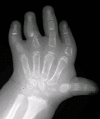
  • Hands may exhibit typical features (Figure 2):
    • Hitchhiker thumb with ulnar deviation of the fingers
    • Shortness of the first metacarpal
    • Delta-shaped proximal and middle phalanges
    • Symphalangism
    • In some severe cases, ossification of two to three carpal bones in the newborn, simulating advanced skeletal age
Figure 2. . Radiograph of the hand of a child, age three years, with diastrophic dysplasia.

Figure 2.

Radiograph of the hand of a child, age three years, with diastrophic dysplasia. The phalanges are short; some show a "delta"-shape deformity. Ossification of the carpal bones is advanced for age, a phenomenon known as "pseudo-acceleration" of the bone (more...)

Establishing the Diagnosis

The diagnosis of DTD is established in a proband with the characteristic clinical and radiographic features described in suggestive findings and/or biallelic pathogenic (or likely pathogenic) variants in SLC26A2 identified by molecular genetic testing (see Table 1).

Note: (1) Per ACMG/AMP variant interpretation guidelines, the terms "pathogenic variants" and "likely pathogenic variants" are synonymous in a clinical setting, meaning that both are considered diagnostic and both can be used for clinical decision making [Richards et al 2015]. Reference to "pathogenic variants" in this section is understood to include any likely pathogenic variants. (2) Identification of biallelic SLC26A2 variants of uncertain significance (or of one known SLC26A2 pathogenic variant and one SLC26A2 variant of uncertain significance) does not establish or rule out the diagnosis.

Molecular genetic testing approaches can include a combination of gene-targeted testing (single-gene testing, multigene panel) and comprehensive genomic testing (exome sequencing, genome sequencing) depending on the phenotype and/or clinical context.

Gene-targeted testing requires that the clinician determine which gene(s) are likely involved, whereas genomic testing does not. Individuals with the distinctive findings described in Suggestive Findings are likely to be diagnosed using gene-targeted testing (see Option 1), whereas those with a phenotype indistinguishable from many other skeletal dysplasias are more likely to be diagnosed using genomic testing (see Option 2).

Option 1

Single-gene testing. Sequence analysis of SLC26A2 detects small intragenic deletions/insertions and missense, nonsense, and splice site variants. Typically, if only one or no variant is detected by the sequencing method used, the next step is to perform gene-targeted deletion/duplication analysis to detect exon and whole-gene deletions or duplications; however, to date such variants have not been identified as a cause of this disorder.

A multigene panel that includes SLC26A2 and other genes of interest (see Differential Diagnosis) is most likely to identify the genetic cause of the condition while limiting identification of variants of uncertain significance and pathogenic variants in genes that do not explain the underlying phenotype. Note: (1) The genes included in the panel and the diagnostic sensitivity of the testing used for each gene vary by laboratory and are likely to change over time. (2) Some multigene panels may include genes not associated with the condition discussed in this GeneReview. (3) In some laboratories, panel options may include a custom laboratory-designed panel and/or custom phenotype-focused exome analysis that includes genes specified by the clinician. (4) Methods used in a panel may include sequence analysis, deletion/duplication analysis, and/or other non-sequencing-based tests.

For an introduction to multigene panels click here. More detailed information for clinicians ordering genetic tests can be found here.

Option 2

When the phenotype is not clearly distinguishable from other skeletal dysplasias, comprehensive genomic testing, which does not require the clinician to determine which gene is likely involved, is the best option. Exome sequencing is most commonly used; genome sequencing is also possible.

For an introduction to comprehensive genomic testing click here. More detailed information for clinicians ordering genomic testing can be found here.

Table 1.

Molecular Genetic Testing Used in Diastrophic Dysplasia (DTD)

Gene 1MethodProportion of Pathogenic Variants 2 Detectable by Method
SLC26A2 Sequence analysis 3>90% 4
Gene-targeted deletion/duplication analysis 5None reported 6
1.
2.

See Molecular Genetics for information on variants detected in this gene.

3.

Sequence analysis detects variants that are benign, likely benign, of uncertain significance, likely pathogenic, or pathogenic. Variants may include small intragenic deletions/insertions and missense, nonsense, and splice site variants; typically, exon or whole-gene deletions/duplications are not detected. For issues to consider in interpretation of sequence analysis results, click here.

4.

The four most common SLC26A2 pathogenic variants (p.Arg279Trp, c.-26+2T>C (IVS1+2T>C), p.Arg178Ter, and p.Cys653Ser) account for approximately 65% of disease alleles in DTD. Sequence analysis identifies 90% of alleles in individuals with radiologic and histologic features of DTD [Rossi & Superti-Furga 2001].

5.

Gene-targeted deletion/duplication analysis detects intragenic deletions or duplications. Methods used may include a range of techniques such as quantitative PCR, long-range PCR, multiplex ligation-dependent probe amplification (MLPA), and a gene-targeted microarray designed to detect single-exon deletions or duplications.

6.

Data derived from the subscription-based professional view of Human Gene Mutation Database [Stenson et al 2020]

Other Testing

Histopathology testing. The histopathology of cartilage is similar to that seen in SLC26A2-related atelosteogenesis and achondrogenesis type 1B (ACG1B), as it reflects the paucity of sulfated proteoglycans in cartilage matrix. It shows an abnormal extracellular matrix with threads of fibrillar material between cystic acellular areas and areas of normal cellularity. Some chondrocytes appear surrounded by lamellar material forming concentric rings; in some cases, these are indistinguishable from the collagen rings typical of ACG1B. The growth plate shows disruption of column formation and hypertrophic zones with irregular invasion of the metaphyseal capillaries and fibrosis. These cartilage matrix abnormalities are present in long bones as well as in tracheal, laryngeal, and peribronchial cartilage, whereas intramembranous bone shows no ossification abnormalities [Superti-Furga 2001, Superti-Furga 2002].

Biochemical testing. The incorporation of sulfate into macromolecules can be studied in cultured chondrocytes and/or skin fibroblasts through double labeling with 3H-glycine and 35S-sodium sulfate. After incubation with these compounds and purification, the electrophoretic analysis of medium proteoglycans reveals a lack of sulfate incorporation, which can be observed even in total macromolecules.

Note: (1) The determination of sulfate uptake is cumbersome and not used for diagnostic purposes. (2) The sulfate incorporation assay in cultured skin fibroblasts (or chondrocytes) is recommended only in the rare instance in which the diagnosis of DTD is strongly suspected but molecular genetic testing fails to detect SLC26A2 pathogenic variants [Rossi et al 2003].

Clinical Characteristics

Clinical Description

Neonatal respiratory compromise. Neonates with diastrophic dysplasia (DTD) may experience respiratory insufficiency because of the small rib cage and tracheal instability and collapsibility. Mechanical ventilation is required in a significant proportion of infants. Mortality in the first months of life is increased, mainly because of respiratory complications such as pneumonia, including aspiration pneumonia.

Musculoskeletal manifestations. The tendons, ligaments, and joint capsules are tighter and shorter than normal, causing restricted joint mobility. Peltonen et al [2003] reported a high prevalence of congenital aplasia of menisci and cruciate ligaments within the knee joints. Pretibial dimples may be present, possibly a consequence of reduced intrauterine movement.

Joint contractures and spine deformity tend to worsen with age. Painful degenerative arthrosis of the hip is common in young adults. Anterior tilting of the pelvis may occur as a consequence and exacerbate the lumbar lordosis. The spine frequently develops excessive lumbar lordosis, thoracolumbar kyphosis, and scoliosis. In anteroposterior radiographs of the lumbar spine, a decrease of the vertebral interpedicular distance is almost invariably observed; however, related neurologic symptoms are only rarely observed [Remes et al 2004, Shafi et al 2023].

The knee may be unstable in childhood; flexion contractures develop with progressive valgus deformity and lateral positioning of the patella. The development and position of the patella may determine whether contraction of the quadriceps muscle results in extension of the knee or paradoxic flexion of the knee. If paradoxic flexion occurs, severe difficulty with walking results [Remes et al 2004].

Because of foot deformities (clubfoot) and shortened tendons, many adults with DTD are unable to place their heels on the ground, and thus stand solely on their metatarsals and toes.

Brachydactyly, ulnar deviation, phalangeal synostosis, and ankylosis of the fingers with significant disability may be observed. Phalangeal synostosis, usually between proximal and middle phalanges, develops in those fingers that have an abnormal phalangeal patterning at birth, including so-called delta-shaped phalanges that usually lack a proper joint space. Often, newborns with DTD lack phalangeal flexion creases (see Figure 1), a sign of marked reduction of joint motion already present at early developmental stages. The thumb may be placed more proximally than usual and may be hypotonic and thus weak. As a consequence, some individuals may have difficulty opposing the thumb and the index finger to accomplish a pincer grasp. In older children and adults, ulnar deviation of the second finger frequently occurs together with radial deviation of the fifth finger (clinodactyly), giving a characteristic "brackets" appearance.

In addition to the skeletal abnormalities, a mild degree of muscular hypoplasia of the thighs and legs is common.

Facial features. The forehead is broad with a high anterior hairline; the palpebral fissures may be downslanting; the nose is long and thin with hypoplastic alae nasi; the facial tissues are tight; the mouth is small, and the mandible normally developed. Cystic ear swelling is frequent and can be associated with inflammation and pain [Cushing et al 2011].

Adult stature ranged between 100 and 140 cm in an early review of Americans and Europeans with DTD. A 1982 study reported a mean adult height of 118 cm [Horton et al 1982]; while a study of Finnish individuals with DTD (who are genetically homogeneous at the SLC26A2 locus) revealed a mean adult height of 136 cm for males and 129 cm for females [Mäkitie & Kaitila 1997]. The discrepancy in mean height between the older studies and the later Finnish study may be the result of variant heterogeneity or may reflect bias of ascertainment of more severely affected individuals in the older studies. It must be noted that the usefulness of such growth curves in predicting adult height is limited by the occurrence of many different allelic combinations.

Neurologic complications may occur, particularly in the cervical region. Cervical kyphosis is seen in lateral radiographs in most newborns; in most children, the kyphosis becomes less severe over the first three to five years of life, but in some instances severe cervical kyphosis may lead to spinal cord compression, either spontaneously or during endotracheal intubation, which requires hyperextension of the neck. A newborn with DTD and severe cervical kyphosis died immediately after birth of respiratory insufficiency; autopsy revealed neuronal degeneration and gliosis of the cervical spinal cord that had developed before birth.

MRI findings have confirmed that in individuals with DTD, the foramen magnum is of normal size but the cervical spinal canal is narrowed. Individual cervical vertebral bodies (usually C3 to C5) may be hypoplastic, but the frequently observed kyphosis is not explained by changes of the vertebral bodies and may thus be the consequence of abnormal intervertebral disks. The rate of spontaneous correction of cervical kyphosis is rather high. MRI studies have shown a peculiar signal anomaly of intervertebral disks, suggesting reduced water content. This anomaly may be the consequence of reduced proteoglycan sulfation.

Cervical spina bifida occulta has been frequently reported in individuals with DTD.

Mental development and intelligence are normal; numerous individuals affected by DTD attain high academic and social recognition or success in the arts.

Hearing loss is unusual in individuals with DTD, although it may be overestimated if studies are based on small cohorts [Tunkel et al 2012].

Vision defects are seldom observed, although a tendency toward myopia has been reported.

Genotype-Phenotype Correlations

Genotype-phenotype correlations indicate that the amount of residual activity of the sulfate transporter modulates the phenotype in this spectrum of disorders, which extends from lethal achondrogenesis type 1B (ACG1B) to SLC26A2-related multiple epiphyseal dysplasia (SLC26A2-MED). Homozygosity or compound heterozygosity for pathogenic variants predicting stop codons or structural pathogenic variants in transmembrane domains of the sulfate transporter are associated with ACG1B, while pathogenic variants located in extracellular loops, in the cytoplasmic tail of the protein, or in the regulatory 5'-flanking region of the gene result in less severe phenotypes [Superti-Furga et al 1996, Karniski 2001, Maeda et al 2006].

The pathogenic variant p.Arg279Trp is the most common SLC26A2 variant found outside of Finland (45% of alleles); it results in the mild SLC26A2-MED phenotype when homozygous and mostly in the DTD and SLC26A2-related atelosteogenesis phenotypes when found in the compound heterozygous state [Barbosa et al 2011].

Pathogenic variant p.Arg178Ter is the second most common variant (9% of alleles) and is associated with a more severe DTD phenotype or even the perinatal-lethal SLC26A2-related atelosteogenesis phenotype, particularly when combined in trans with the p.Arg279Trp variant.

Pathogenic variants p.Cys653Ser and c.-26+2T>C are the third most common variants (8% of alleles).

Pathogenic variant p.Cys653Ser results in SLC26A2-MED when homozygous and in SLC26A2-MED or DTD when present in trans with other pathogenic variants [Czarny-Ratajczak et al 2010].

Pathogenic variant c.-26+2T>C is sometimes referred to as the "Finnish" variant because it is much more frequent in Finland than in the remainder of the world population. It produces low levels of correctly spliced mRNA and results in DTD when homozygous. It is the only variant that has been identified in all four SLC26A2-related dysplasias, in compound heterozygosity with mild (SLC26A2-MED and DTD) or severe (SLC26A2-related atelosteogenesis and ACG1B) alleles [Dwyer et al 2010].

The same pathogenic variants found in the ACG1B phenotype can also be found in the milder phenotypes (SLC26A2-related atelosteogenesis and DTD) if the second allele is a relatively mild variant. Indeed, missense variants located outside of the transmembrane domain of the sulfate transporter are often associated with a residual activity that can "rescue" the effect of a null allele [Rossi & Superti-Furga 2001].

Nomenclature

DTD was recognized as a distinct entity by Lamy & Maroteaux [1960]. At that time, they described a disorder that "resembled achondroplasia in the newborn period but had a quite distinct evolution." The name was chosen to indicate the "twisted" appearance of the spine and limbs in severely affected individuals. The clinical and radiographic features of diastrophic dysplasia are so characteristic that no other name has been associated with the condition.

The existence of clinical variability was recognized early; instances of "severe" or "lethal" DTD are now classified as SLC26A2-related atelosteogenesis, while milder cases ‒ once termed "diastrophic variant" ‒ are now classified as SLC26A2-MED.

In the 2023 revised Nosology of Genetic Skeletal Disorders [Unger et al 2023], DTD is referred to as SLC26A2-related diastrophic dysplasia and is included in the sulfation disorders group.

Prevalence

No reliable data exist regarding the prevalence of DTD. In the experience of several genetic and metabolic centers that can compare its incidence with that of other genetic diseases, DTD disorders are generally believed to be in the range of approximately 1:100,000.

Differential Diagnosis

Differential diagnosis in the prenatal period must include phenotypes in the SLC26A2 skeletal disorder spectrum (see Table 2), other skeletal dysplasias, and conditions with reduced length and/or contractures (Table 3).

Note: The finding of radially deviated thumbs ("hitchhiker thumbs") is suggestive of diastrophic dysplasia (DTD), although not quite pathognomonic.

Table 3.

Genes of Interest in the Differential Diagnosis of Diastrophic Dysplasia

Gene(s)DisorderMOIFeatures Overlapping w/DTD
BPNT2
(IMPAD1)
Chondrodysplasia & abnormal joint development (OMIM 614078)ARPremature carpal ossification & digital malformations in newborns & infants
CANT1
XYLT1
Desbuquois dysplasia (OMIM PS251450)AR
ERGIC1
LGI4
SCYL2
SYNE1
TOR1A
Arthrogryposis multiplex congenita (See SYNE1 Deficiency & OMIM PS617468.)ARCongenital contractures w/mild skeletal anomalies
FLNA XL otopalatodigital spectrum disorders XLPremature carpal ossification & digital malformations in newborns & infants
FLNB Larsen syndrome / atelosteogenesis 1 spectrum (See FLNB Disorders.)AD
FZD2
GPC6
Omodysplasia (OMIM PS258315)AD
AR
Contractures & mesomelic limb shortening reminiscent of DTD

AD = autosomal dominant; AR = autosomal recessive; DTD = diastrophic dysplasia; MOI = mode of inheritance; XL = X-linked

Management

Evaluations Following Initial Diagnosis

To establish the extent of disease and needs in an individual diagnosed with diastrophic dysplasia (DTD), the evaluations summarized in Table 4 (if not performed as part of the evaluation that led to the diagnosis) are recommended.

Table 4.

Recommended Evaluations Following Initial Diagnosis in Individuals with Diastrophic Dysplasia

System/ConcernEvaluationComment
Respiratory Check respiratory rate & for signs of indrawing.Refer to pediatric pulmonologist if any concerns.
Musculoskeletal Cervical films (antero-posterior, lateral, & in flexion-extension)
Complete skeletal surveyIf not performed prior to diagnosis
Orthopedic referral
PT consultation
Genetic
counseling
By genetics professionals 1To inform affected persons & families re nature, MOI, & implications of DTD to facilitate medical & personal decision making

DTD = diastrophic dysplasia; MOI = mode of inheritance; PT = physical therapy

1.

Medical geneticist, certified genetic counselor, certified advanced genetic nurse

Treatment of Manifestations

Table 5.

Treatment of Manifestations in Individuals with Diastrophic Dysplasia

Manifestation/
Concern
TreatmentConsiderations/Other
Cervical
kyphosis
Cervical spine surgery in infancy may be limited to those w/clinical or neurophysiologic evidence of spinal cord impingement.The rate of spontaneous correction is rather high.
Contractures
  • PT may prevent early joint contractures.
  • In children, maintain joint positioning/ mobility as much as possible by physical means (PT & casting, e.g., for clubfeet)
Tightness of joint capsules & ligaments makes correction by casting or other physical means difficult.
Clubfoot Surgical correction is indicated when foot deformity makes ambulation impossible.
  • Undertake surgery w/caution, as deformities tend to recur.
  • Simple tenotomy does not suffice, & more extensive plasty of tarsal bones may be needed [Weiner et al 2008].
Scoliosis Treatment per orthopedic specialist 1
  • Postpubertal surgical correction is best in most cases [Jalanko et al 2009].
  • Surgery before puberty may be helpful for those w/severe spinal deformity → respiratory compromise or neurologic signs.
Premature
degenerative
arthrosis
ArthroplastyTotal arthroplasty of hips & knees ↓ pain & ↑ mobility in a group of adult Finnish persons [Helenius et al 2003a, Helenius et al 2003b]. The authors concluded that arthroplasty is indicated in "relatively young adults" w/DTD.
Cystic ear
swelling
Conservative approach Cushing et al [2011]

DTD = diastrophic dysplasia; PT = physical therapy

1.

Indications for surgical correction of have not been established, nor have criteria to define a successful surgical outcome [Matsuyama et al 1999, Remes et al 2001]. It should be noted that surgical series are inevitably biased toward more severely affected individuals. The key issue seems to be the early identification of those individuals at risk for rapid increase in scoliotic curvature.

Surveillance

Table 6.

Recommended Surveillance for Individuals with Diastrophic Dysplasia

System/ConcernEvaluationFrequency
Musculoskeletal
  • Monitor spinal curvature to prevent neurologic complications & joint contractures.
  • Physical medicine, OT/PT assessment of mobility, self-help skills
Annually

OT = occupational therapy; PT = physical therapy

Agents/Circumstances to Avoid

Obesity places an excessive load on the large weight-bearing joints and thus should be avoided.

Evaluation of Relatives at Risk

See Genetic Counseling for issues related to testing of at-risk relatives for genetic counseling purposes.

Pregnancy Management

Although not specific to DTD, women with severe kyphoscoliosis may experience complications related to thoracic compression in later stages of pregnancy and need to be monitored closely. Kyphoscoliosis can also complicate the use of spinal anesthetics; thus, consultation with an anesthesiologist prior to delivery is advisable.

See MotherToBaby for further information on medication use during pregnancy.

Therapies Under Investigation

Search ClinicalTrials.gov in the US and EU Clinical Trials Register in Europe for access to information on clinical studies for a wide range of diseases and conditions. Note: There may not be clinical trials for this disorder.

Genetic Counseling

Genetic counseling is the process of providing individuals and families with information on the nature, mode(s) of inheritance, and implications of genetic disorders to help them make informed medical and personal decisions. The following section deals with genetic risk assessment and the use of family history and genetic testing to clarify genetic status for family members; it is not meant to address all personal, cultural, or ethical issues that may arise or to substitute for consultation with a genetics professional. —ED.

Mode of Inheritance

Diastrophic dysplasia (DTD) is inherited in an autosomal recessive manner.

Risk to Family Members

Parents of a proband

  • The parents of an affected child are obligate heterozygotes (i.e., presumed to be carriers of one SLC26A2 pathogenic variant based on family history).
  • If a molecular diagnosis has been established in the proband, molecular genetic testing is recommended for the parents of a proband to confirm that both parents are heterozygous for an SLC26A2 pathogenic variant and to allow reliable recurrence risk assessment. If a pathogenic variant is detected in only one parent and parental identity testing has confirmed biological maternity and paternity, the following possibilities should be considered:
  • Heterozygotes (carriers) are usually asymptomatic and have normal stature. There is no evidence that they are at increased risk for degenerative joint disease.

Sibs of a proband

  • If both parents are known to be heterozygous for an SLC26A2 pathogenic variant, each sib of an affected individual has at conception a 25% chance of being affected, a 50% chance of being an asymptomatic carrier, and a 25% chance of being unaffected and not a carrier.
  • Heterozygotes (carriers) are usually asymptomatic and have normal stature.

Offspring of a proband. The offspring of an individual with DTD are obligate heterozygotes (carriers) for a pathogenic variant in SLC26A2.

Other family members. Each sib of the proband's parents is at a 50% risk of being a carrier of an SLC26A2 pathogenic variant.

Carrier Detection

Carrier testing for at-risk relatives requires prior identification of the SLC26A2 pathogenic variants in the family.

Carrier detection in reproductive partners of a heterozygous individual is possible.

Related Genetic Counseling Issues

Family planning

  • The optimal time for determination of genetic risk and discussion of the availability of prenatal/preimplantation genetic testing is before pregnancy.
  • It is appropriate to offer genetic counseling (including discussion of potential risks to offspring and reproductive options) to young adults who are affected, are carriers, or are at risk of being carriers.

DNA banking. Because it is likely that testing methodology and our understanding of genes, pathogenic mechanisms, and diseases will improve in the future, consideration should be given to banking DNA from probands in whom a molecular diagnosis has not been confirmed (i.e., the causative pathogenic mechanism is unknown).

Prenatal Testing and Preimplantation Genetic Testing

High-Risk Pregnancies

Molecular genetic testing. Once the SLC26A2 pathogenic variants have been identified in an affected family member, prenatal and preimplantation genetic testing are possible.

Ultrasound examination. Most fetuses with DTD will have ultrasound-detectable anomalies. However, this should not be relied on as a diagnostic method to exclude a recurrence. See also Low-Risk Pregnancies, Routine ultrasound examination, Note.

Low-Risk Pregnancies

Routine ultrasound examination. Routine prenatal ultrasound examination may identify short fetal limbs, polyhydramnios, and/or small thorax, raising the possibility of DTD in a fetus not known to be at risk. The finding of radially deviated thumbs ("hitchhiker thumbs") is suggestive, although never pathognomonic, of DTD. Subtle findings on ultrasound examination may be recognizable in the first trimester, but in low-risk pregnancies the diagnosis of skeletal dysplasia is usually not made until the second trimester.

Note: While several reports of "successful" early ultrasonographic identification of DTD have been published, the literature is heavily biased toward positive cases [Tongsong et al 2002, Severi et al 2003, Wax et al 2003]. In the authors' experience, only a minority of fetuses with DTD in low-risk pregnancies are identified correctly by ultrasound examination, most cases being diagnosed as unspecific skeletal dysplasia or some other skeletal condition.

Molecular genetic testing. Molecular analysis for suspicion of DTD in the prenatal context should be done via a comprehensive panel (skeletal dysplasia and arthrogryposis), exome sequencing, or genome sequencing.

Resources

GeneReviews staff has selected the following disease-specific and/or umbrella support organizations and/or registries for the benefit of individuals with this disorder and their families. GeneReviews is not responsible for the information provided by other organizations. For information on selection criteria, click here.

Molecular Genetics

Information in the Molecular Genetics and OMIM tables may differ from that elsewhere in the GeneReview: tables may contain more recent information. —ED.

Table A.

Diastrophic Dysplasia: Genes and Databases

GeneChromosome LocusProteinHGMDClinVar
SLC26A2 5q32 Sulfate transporter SLC26A2 SLC26A2

Data are compiled from the following standard references: gene from HGNC; chromosome locus from OMIM; protein from UniProt. For a description of databases (Locus Specific, HGMD, ClinVar) to which links are provided, click here.

Table B.

OMIM Entries for Diastrophic Dysplasia (View All in OMIM)

222600DIASTROPHIC DYSPLASIA; DTD
606718SOLUTE CARRIER FAMILY 26 (SULFATE TRANSPORTER), MEMBER 2; SLC26A2

Molecular Pathogenesis

SLC26A2 pathogenic variants are responsible for the family of chondrodysplasias including achondrogenesis 1B (ACG1B), SLC26A2-related atelosteogenesis, diastrophic dysplasia (DTD), and SLC26A2-related multiple epiphyseal dysplasia (SLC26A2-MED). Impaired activity of the sulfate transporter in chondrocytes and fibroblasts results in the synthesis of proteoglycans that are not sulfated or are insufficiently sulfated [Rossi et al 1998, Satoh et al 1998], most likely because of intracellular sulfate depletion [Rossi et al 1996, Gualeni et al 2010]. Undersulfation of proteoglycans affects the composition of the extracellular matrix and leads to impairment of proteoglycan deposition, which is necessary for proper enchondral bone formation [Corsi et al 2001, Forlino et al 2005, Dawson 2011]. The clinical severity can be correlated with the residual activities of the sulfate transporter resulting from different pathogenic variants [Rossi et al 1996, Rossi et al 1997, Corsi et al 2001, Rossi & Superti-Furga 2001, Rossi et al 2003, Karniski 2004, Maeda et al 2006].

In a Xenopus oocyte model, the p.Arg178Ter pathogenic variant was shown to abolish sulfate transporter activity, and the p.Val341del pathogenic variant showed detectable but very low activity (17% of the wild type) of sulfate transporter [Karniski 2001]. The same variants associated in some individuals with the ACG1B phenotype can be found in individuals with a milder phenotype (SLC26A2-related atelosteogenesis and DTD) if the second allele is a relatively mild variant. Indeed, missense variants located outside the transmembrane domain of the sulfate transporter are often associated with residual activity that can "rescue" the effect of a null allele. Other conclusions from the Xenopus study are at odds with consistent clinical observations, the discrepancy probably being the result of temperature and cellular processing differences between Xenopus oocytes and the human (20°C vs 37°C) [Superti-Furga et al 1996, Rossi & Superti-Furga 2001, Superti-Furga 2001, Superti-Furga 2002]. Similar studies conducted in mammalian cells [Karniski 2004] have produced results that are much more consistent with clinical genotype-phenotype correlations. These studies have essentially confirmed predictions that ACG1B-causing variants are associated with no residual transport activity, while the milder phenotypes result from either different combinations of "null" variants with other alleles that allow for some residual activity or from two variants with residual activity. Original observations were: (1) intracellular retention of the sulfate transporter protein with the variant p.Gly678Val and (2) abnormal molecular weight of sulfate transporter with p.Gln454Pro, possibly indicating protease sensitivity or aberrant glycosylation.

Mechanism of disease causation. Loss of function

Table 7.

Notable SLC26A2 Pathogenic Variants

Reference SequencesDNA Nucleotide
Change
(Alias 1)
Predicted
Protein Change
(Alias 1)
Comment [Reference]
NM_000112​.3 c.-26+2T>C
(IVS1+2T>C)
--Founder variant in Finland; only variant that has been identified in all 4 SLC26A2-related dysplasias, in compound heterozygosity w/mild (SLC26A2-MED & DTD) or severe (SLC26A2-related atelosteogenesis & ACG1B) alleles [Dwyer et al 2010]
NM_000112​.3
NP_000103​.2
c.532C>T
(559C>T)
p.Arg178TerSecond most common variant (9% of alleles); assoc w/more severe DTD phenotype or perinatal-lethal SLC26A2-related atelosteogenesis, esp when combined in trans w/p.Arg279Trp variant
c.835C>T
(c.862C>T)
p.Arg279TrpMost common variant found outside of Finland (45% of alleles); mild SLC26A2-MED when homozygous & mostly DTD & SLC26A2-related atelosteogenesis when found in compound heterozygous state [Barbosa et al 2011]
c.1020_1022delTGT
(1045-1047delGTT)
p.Val341del
(Val340del)
See Molecular Pathogenesis.
c.1361A>C
(1388A>C)
p.Gln454ProSee Molecular Pathogenesis.
c.1957T>A
(1984T>A)
p.Cys653SerThird most common variant (8% of alleles); SLC26A2-MED when homozygous & SLC26A2-MED or DTD when present in trans w/other pathogenic variants [Czarny-Ratajczak et al 2010]
c.2033G>T
(2060G>T)
p.Gly678ValSee Molecular Pathogenesis.

ACG1B = achondrogenesis 1B; DTD = diastrophic dysplasia; MED = multiple epiphyseal dysplasia

Variants listed in the table have been provided by the authors. GeneReviews staff have not independently verified the classification of variants.

GeneReviews follows the standard naming conventions of the Human Genome Variation Society (varnomen​.hgvs.org). See Quick Reference for an explanation of nomenclature.

1.

Variant designation that does not conform to current naming conventions

Chapter Notes

Author History

Diana Ballhausen, MD; Lausanne University Hospital (2013-2021)
Luisa Bonafé, MD, PhD; Lausanne University Hospital (2004-2021)
Lauréane Mittaz-Crettol, PhD; Lausanne University Hospital (2013-2021)
Andrea Superti-Furga, MD (2004-present)
Sheila Unger, MD (2021-present)

Revision History

  • 16 March 2023 (sw) Revision: "SLC26A2-Related Diastrophic Dysplasia" added as synonym; Nomenclature updated
  • 23 December 2021 (sw) Comprehensive update posted live
  • 18 July 2013 (me) Comprehensive update posted live
  • 12 June 2007 (me) Update posted live
  • 15 November 2004 (me) Review posted live
  • 17 February 2004 (asf) Original submission

References

Literature Cited

  • Barbosa M, Sousa AB, Medeira A, Lourenço T, Saraiva J, Pinto-Basto J, Soares G, Fortuna AM, Superti-Furga A, Mittaz L, Reis-Lima M, Bonafé L. Clinical and molecular characterization of Diastrophic Dysplasia in the Portuguese population. Clin Genet. 2011;80:550–7. [PubMed: 21155763]
  • Corsi A, Riminucci M, Fisher LW, Bianco P. Achondrogenesis type IB: agenesis of cartilage interterritorial matrix as the link between gene defect and pathological skeletal phenotype. Arch Pathol Lab Med. 2001;125:1375–8. [PubMed: 11570921]
  • Cushing SL, Swanson RL, Sie KCY. Prevention of auricular deformity in children with diastrophic dysplasia. Int J Pediatr Otorhinolaryngol. 2011;75:713–5. [PubMed: 21414669]
  • Czarny-Ratajczak M, Bieganski T, Rogala P, Glowacki M, Trzeciak T, Kozlowski K. New intermediate phenotype between MED and DD caused by compound heterozygous mutations in the DTDST gene. Am J Med Genet A. 2010;152A:3036–42. [PubMed: 21077204]
  • Dawson PA. Sulfate in fetal development. Semin Cell Dev Biol. 2011;22:653–9. [PubMed: 21419855]
  • Dwyer E, Hyland J, Modaff P, Pauli RM. Genotype-phenotype correlation in DTDST dysplasias: Atelosteogenesis type II and diastrophic dysplasia variant in one family. Am J Med Genet A. 2010;152A:3043–50. [PubMed: 21077202]
  • Forlino A, Piazza R, Tiveron C, Della Torre S, Tatangelo L, Bonafé L, Gualeni B, Romano A, Pecora F, Superti-Furga A, Cetta G, Rossi A. A diastrophic dysplasia sulfate transporter (SLC26A2) mutant mouse: morphological and biochemical characterization of the resulting chondrodysplasia phenotype. Hum Mol Genet. 2005;14:859–71. [PubMed: 15703192]
  • Gualeni B, Facchini M, De Leonardis F, Tenni R, Cetta G, Viola M, Passi A, Superti-Furga A, Forlino A, Rossi A. Defective proteoglycan sulfation of the growth plate zones causes reduced chondrocyte proliferation via an altered Indian hedgehog signalling. Matrix Biol. 2010;29:453–60. [PubMed: 20470884]
  • Helenius I, Remes V, Lohman M, Tallroth K, Poussa M, Helenius M, Paavilainen T. Total knee arthroplasty in patients with diastrophic dysplasia. J Bone Joint Surg Am. 2003a;85:2097–102. [PubMed: 14630837]
  • Helenius I, Remes V, Tallroth K, Peltonen J, Poussa M, Paavilainen T. Total hip arthroplasty in diastrophic dysplasia. J Bone Joint Surg Am. 2003b;85:441–7. [PubMed: 12637429]
  • Horton WA, Hall JG, Scott CI, Pyeritz RE, Rimoin DL. Growth curves for height for diastrophic dysplasia, spondyloepiphyseal dysplasia congenita, and pseudoachondroplasia. Am J Dis Child. 1982;136:316–9. [PubMed: 6803579]
  • Jalanko T, Remes V, Peltonen J, Poussa M, Helenius I. Treatment of spinal deformities in patients with diastrophic dysplasia: a long-term, population based, retrospective outcome study. Spine. 2009;34:2151–7. [PubMed: 19752701]
  • Jónsson H, Sulem P, Kehr B, Kristmundsdottir S, Zink F, Hjartarson E, Hardarson MT, Hjorleifsson KE, Eggertsson HP, Gudjonsson SA, Ward LD, Arnadottir GA, Helgason EA, Helgason H, Gylfason A, Jonasdottir A, Jonasdottir A, Rafnar T, Frigge M, Stacey SN, Th Magnusson O, Thorsteinsdottir U, Masson G, Kong A, Halldorsson BV, Helgason A, Gudbjartsson DF, Stefansson K. Parental influence on human germline de novo mutations in 1,548 trios from Iceland. Nature. 2017;549:519–22. [PubMed: 28959963]
  • Karniski LP. Mutations in the diastrophic dysplasia sulfate transporter (DTDST) gene: correlation between sulfate transport activity and chondrodysplasia phenotype. Hum Mol Genet. 2001;10:1485–90. [PubMed: 11448940]
  • Karniski LP. Functional expression and cellular distribution of diastrophic dysplasia sulfate transporter (DTDST) gene mutations in HEK cells. Hum Mol Genet. 2004;13:2165–71. [PubMed: 15294877]
  • Lamy M, Maroteaux P. Diastrophic nanism. Presse Med. 1960;68:1977–80. [PubMed: 13758600]
  • Maeda K, Miyamoto Y, Sawai H, Karniski LP, Nakashima E, Nishimura G, Ikegawa S. A compound heterozygote harboring novel and recurrent DTDST mutations with intermediate phenotype between atelosteogenesis type II and diastrophic dysplasia. Am J Med Genet A. 2006;140:1143–7. [PubMed: 16642506]
  • Mäkitie O, Kaitila I. Growth in diastrophic dysplasia. J Pediatr. 1997;130:641–6. [PubMed: 9108864]
  • Matsuyama Y, Winter RB, Lonstein JE. The spine in diastrophic dysplasia. The surgical arthrodesis of thoracic and lumbar deformities in 21 patients. Spine. 1999;24:2325–31. [PubMed: 10586456]
  • Peltonen J, Remes V, Tervahartiala P. Early degeneration of the knee in diastrophic dysplasia: an MRI study. J Pediatr Orthop. 2003;23:722–6. [PubMed: 14581774]
  • Remes V, Poussa M, Lönnqvist T, Puusa A, Tervahartiala P, Helenius I, Peltonen J. Walking ability in patients with diastrophic dysplasia: a clinical, electroneurophysiological, treadmill, and MRI analysis. J Pediatr Orthop. 2004;24:546–51. [PubMed: 15308906]
  • Remes V, Poussa M, Peltonen J. Scoliosis in patients with diastrophic dysplasia: a new classification. Spine. 2001;26:1689–97. [PubMed: 11474356]
  • Rossi A, Bonaventure J, Delezoide AL, Cetta G, Superti-Furga A. Undersulfation of proteoglycans synthesized by chondrocytes from a patient with achondrogenesis type 1B homozygous for an L483P substitution in the diastrophic dysplasia sulfate transporter. J Biol Chem. 1996;271:18456–64. [PubMed: 8702490]
  • Rossi A, Bonaventure J, Delezoide AL, Superti-Furga A, Cetta G. Undersulfation of cartilage proteoglycans ex vivo and increased contribution of amino acid sulfur to sulfation in vitro in McAlister dysplasia/atelosteogenesis type 2. Eur J Biochem. 1997;248:741–7. [PubMed: 9342225]
  • Rossi A, Cetta G, Piazza R, Bonaventure J, Steinmann B, Supereti-Furga A. In vitro proteoglycan sulfation derived from sulfhydryl compounds in sulfate transporter chondrodysplasias. Pediatr Pathol Mol Med. 2003;22:311–21. [PubMed: 14692227]
  • Rossi A, Kaitila I, Wilcox WR, Rimoin DL, Steinmann B, Cetta G, Superti-Furga A. Proteoglycan sulfation in cartilage and cell cultures from patients with sulfate transporter chondrodysplasias: relationship to clinical severity and indications on the role of intracellular sulfate production. Matrix Biol. 1998;17:361–9. [PubMed: 9822202]
  • Rossi A, Superti-Furga A. Mutations in the diastrophic dysplasia sulfate transporter (DTDST) gene (SLC26A2): 22 novel mutations, mutation review, associated skeletal phenotypes, and diagnostic relevance. Hum Mutat. 2001;17:159–71. [PubMed: 11241838]
  • Satoh H, Susaki M, Shukunami C, Iyama K, Negoro T, Hiraki Y. Functional analysis of diastrophic dysplasia sulfate transporter. Its involvement in growth regulation of chondrocytes mediated by sulfated proteoglycans. J Biol Chem. 1998;273:12307–15. [PubMed: 9575183]
  • Severi FM, Bocchi C, Sanseverino F, Petraglia F. Prenatal ultrasonographic diagnosis of diastrophic dysplasia at 13 weeks of gestation. J Matern Fetal Neonatal Med. 2003;13:282–4. [PubMed: 12854932]
  • Shafi K, Lovecchio F, Sava M, Steinhaus M, Samuel A, Carter E, Lebl D, Farmer J, Raggio C. Complications and revisions after spine surgery in patients with skeletal dysplasia: have we improved? Global Spine J. 2023;13:268–75. [PMC free article: PMC9972265] [PubMed: 33596686]
  • Stenson PD, Mort M, Ball EV, Chapman M, Evans K, Azevedo L, Hayden M, Heywood S, Millar DS, Phillips AD, Cooper DN. The Human Gene Mutation Database (HGMD®): optimizing its use in a clinical diagnostic or research setting. Hum Genet. 2020;139:1197–207. [PMC free article: PMC7497289] [PubMed: 32596782]
  • Superti-Furga A. Defects in sulfate metabolism and skeletal dysplasias. In: Scriver CR, Beaudet AL, Sly WS, Valle D, Vogelstein B, Childs B, eds. The Metabolic and Molecular Bases of Inherited Disease. 8 ed. New York, NY: McGraw-Hill; 2001:5189-201.
  • Superti-Furga A. Skeletal dysplasias related to defects in sulfate metabolism. In: Royce P, Steinmann B, eds. Connective Tissue and Its Heritable Disorders. 2 ed. New York, NY: Wiley-Liss, Inc; 2002:939-60.
  • Superti-Furga A, Rossi A, Steinmann B, Gitzelmann R. A chondrodysplasia family produced by mutations in the diastrophic dysplasia sulfate transporter gene: genotype/phenotype correlations. Am J Med Genet. 1996;63:144–7. [PubMed: 8723100]
  • Tongsong T, Wanapirak C, Sirichotiyakul S, Chanprapaph P. Prenatal sonographic diagnosis of diastrophic dwarfism. J Clin Ultrasound. 2002;30:103–5. [PubMed: 11857516]
  • Tunkel D, Alade Y, Kerbavaz R, Smith B, Rose-Hardison D, Hoover-Fong J. Hearing loss in skeletal dysplasia patients. Am J Med Genet A. 2012;158A:1551–5. [PubMed: 22628261]
  • Unger S, Ferreira CR, Mortier GR, Ali H, Bertola DR, Calder A, Cohn DH, Cormier-Daire V, Girisha KM, Hall C, Krakow D, Makitie O, Mundlos S, Nishimura G, Robertson SP, Savarirayan R, Sillence D, Simon M, Sutton VR, Warman ML, Superti-Furga A. Nosology of genetic skeletal disorders: 2023 revision. Am J Med Genet A. 2023. Epub ahead of print. [PMC free article: PMC10081954] [PubMed: 36779427]
  • Wax JR, Carpenter M, Smith W, Grimes C, Pinette MG, Blackstone J, Cartin A. Second-trimester sonographic diagnosis of diastrophic dysplasia: report of 2 index cases. J Ultrasound Med. 2003;22:805–8. [PubMed: 12901408]
  • Weiner DS, Jonah D, Kopits S. The 3-dimensional configuration of the typical hip and knee in diastrophic dysplasia. J Pediatr Orthop. 2008;28:60–7. [PubMed: 20502243]
Copyright © 1993-2024, University of Washington, Seattle. GeneReviews is a registered trademark of the University of Washington, Seattle. All rights reserved.

GeneReviews® chapters are owned by the University of Washington. Permission is hereby granted to reproduce, distribute, and translate copies of content materials for noncommercial research purposes only, provided that (i) credit for source (http://www.genereviews.org/) and copyright (© 1993-2024 University of Washington) are included with each copy; (ii) a link to the original material is provided whenever the material is published elsewhere on the Web; and (iii) reproducers, distributors, and/or translators comply with the GeneReviews® Copyright Notice and Usage Disclaimer. No further modifications are allowed. For clarity, excerpts of GeneReviews chapters for use in lab reports and clinic notes are a permitted use.

For more information, see the GeneReviews® Copyright Notice and Usage Disclaimer.

For questions regarding permissions or whether a specified use is allowed, contact: ude.wu@tssamda.

Bookshelf ID: NBK1350PMID: 20301524

Views

Tests in GTR by Gene

Related information

  • MedGen
    Related information in MedGen
  • OMIM
    Related OMIM records
  • PMC
    PubMed Central citations
  • PubMed
    Links to PubMed
  • Gene
    Locus Links

Similar articles in PubMed

See reviews...See all...

Recent Activity

Your browsing activity is empty.

Activity recording is turned off.

Turn recording back on

See more...