U.S. flag

An official website of the United States government

NCBI Bookshelf. A service of the National Library of Medicine, National Institutes of Health.

Adam MP, Feldman J, Mirzaa GM, et al., editors. GeneReviews® [Internet]. Seattle (WA): University of Washington, Seattle; 1993-2024.

Cover of GeneReviews®

GeneReviews® [Internet].

Show details

Cerebrotendinous Xanthomatosis

, MD and , DSci.

Author Information and Affiliations

Initial Posting: ; Last Update: March 17, 2022.

Estimated reading time: 21 minutes

Summary

Clinical characteristics.

Cerebrotendinous xanthomatosis (CTX) is a lipid storage disease characterized by infantile-onset diarrhea, childhood-onset cataract, adolescent- to young adult-onset tendon xanthomas, and adult-onset progressive neurologic dysfunction (dementia, psychiatric disturbances, pyramidal and/or cerebellar signs, dystonia, atypical parkinsonism, peripheral neuropathy, and seizures). Chronic diarrhea from infancy and/or neonatal cholestasis may be the earliest clinical manifestation. In approximately 75% of affected individuals, cataracts are the first finding, often appearing in the first decade of life. Xanthomas appear in the second or third decade; they occur on the Achilles tendon, the extensor tendons of the elbow and hand, the patellar tendon, and the neck tendons. Xanthomas have been reported in the lung, bones, and central nervous system. Some individuals show cognitive impairment from early infancy, whereas the majority have normal or only slightly impaired intellectual function until puberty; dementia with slow deterioration in intellectual abilities occurs in the third decade in more than 50% of individuals. Neuropsychiatric symptoms such as behavioral changes, hallucinations, agitation, aggression, depression, and suicide attempts may be prominent. Pyramidal signs (i.e., spasticity) and/or cerebellar signs almost invariably become evident between ages 20 and 30 years.

The biochemical abnormalities that distinguish CTX from other conditions with xanthomas include high plasma and tissue cholestanol concentration, normal-to-low plasma cholesterol concentration, decreased chenodeoxycholic acid (CDCA), increased concentration of bile alcohols and their glyconjugates, and increased concentrations of cholestanol and apolipoprotein B in cerebrospinal fluid.

Diagnosis/testing.

The diagnosis of CTX is established in a proband with suggestive findings and biallelic pathogenic variants in CYP27A1 identified on molecular genetic testing.

Management.

Treatment of manifestations: Long-term treatment with CDCA normalizes plasma and CSF concentration of cholestanol and improves neurophysiologic findings. Inhibitors of HMG-CoA reductase alone or in combination with CDCA are also effective in decreasing cholestanol concentration and improving clinical signs; however, they may induce muscle damage. Cholic acid treatment decreases cholestanol levels and improves neurologic symptoms in the few individuals in whom it has been tried and may be useful in those who experience side effects with CDCA treatments. Cataract extraction is typically required in at least one eye by age 50 years. Epilepsy, spasticity, and parkinsonism are treated symptomatically.

Prevention of primary manifestations: Early treatment with CDCA in presymptomatic individuals appears to prevent clinical manifestations.

Surveillance: Annual cholestanol plasma concentration, neurologic and neuropsychological evaluation, brain MRI, echocardiogram, and assessment of bone density.

Agents/circumstances to avoid: Caution has been suggested with statins.

Evaluation of relatives at risk: It is appropriate to clarify the genetic status of apparently asymptomatic older and younger at-risk relatives of an affected individual in order to identify as early as possible those who would benefit from prompt initiation of CDCA treatment and surveillance.

Pregnancy management: Treatment with CDCA should not be interrupted during pregnancy.

Genetic counseling.

CTX is inherited in an autosomal recessive manner. If both parents are known to be heterozygous for a CYP27A1 pathogenic variant, each sib of an affected individual has at conception a 25% chance of being affected, a 50% chance of being a carrier, and a 25% chance of inheriting neither of the familial pathogenic variants. Carrier testing for at-risk family members and prenatal and preimplantation genetic testing are possible if both CYP27A1 pathogenic variants in the family are known.

Diagnosis

A consensus paper on the diagnostic criteria and management of cerebrotendinous xanthomatosis (CTX) has been published [Stelten et al 2021a] (full text).

Suggestive Findings

CTX, a lipid storage disease, should be suspected in individuals with the following clinical, laboratory, imaging, and family history findings.

Clinical findings

  • Neonatal cholestasis
  • Infantile-onset diarrhea
  • Childhood-onset cataract
  • Adolescent- to young adult-onset tendon xanthomas (Figure 1)
  • Adult-onset progressive neurologic dysfunction (dementia, psychiatric disturbances, pyramidal and/or cerebellar signs, and seizures)
Figure 1. . Different localization and severity of tendon xanthomas in CTX.

Figure 1.

Different localization and severity of tendon xanthomas in CTX. Besides the classic xanthomas of the Achilles tendon (A), xanthomas of the patellar tendon (B), the extensor tendons of the hand (C), and the extensor tendons of the elbow (D) have been observed. (more...)

Laboratory findings

  • High plasma and tissue cholestanol concentration (Table 1)
  • Normal-to-low plasma cholesterol concentration
  • Markedly decreased formation of chenodeoxycholic acid as a result of impaired primary bile acid synthesis
  • Increased concentration of bile alcohols and their glyconjugates in bile, urine, and plasma (Table 1)
  • Increased concentration of cholestanol and apolipoprotein B in cerebrospinal fluid
  • Increased plasma lactate concentration

Table 1.

Biochemical Abnormalities in Cerebrotendinous Xanthomatosis

AnalyteSourceConcentration
In CTXNormal
CholestanolPlasma & tissue≤5-10x normal330±30 µg/dL
Bile alcoholsUrine14,000±3500 nmol/LNot detectable
Plasma≤500-1000x normal8.48±3.67

Brain imaging

  • Bilateral hyperintensity of the dentate nuclei and cerebral and cerebellar white matter (Figure 2) on brain MRI. Additional changes on brain CT and MRI include diffuse brain and cerebellar atrophy, white matter signal alterations, and bilateral focal cerebellar lesions.
  • Increased brain lactate and decreased n-acetylaspartate concentration (by MR spectroscopy)
Figure 2.

Figure 2.

MRI findings in three persons with CTX A. Signal alterations of cerebral peduncle

Family history is consistent with autosomal recessive inheritance (e.g., affected sibs and/or parental consanguinity). Absence of a known family history does not preclude the diagnosis.

Establishing the Diagnosis

The diagnosis of CTX is established in a proband with suggestive findings and biallelic pathogenic variants in CYP27A1 identified on molecular genetic testing (see Table 2).

Note: Identification of biallelic CYP27A1 variants of uncertain significance (or identification of one known CYP27A1 pathogenic variant and one CYP27A1 variant of uncertain significance) does not establish or rule out the diagnosis of this disorder.

Molecular genetic testing approaches can include a combination of gene-targeted testing (single-gene testing, multigene panel) and comprehensive genomic testing (exome sequencing, genome sequencing) depending on the phenotype.

Gene-targeted testing requires that the clinician determine which gene(s) are likely involved, whereas genomic testing does not. Individuals with the distinctive findings described in Suggestive Findings are likely to be diagnosed using gene-targeted testing (see Option 1), whereas those in whom the diagnosis of CTX has not been considered are more likely to be diagnosed using genomic testing (see Option 2).

Option 1

Single-gene testing. Sequence analysis of CYP27A1 is performed first to detect small intragenic deletions/insertions and missense, nonsense, and splice site variants. Note: Depending on the sequencing method used, single-exon, multiexon, or whole-gene deletions/duplications may not be detected. If only one or no variant is detected by the sequencing method used, the next step is to perform gene-targeted deletion/duplication analysis to detect exon and whole-gene deletions or duplications.

A multigene panel that includes CYP27A1 and other genes of interest (see Differential Diagnosis) is most likely to identify the genetic cause of the condition while limiting identification of variants of uncertain significance and pathogenic variants in genes that do not explain the underlying phenotype. Note: (1) The genes included in the panel and the diagnostic sensitivity of the testing used for each gene vary by laboratory and are likely to change over time. (2) Some multigene panels may include genes not associated with the condition discussed in this GeneReview. (3) In some laboratories, panel options may include a custom laboratory-designed panel and/or custom phenotype-focused exome analysis that includes genes specified by the clinician. (4) Methods used in a panel may include sequence analysis, deletion/duplication analysis, and/or other non-sequencing-based tests.

For an introduction to multigene panels click here. More detailed information for clinicians ordering genetic tests can be found here.

Option 2

Comprehensive genomic testing does not require the clinician to determine which gene is likely involved. Exome sequencing is most commonly used; genome sequencing is also possible.

For an introduction to comprehensive genomic testing click here. More detailed information for clinicians ordering genomic testing can be found here.

Table 2.

Molecular Genetic Testing Used in Cerebrotendinous Xanthomatosis

Gene 1MethodProportion of Pathogenic Variants 2 Detectable by Method
CYP27A1 Sequence analysis 399% 4
Gene-targeted deletion/duplication analysis 51% 4
1.
2.

See Molecular Genetics for information on variants detected in this gene.

3.

Sequence analysis detects variants that are benign, likely benign, of uncertain significance, likely pathogenic, or pathogenic. Variants may include small intragenic deletions/insertions and missense, nonsense, and splice site variants; typically, exon or whole-gene deletions/duplications are not detected. For issues to consider in interpretation of sequence analysis results, click here.

4.

Data derived from the subscription-based professional view of Human Gene Mutation Database and Ensembl 105: Dec 2021 [Stenson et al 2020, Howe et al 2021]

5.

Gene-targeted deletion/duplication analysis detects intragenic deletions or duplications. Methods used may include a range of techniques such as quantitative PCR, long-range PCR, multiplex ligation-dependent probe amplification (MLPA), and a gene-targeted microarray designed to detect single-exon deletions or duplications.

Clinical Characteristics

Clinical Description

Cerebrotendinous xanthomatosis (CTX) is a lipid storage disease characterized by infantile-onset diarrhea, childhood-onset cataract, adolescent- to young adult-onset tendon xanthomas, and adult-onset progressive neurologic dysfunction (dementia, psychiatric disturbances, pyramidal and/or cerebellar signs, dystonia, atypical parkinsonism, peripheral neuropathy, and seizures). Intrafamilial variability is considerable. A suspicion index for diagnosis has been reported based on clinical and laboratory findings [Mignarri et al 2014].

Table 3.

Cerebrotendinous Xanthomatosis: Frequency of Select Features

Feature% of Persons w/Feature
Infantile-onset diarrhea40%
Childhood-onset cataract89%
Adolescent- to young adult-onset tendon xanthomas78%
Cardiovascular findings25%
Osteopenia67%
Adult-onset progressive neurologic dysfunctionIntellectual disability60%
Psychiatric disturbances44%
Ataxia36%
Spastic paraparesis64%
Parkinsonism9%
Peripheral neuropathy70%
Seizures33%

Gastrointestinal and hepatic findings. Chronic diarrhea from infancy, even in the neonatal period, may be the earliest clinical manifestation of CTX [Cruysberg 2002, Gong et al 2017]. Gallstones have been reported on occasion. Neonatal cholestasis has been identified as a presenting manifestation of CTX [Zhang et al 2021]. Cases with fatal cholestasis [von Bahr et al 2005] and infantile hepatitis in infancy [Clayton et al 2002] have been also reported.

Eye. In approximately 75% of affected individuals, cataracts are the first finding, often appearing in the first decade of life. In 25% of individuals, cataracts are first observed after age 40 years. Cataracts may be visually significant opacities requiring lensectomy or visually insignificant cortical opacities. The appearance can include irregular cortical opacities, anterior polar cataracts, and dense posterior subcapsular cataracts [Cruysberg et al 1995]. Among large study groups of individuals with juvenile-onset cataracts, CTX was diagnosed in 1.8% in the United States [Freedman et al 2019] and 1.55% in Turkey [Atilla et al 2021].

Other findings include palpebral xanthelasmas, optic nerve atrophy and proptosis, paleness of the optic disk, premature retinal senescence with retinal vessel sclerosis, cholesterol-like deposits along vascular arcades, and myelinated nerve fibers [Dotti et al 2001].

Khan et al [2013] reported the unique finding of fleck lenticular opacities in three children with CTX; these affected children also had capsular opacities (posterior only or posterior and anterior) that caused visual symptoms.

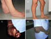
Xanthomas appear in the second or third decade. In addition to the classic xanthomas of the Achilles tendon, xanthomas also occur on the extensor tendons of the elbow and hand, the patellar tendon (see Figure 1), and the neck tendons. Xanthomas have been reported in the lung, bones, and central nervous system [Brienza et al 2015].

Cardiovascular system. Premature atherosclerosis and coronary artery disease have been reported [Valdivielso et al 2004, Androdias et al 2012], as has lipomatous hypertrophy of the atrial septum [Dotti et al 1998, Frih-Ayed et al 2005].

Skeleton. Bone involvement is characterized by granulomatous lesions in the lumbar vertebrae and femur, osteoporosis and increased risk of bone fractures, and impaired adsorption of radiocalcium, which improves with chenodeoxycholic acid treatment [Martini et al 2013]. Osteoporosis is evident by total body densitometry in untreated individuals. Individuals may have marked thoracic kyphosis.

Premature aging. Early-onset cataract, osteopenia with bone fractures and loss of teeth, atherosclerosis, and neurologic impairment with dementia and/or parkinsonism (associated with the characteristic facies) suggest a generalized premature aging process [Dotti et al 1991].

Neurologic Signs

Intellectual disability or dementia following slow deterioration in intellectual abilities occurs in the third decade in more than 50% of individuals [Verrips et al 2000a]. Some individuals show cognitive impairment from early infancy, whereas the majority have normal or only slightly impaired intellectual function until puberty. In the spinal form, mainly characterized by myelopathy and spastic paraparesis, intellect is almost always normal.

Neuropsychiatric symptoms including behavioral changes, hallucinations, agitation, aggression, depression, and suicide attempts may be prominent [Fraidakis 2013].

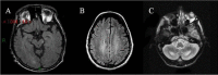
Pyramidal signs (i.e., spasticity) and/or cerebellar signs are almost invariably present between ages 20 and 30 years. The clinical findings are related to the primary involvement of corticospinal tracts, subcortical white matter, dentate nuclei, and cerebellum cortex involvement that is evident on MRI [Dotti et al 1994, Inglese et al 2003, Mignarri et al 2017, Rosini et al 2017, Catarino et al 2018, Makary et al 2018].

Some individuals present with a spinal form, in which progressive spastic paraparesis is the main clinical concern [Nicholls et al 2015, Catarino et al 2018].

Extrapyramidal manifestations can be considered a late disease manifestation, with parkinsonism the most frequently reported, followed by dystonia, myoclonus, and postural tremor. In a recent review of 79 individuals with CTX, the mean age at onset of a movement disorder was 40±12 years (median 40, range 13-62 years). Movement disorders were found to be mixed in 23% of individuals and were usually part of a complex clinical picture, rather than a prominent finding. Still, in 18% of individuals, a movement disorder was the presenting manifestation [Stelten et al 2019].

Seizures are reported in approximately 50% of individuals with CTX [Pedroso et al 2012].

Peripheral neuropathy is evident on electrophysiologic studies [Ginanneschi et al 2013, Zhang et al 2020], which reveal decreased nerve conduction velocities and abnormalities in somatosensory, motor, brain stem, and visual evoked potentials. Clinical manifestations related to peripheral nerve involvement are distal muscle atrophy and pes cavus. Sensory abnormalities are rarely described.

Heterozygotes

Heterozygotes are generally asymptomatic; however, clinical findings have been reported in heterozygotes ranging from an increased incidence of cardiovascular disorders to gallstones [Author, personal observation].

Genotype-Phenotype Correlations

No genotype-phenotype correlations for CYP27A1 have been identified.

Nomenclature

Terms used in the past for CTX and no longer in use include the following:

  • Cerebral cholesterinosis
  • Cerebrotendinous cholesterosis
  • Van Bogaert-Scherer-Epstein syndrome

Prevalence

More than 400 individuals with CTX have been reported worldwide [Stelten et al 2021a], with larger groups of affected individuals being reported in the medical literature from Italy, the Netherlands, Germany, Japan, China, Turkey, Israel, and Spain.

Differential Diagnosis

Selected monogenic disorders that may present with clinical features similar to those of cerebrotendinous xanthomatosis are summarized in Table 4.

Table 4.

Selected Monogenic Disorders in the Differential Diagnosis of Cerebrotendinous Xanthomatosis

FeatureGenetic DisorderGene(s)MOI
Chronic
diarrhea
Congenital diarrhea (OMIM PS214700) DGAT1
EPCAM
GUCY2C
MYO5B
NEUROG3
PERCC1
PLVAP
SLC26A3
SLC9A3
SPINT2
STX3
WNT2B
AR
AD 1
Neonatal
cholestasis 2
Alagille syndrome JAG1
NOTCH2
AD
Dubin-Johnson syndrome (OMIM 237500) ABCC2 AR
Neonatal intrahepatic cholestasis caused by citrin deficiency SLC25A13 AR
Progressive familial intrahepatic cholestasis (see ATP8B1 deficiency & OMIM PS211600) ABCB4
ABCB11
ATP8B1
NR1H4
SLC51A
TJP2
AR
Juvenile
cataracts
Myotonic dystrophy type 1 DMPK AD
Xanthomas Sitosterolemia. Note: Tendon xanthomas or tuberous (i.e., planar) xanthomas can occur in childhood & in unusual locations (heels, knees, elbows, & buttocks). ABCG5
ABCG8
AR
Familial hypercholesterolemia (FH). Note: Common locations of xanthomas incl around eyelids, tendons of elbows, hands, knees, & feet, particularly Achilles tendon. Interdigital xanthomas occur in persons w/homozygous FH. APOB
LDLR
PCSK9
AD
Spastic
paraplegia
See Hereditary Spastic Paraplegia Overview.>80 genesAD
AR
XL
Mat
Ataxia See Hereditary Ataxia Overview.>130 genesAD
AR
XL
Intellectual
disability
See OMIM Autosomal Dominant, Autosomal Recessive, Nonsyndromic X-Linked, & Syndromic X-Linked Intellectual Developmental Disorder Phenotypic Series.>200 genesAD
AR
XL
Genetic
leukoenceph-
alopathies
See Vanderver [2016].>100 genesAD
AR
XL
Mat

AD = autosomal dominant; AR = autosomal recessive; Mat = maternal; MOI = mode of inheritance; XL = X-linked

1.

Inheritance is autosomal recessive with the exception of GUCY2C-related diarrhea, which is inherited in an autosomal dominant manner.

2.

Management

A clinical practice guideline on the diagnosis, treatment, and management of cerebrotendinous xanthomatosis (CTX) has been published, based on expert opinion collected with the Delphi method [Stelten et al 2021a] (full text).

Evaluations Following Initial Diagnosis

To establish the extent of disease and needs in an individual diagnosed with CTX, the evaluations summarized in Table 5 (if not performed as part of the evaluation that led to the diagnosis) are recommended.

Table 5.

Recommended Evaluations Following Initial Diagnosis in Individuals with Cerebrotendinous Xanthomatosis

System/ConcernEvaluationComment
↑ cholestanol level Lab testing of lipids incl plasma cholestanol level
Peripheral neuropathy EMG & NCV studies as baseline
Cardiologic concerns Cardiac eval incl EKG & echocardiogram
Osteoporosis Bone density study
Cataracts Ophthalmologic eval
Neurologic &
behavioral concerns
Baseline neurologic & neuropsychiatric eval
Genetic counseling By genetics professionals 1To inform affected persons & their families re nature, MOI, & implications of CTX to facilitate medical & personal decision making
Family support
& resources
Assess need for:

MOI = mode of inheritance; NCV = nerve conduction velocity

1.

Medical geneticist, certified genetic counselor, certified advanced genetic nurse

Treatment of Manifestations

Table 6.

Treatment of Manifestations in Individuals with Cerebrotendinous Xanthomatosis

Manifestation/ConcernTreatmentConsiderations/Other
↑ cholestanol assoc w/neurologic issues & osteoporosis
  • Long-term treatment w/CDCA 1: 750 mg/day in adults; 10-20 mg/kg/day in children
  • Should be started early as effect of therapy depends largely on extent of irreversible structural damage to axons.
  • Normalizes plasma & CSF concentration of cholestanol by suppressing cholestanol biosynthesis.
  • Improves neurophysiologic findings (normalization of NCVs & stabilization; slow & continuous improvement of MEPs & SEPs).
  • Also improves osteoporosis.
Inhibitors of HMG-CoA reductase (statins such as simvastatin & pravastatin) can be used as alternative treatment alone or in combination w/CDCA.Caution required when using these drugs: may induce muscle damage or even rhabdomyolysis
  • Cholic acid treatment has been used in a few persons 2 & is assoc w/↓ of cholestanol level & improvement of neurologic symptoms.
  • Such therapy may be useful in those who experience side effects w/CDCA treatments.
Cataracts Surgical cataract extractionTypically required in at least 1 eye by age 50 yrs
Epilepsy Symptomatic treatments
Spasticity
Parkinsonism Persons w/CTX & parkinsonism are poorly responsive to levodopa.

CDCA = chenodeoxycholic acid; CSF = cerebrospinal fluid; MEP = motor evoked potential; NCV = nerve conduction velocity, SEP = sensory evoked potential

1.

Verrips et al [2020] highlighted the efficacy and safety of therapeutic treatment with CDCA through two retrospective studies.

2.

Prevention of Primary Manifestations

Early treatment with chenodeoxycholic acid (CDCA) in presymptomatic individuals appears to prevent clinical manifestations (see Treatment of Manifestations) [Degrassi et al 2020].

Surveillance

Table 7.

Recommended Surveillance for Individuals with Cerebrotendinous Xanthomatosis

System/ConcernEvaluationFrequency
↑ cholestanol levels Cholestanol plasma concentrationAnnually
Neurologic &
neuropsychologic issues
Neurologic & neuropsychologic eval
Brain MRI
Cardiac concerns Echocardiogram
Osteoporosis Bone density eval

Agents/Circumstances to Avoid

Caution in the use of statins has been suggested [Federico & Dotti 2001]. See Table 6.

Evaluation of Relatives at Risk

It is appropriate to clarify the genetic status of apparently asymptomatic older and younger at-risk relatives of an affected individual in order to identify as early as possible those who would benefit from prompt initiation of CDCA treatment and surveillance. Early treatment with CDCA in presymptomatic individuals appears to prevent clinical manifestations (see Treatment of Manifestations). Evaluations can include:

  • Molecular genetic testing if the CYP27A1 pathogenic variants in the family are known;
  • Biochemical testing including cholestanol plasma concentration if the pathogenic variants in the family are not known.

See Genetic Counseling for issues related to testing of at-risk relatives for genetic counseling purposes.

Pregnancy Management

Treatment with CDCA should not be interrupted during pregnancy.

Therapies Under Investigation

Gene therapies are under investigation in a mouse model of CTX [Lumbreras et al 2021].

Search ClinicalTrials.gov in the US and EU Clinical Trials Register in Europe for access to information on clinical studies for a wide range of diseases and conditions.

Genetic Counseling

Genetic counseling is the process of providing individuals and families with information on the nature, mode(s) of inheritance, and implications of genetic disorders to help them make informed medical and personal decisions. The following section deals with genetic risk assessment and the use of family history and genetic testing to clarify genetic status for family members; it is not meant to address all personal, cultural, or ethical issues that may arise or to substitute for consultation with a genetics professional. —ED.

Mode of Inheritance

Cerebrotendinous xanthomatosis (CTX) is inherited in an autosomal recessive manner.

Risk to Family Members

Parents of a proband

  • The parents of an affected child are presumed to be heterozygous for a CYP27A1 pathogenic variant.
  • If a molecular diagnosis has been established in the proband, molecular genetic testing is recommended for the parents of the proband to confirm that both parents are heterozygous for a CYP27A1 pathogenic variant and to allow reliable recurrence risk assessment.
  • If a pathogenic variant is detected in only one parent and parental identity testing has confirmed biological maternity and paternity, it is possible that one of the pathogenic variants identified in the proband occurred as a de novo event in the proband or as a postzygotic de novo event in a mosaic parent [Jónsson et al 2017]. If the proband appears to have homozygous pathogenic variants (i.e., the same two pathogenic variants), additional possibilities to consider include:
  • Heterozygotes (carriers) are generally asymptomatic; however, clinical findings have been reported in heterozygotes ranging from an increased incidence of cardiovascular disorders to gallstones [Author, personal observation].

Sibs of a proband

  • If both parents are known to be heterozygous for a CYP27A1 pathogenic variant, each sib of an affected individual has at conception a 25% chance of being affected, a 50% chance of being a carrier, and a 25% chance of inheriting neither of the familial pathogenic variants.
  • Significant clinical variability may be observed between affected family members: a sib with biallelic pathogenic variants may experience less severe manifestations with later onset of neurologic signs than the proband [Guenzel et al 2021, Stelten et al 2021b] and may not develop xanthomas [Verrips et al 2000b, Gelzo et al 2021].
  • Heterozygotes (carriers) are generally asymptomatic; however, clinical findings have been reported in heterozygotes ranging from an increased incidence of cardiovascular disorders to gallstones [Author, personal observation].

Offspring of a proband

  • The offspring of an individual with CTX are obligate heterozygotes for a CYP27A1 pathogenic variant.
  • If the reproductive partner of a proband is heterozygous for a CYP27A1 pathogenic variant – a situation more likely to be seen in Israel or Morocco and/or in reproductive partners of Druze heritage, due to a founder effect – offspring are at risk of inheriting biallelic CYP27A1 pathogenic variants and being affected with CTX.

Other family members. Each sib of the proband's parents is at a 50% risk of being a carrier of a CYP27A1 pathogenic variant.

Carrier (Heterozygote) Detection

Carrier testing for at-risk relatives requires prior identification of the CYP27A1 pathogenic variants in the family.

Related Genetic Counseling Issues

See Management, Evaluation of Relatives at Risk for information on evaluating at-risk relatives for the purpose of early diagnosis and treatment.

Family planning

  • The optimal time for determination of genetic risk and discussion of the availability of prenatal/preimplantation genetic testing is before pregnancy.
  • It is appropriate to offer genetic counseling (including discussion of potential risks to offspring and reproductive options) to young adults who are affected, are carriers, or are at risk of being carriers.

DNA banking. Because it is likely that testing methodology and our understanding of genes, pathogenic mechanisms, and diseases will improve in the future, consideration should be given to banking DNA from probands in whom a molecular diagnosis has not been confirmed (i.e., the causative pathogenic mechanism is unknown).

Prenatal Testing and Preimplantation Genetic Testing

Once the CYP27A1 pathogenic variants have been identified in an affected family member, prenatal and preimplantation genetic testing are possible.

Differences in perspective may exist among medical professionals and within families regarding the use of prenatal testing. While most centers would consider use of prenatal testing to be a personal decision, discussion of these issues may be helpful.

Resources

GeneReviews staff has selected the following disease-specific and/or umbrella support organizations and/or registries for the benefit of individuals with this disorder and their families. GeneReviews is not responsible for the information provided by other organizations. For information on selection criteria, click here.

Molecular Genetics

Information in the Molecular Genetics and OMIM tables may differ from that elsewhere in the GeneReview: tables may contain more recent information. —ED.

Table A.

Cerebrotendinous Xanthomatosis: Genes and Databases

GeneChromosome LocusProteinLocus-Specific DatabasesHGMDClinVar
CYP27A1 2q35 Sterol 26-hydroxylase, mitochondrial CYP27A1 @ LOVD CYP27A1 CYP27A1

Data are compiled from the following standard references: gene from HGNC; chromosome locus from OMIM; protein from UniProt. For a description of databases (Locus Specific, HGMD, ClinVar) to which links are provided, click here.

Table B.

OMIM Entries for Cerebrotendinous Xanthomatosis (View All in OMIM)

213700CEREBROTENDINOUS XANTHOMATOSIS; CTX
606530CYTOCHROME P450, SUBFAMILY XXVIIA, POLYPEPTIDE 1; CYP27A1

Molecular Pathogenesis

Cerebrotendinous xanthomatosis (CTX) is caused by biallelic pathogenic variants in CYP27A1. Many of the reported pathogenic variants involve splice sites and are predicted to affect mRNA stability or lead to the formation of abnormal mRNA with translation products that are devoid of an adrenodoxin-binding region and/or the heme-binding site, important for enzyme activity. Other pathogenic variants are predicted to result in truncated peptides devoid of function. The associated deficiency of a functional mitochondrial enzyme sterol 27-hydroxylase causes cholestanol and cholesterol accumulation in virtually every tissue.

Mechanism of disease causation. CTX occurs via a loss-of-function mechanism.

Table 8.

Notable CYP27A1 Pathogenic Variants

Reference SequencesDNA Nucleotide
Change
Predicted
Protein Change
Comment [Reference]
NM_000784​.4

NP_000775​.1

c.355delCp.(Arg119GlyfsTer24)Founder variant in Israeli Druze [Falik-Zaccai et al 2008]
c.1183C>Tp.Arg395CysCommon pathogenic variant [Cali et al 1991; Authors, personal observation]

Variants listed in the table have been provided by the authors. GeneReviews staff have not independently verified the classification of variants.

GeneReviews follows the standard naming conventions of the Human Genome Variation Society (varnomen​.hgvs.org). See Quick Reference for an explanation of nomenclature.

Chapter Notes

Author Notes

Antonio Federico is Emeritus professor of Clinical Neurology, Department of Medicine, Surgery and Neurosciences, Medical School, University of Siena, Siena Italy.

Gian Nicola Gallus is a postdoctoral fellow in Molecular Biology, Department of Medicine, Surgery and Neurosciences, Medical School, University of Siena, Siena, Italy.

Acknowledgments

We acknowledge all our colleagues who in the last 40 years have collaborated with us in the investigation of this condition: Prof GC Guazzi, Prof N De Stefano, Prof A Malandrini, Dr C Battisti, Dr E Cardaioli, Dr P Formichi, Dr S Bianchi, Dr A Rufa, Dr F Sicurelli; particular thanks to Prof MT Dotti and Dr A Mignarri, and all the physicians who referred patients from Italy and abroad. We also acknowledge all our patients.

Author History

Maria Teresa Dotti, MD; University of Siena (2003-2022)
Antonio Federico, MD (2003-present)
Gian Nicola Gallus, DSci (2003-present)

Revision History

  • 17 March 2022 (ha) Comprehensive update posted live
  • 14 April 2016 (ma) Comprehensive update posted live
  • 1 August 2013 (me) Comprehensive update posted live
  • 20 October 2011 (cd) Revision: deletion/duplication analysis available clinically
  • 16 November 2010 (me) Comprehensive update posted live
  • 25 January 2008 (cd) Revision: prenatal testing available
  • 7 February 2006 (me) Comprehensive update posted live
  • 16 July 2003 (me) Review posted live
  • 18 December 2002 (af) Original submission

References

Published Gidelines / Consensus Statements

  • Stelten BML, Dotti MT, Verrips A, Elibol B, Falik-Zaccai TC, Hanman K, Mignarri A, Sithole B, Steiner RD, Verma S, Yahalom G, Zubarioglu T, Mochel F, Federico A (2021a) Expert opinion on diagnosing, treating and managing patients with cerebrotendinous xanthomatosis (CTX): a modified Delphi study. Available online. 2021. Accessed 3-9-22. [PMC free article: PMC8349076] [PubMed: 34362411]

Literature Cited

  • Androdias G, Vukusic S, Gignoux L, Boespflug-Tanguy O, Acquaviva C, Zabot MT, Couvert P, Carrie A, Confavreux C, Labauge P. Leukodystrophy with a cerebellar cystic aspect and intracranial atherosclerosis: an atypical presentation of cerebrotendinous xanthomatosis. J Neurol. 2012;259:364–6. [PubMed: 21769531]
  • Atilla H, Coskun T, Elibol B, Kadayifcilar S, Altinel S, et al. Prevalence of cerebrotendinous xanthomatosis in cases with idiopathic bilateral juvenile cataract in ophthalmology clinics in Turkey. J AAPOS. 2021;25:269. [PubMed: 34600103]
  • Brienza M, Fiermonte G, Cambieri C, Mignarri A, Dotti MT, Fiorelli M. Enlarging brain xanthomas in a patient with cerebrotendinous xanthomatosis. J Inherit Metab Dis. 2015;38:981–2. [PubMed: 25567502]
  • Cali JJ, Hsieh CL, Francke U, Russell DW. Mutations in the bile acid biosynthetic enzyme sterol 27-hydroxylase underlie cerebrotendinous xanthomatosis. J Biol Chem. 1991;266:7779–83. [PMC free article: PMC4449724] [PubMed: 2019602]
  • Catarino CB, Vollmar C, Küpper C, Seelos K, Gallenmüller C, Bartkiewicz J, Biskup S, Hörtnagel K, Klopstock T. Brain diffusion tensor imaging changes in cerebrotendinous xanthomatosis reversed with treatment. J Neurol. 2018;265:388–393. [PubMed: 29260356]
  • Clayton PT, Verrips A, Sistermans E, Mann A, Mieli-Vergani G, Wevers R. Mutations in the sterol 27-hydroxylase gene (CYP27A) cause hepatitis of infancy as well as cerebrotendinous xanthomatosis. J Inherit Metab Dis. 2002;25:501–13. [PubMed: 12555943]
  • Cruysberg JR. Cerebrotendinous xanthomatosis: juvenile cataract and chronic diarrhea before the onset of neurologic disease. Arch Neurol. 2002;59:1975. [PubMed: 12470193]
  • Cruysberg JR, Wevers RA, van Engelen BG, Pinckers A, van Spreeken A, Tolboom JJ. Ocular and systemic manifestations of cerebrotendinous xanthomatosis. Am J Ophthalmol. 1995;120:597–604. [PubMed: 7485361]
  • Degrassi I, Amoruso C, Giordano G, Del Puppo M, Mignarri A, Dotti MT, Naturale M, Nebbia G. Case report: early treatment with chenodeoxycholic acid in cerebrotendinous xanthomatosis presenting as neonatal cholestasis. Front Pediatr. 2020;16:382. [PMC free article: PMC7381104] [PubMed: 32766184]
  • Dotti MT, Federico A, Signorini E, Caputo N, Venturi C, Filosomi G, Guazzi GC. Cerebrotendinous xanthomatosis (van Bogaert-Scherer-Epstein disease): CT and MR findings. AJNR Am J Neuroradiol. 1994;15:1721–6. [PMC free article: PMC8333715] [PubMed: 7847220]
  • Dotti MT, Mondillo S, Plewnia K, Agricola E, Federico A. Cerebrotendinous xanthomatosis: evidence of lipomatous hypertrophy of the atrial septum. J Neurol. 1998;245:723–6. [PubMed: 9808240]
  • Dotti MT, Rufa A, Federico A. Cerebrotendinous xanthomatosis: heterogeneity of clinical phenotype with evidence of previously undescribed ophthalmological findings. J Inherit Metab Dis. 2001;24:696–706. [PubMed: 11804206]
  • Dotti MT, Salen G, Federico A. Cerebrotendinous xanthomatosis as a multisystem disease mimicking premature ageing. Dev Neurosci. 1991;13:371–6. [PubMed: 1817044]
  • Falik-Zaccai TC, Kfir N, Frenkel P, Cohen C, Tanus M, Mandel H, Shihab S, Morkos S, Aaref S, Summar ML, Khayat M. Population screening in a Druze community: the challenge and the reward. Genet Med. 2008;10:903–9. [PubMed: 19092443]
  • Federico A, Dotti MT. Cerebrotendinous xanthomatosis. Neurology. 2001;57:1743. [PubMed: 11706139]
  • Fraidakis MJ. Psychiatric manifestations in cerebrotendinous xanthomatosis. Transl Psychiatry. 2013;3:e302. [PMC free article: PMC3784765] [PubMed: 24002088]
  • Freedman SF, Brennand C, Chiang J, DeBarber A, Del Monte MA, Duell PB, Fiorito J, Marshall R. Prevalence of cerebrotendinous xanthomatosis among patients diagnosed with acquired juvenile-onset idiopathic bilateral cataracts. JAMA Ophthalmol. 2019;137:1312–16. [PMC free article: PMC6753501] [PubMed: 31536098]
  • Frih-Ayed M, Boughammoura-Bouatay A, Ben Hamda K, Chebel S, Ben Farhat M. Rev Med Interne. 2005;26:992–3. [Hypertrophy of the atrial septum in the cerebrotendinous xanthomatosis] [PubMed: 16236394]
  • Gelzo M, Di Taranto MD, Bisecco A, D'Amico A, Capuano R, Giacobbe C, Caputo M, Cirillo M, Tedeschi G, Fortunato G, Corso G. A case of cerebrotendinous xanthomatosis with spinal cord involvement and without tendon xanthomas: identification of a new mutation of the CYP27A1 gene. Acta Neurol Belg. 2021;121:561–6. [PubMed: 31875301]
  • Ginanneschi F, Mignarri A, Mondelli M, Gallus GN, Del Puppo M, Giorgi S, Federico A, Rossi A, Dotti MT. Polyneuropathy in cerebrotendinous xanthomatosis and response to treatment with chenodeoxycholic acid. J Neurol. 2013;260:268–74. [PubMed: 22878431]
  • Gong JY, Setchell KDR, Zhao J, Zhang W, Wolfe B, Lu Y, Lackner K, Knisely AS, Wang N-L, Hao C-Z, Zhang M-H, Wang J-S. Severe neonatal cholestasis in cerebrotendinous xanthomatosis: genetics, immunostaining, mass spectrometry. J Pediatr Gastroenterol Nutr. 2017;65:561–8. [PubMed: 28937538]
  • Guenzel AJ, DeBarber A, Raymond K, Dhamija R. Familial variability of cerebrotendinous xanthomatosis lacking typical biochemical findings. JIMD Rep. 2021;59:3–9. [PMC free article: PMC8100391] [PubMed: 33977023]
  • Howe KL, Achuthan P, Allen J, Allen J, Alvarez-Jarreta J, Amode MR, Armean IM, Azov AG, Bennett R, Bhai J, Billis K, Boddu S, Charkhchi M, Cummins C, Da Rin FL, Davidson C, Dodiya K, El Houdaigui B, Fatima R, Gall A, Garcia GC, Grego T, Guijarro-Clarke C, Haggerty L, Hemrom A, Hourlier T, Izuogu OG, Juettemann T, Kaikala V, Kay M, Lavidas I, Le T, Lemos D, Gonzalez Martinez J, Marugán JC, Maurel T, McMahon AC, Mohanan S, Moore B, Muffato M, Oheh DN, Paraschas D, Parker A, Parton A, Prosovetskaia I, Sakthivel MP, Salam Ahamed IA, Schmitt BM, Schuilenburg H, Sheppard D. Ensembl 2021. Nucleic Acids Res. 2021;49:D884–D891. [PMC free article: PMC7778975] [PubMed: 33137190]
  • Inglese M, DeStefano N, Pagani E, Dotti MT, Comi G, Federico A, Filippi M. Quantification of brain damage in cerebrotendinous xanthomatosis with magnetization transfer MR imaging. AJNR Am J Neuroradiol. 2003;24:495–500. [PMC free article: PMC7973612] [PubMed: 12637303]
  • Jónsson H, Sulem P, Kehr B, Kristmundsdottir S, Zink F, Hjartarson E, Hardarson MT, Hjorleifsson KE, Eggertsson HP, Gudjonsson SA, Ward LD, Arnadottir GA, Helgason EA, Helgason H, Gylfason A, Jonasdottir A, Jonasdottir A, Rafnar T, Frigge M, Stacey SN, Th Magnusson O, Thorsteinsdottir U, Masson G, Kong A, Halldorsson BV, Helgason A, Gudbjartsson DF, Stefansson K. Parental influence on human germline de novo mutations in 1,548 trios from Iceland. Nature. 2017;549:519–22. [PubMed: 28959963]
  • Khan AO, Aldahmesh MA, Mohamed JY, Alkuraya FS. Juvenile cataract morphology in 3 siblings not yet diagnosed with cerebrotendinous xanthomatosis. Ophthalmology. 2013;120:956–60. [PubMed: 23375591]
  • Lipiński P, Ciara E, Jurkiewicz D, Pollak A, Wypchło M, Płoski R, Cielecka-Kuszyk J, Socha P, Pawłowska J, Jankowska I. Targeted next-generation sequencing in diagnostic approach to monogenic cholestatic liver disorders-single-center experience. Front Pediatr. 2020;8:414. [PMC free article: PMC7393978] [PubMed: 32793533]
  • Lumbreras S, Ricobaraza A, Baila-Rueda L, Gonzalez-Aparicio M, Mora-Jimenez L, Uriarte I, Bunuales M, Avila MA, Monte MJ, Marin JJG, Cenarro A, Gonzalez-Aseguinolaza G, Hernandez-Alcoceba R. Gene supplementation of CYP27A1 in the liver restores bile acid metabolism in a mouse model of cerebrotendinous xanthomatosis. Mol Ther Methods Clin Dev. 2021;22:210–21. [PMC free article: PMC8399082] [PubMed: 34485606]
  • Makary MS, Kisanuki YY, Amin NN, Slone HW. Teaching NeuroImages: Cerebrotendinous xanthomatosis: a rare treatable adult-onset lipid storage disease. Neurology. 2018;90:e637–e638. [PubMed: 29440550]
  • Mandia D, Chaussenot A, Besson G, Lamari F, Castelnovo G, Curot J, Duval F, Giral P, Lecerf JM, Roland D, Pierdet H, Douillard C, Nadjar Y. Cholic acid as a treatment for cerebrotendinous xanthomatosis in adults. J Neurol. 2019;266:2043–50. [PubMed: 31115677]
  • Martini G, Mignarri A, Ruvio M, Valenti R, Franci B, Del Puppo M, Federico A, Nuti R, Dotti MT. Long-term bone density evaluation in cerebrotendinous xanthomatosis: evidence of improvement after chenodeoxycholic acid treatment. Calcif Tissue Int. 2013;92:282–6. [PubMed: 23212544]
  • Mignarri A, Dotti MT, Federico A, De Stefano N, Battaglini M, Grazzini I, Galluzzi P, Monti L. The spectrum of magnetic resonance findings in cerebrotendinous xanthomatosis: redefinition and evidence of new markers of disease progression. J Neurol. 2017;264:862–74. [PubMed: 28324197]
  • Mignarri A, Gallus GN, Dotti MT, Federico A. A suspicion index for early diagnosis and treatment of cerebrotendinous xanthomatosis. J Inherit Metab Dis. 2014;37:421–9. [PubMed: 24442603]
  • Nicholls Z, Hobson E, Martindale J, Shaw PJ. Diagnosis of spinal xanthomatosis by next-generation sequencing: identifying a rare, treatable mimic of hereditary spastic paraparesis. Pract Neurol. 2015;15:280–3. [PubMed: 25862734]
  • Pedroso JL, Pinto WB, Souza PV, Santos LT, Abud IC, Avelino MA, Barsottini OG. Early-onset epilepsy as the main neurological manifestation of cerebrotendinous xanthomatosis. Epilepsy Behav. 2012;24:380–1. [PubMed: 22658436]
  • Pierre G, Setchell K, Blyth J, Preece MA, Chakrapani A, McKiernan P. Prospective treatment of cerebrotendinous xanthomatosis with cholic acid therapy. J Inherit Metab Dis. 2008;31:S241–5. [PubMed: 19125350]
  • Rosini F, Pretegiani E, Mignarri A, Optican LM, Serchi V, De Stefano N, Battaglini M, Monti L, Dotti MT, Federico A, Rufa A. The role of dentate nuclei in human oculomotor control: insights from cerebrotendinous xanthomatosis. J Physiol. 2017;595:3607–20. [PMC free article: PMC5451708] [PubMed: 28168705]
  • Stelten BML, Dotti MT, Verrips A, Elibol B, Falik-Zaccai TC, Hanman K, Mignarri A, Sithole B, Steiner RD, Verma S, Yahalom G, Zubarioglu T, Mochel F, Federico A. Expert opinion on diagnosing, treating and managing patients with cerebrotendinous xanthomatosis (CTX): a modified Delphi study. Orphanet J Rare Dis. 2021a;16:353. [PMC free article: PMC8349076] [PubMed: 34362411]
  • Stelten BML, Raal FJ, Marais AD, Riksen NP, Roeters van Lennep JE, Duell PB, van der Graaf M, Kluijtmans LAJ, Wevers RA, Verrips A. Cerebrotendinous xanthomatosis without neurological involvement. J Intern Med. 2021b;290:1039–47. [PubMed: 33830582]
  • Stelten BML, van de Warrenburg BPC, Wevers RA, Verrips A. Movement disorders in cerebrotendinous xanthomatosis. Parkinsonism Relat Disord. 2019;58:12–16. [PubMed: 30054180]
  • Stenson PD, Mort M, Ball EV, Chapman M, Evans K, Azevedo L, Hayden M, Heywood S, Millar DS, Phillips AD, Cooper DN. The Human Gene Mutation Database (HGMD®): optimizing its use in a clinical diagnostic or research setting. Hum Genet. 2020;139:1197–207. [PMC free article: PMC7497289] [PubMed: 32596782]
  • Valdivielso P, Calandra S, Duran JC, Garuti R, Herrera E, Gonzalez P. Coronary heart disease in a patient with cerebrotendinous xanthomatosis. J Intern Med. 2004;255:680–3. [PubMed: 15147532]
  • Vanderver A. Genetic leukoencephalopathies in adults. Continuum (Minneap Minn). 2016;22:916–42. [PMC free article: PMC5617213] [PubMed: 27261689]
  • Verrips A, Dotti MT, Mignarri A, Stelten BML, Verma S, Federico A. The safety and effectiveness of chenodeoxycholic acid treatment in patients with cerebrotendinous xanthomatosis: two retrospective cohort studies. Neurol Sci. 2020;41:943–9. [PMC free article: PMC7160076] [PubMed: 31863326]
  • Verrips A, van Engelen BG, ter Laak H, Gabreels-Festen A, Janssen A, Zwarts M, Wevers RA, Gabreels FJ. Cerebrotendinous xanthomatosis. Controversies about nerve and muscle: observations in ten patients. Neuromuscul Disord. 2000a;10:407–14. [PubMed: 10899446]
  • Verrips A, van Engelen BG, Wevers RA, van Geel BM, Cruysberg JR, van den Heuvel LP, Keyser A, Gabreels FJ. Presence of diarrhea and absence of tendon xanthomas in patients with cerebrotendinous xanthomatosis. Arch Neurol. 2000b;57:520–4. [PubMed: 10768627]
  • von Bahr S, Björkhem I, Van't Hooft F, Alvelius G, Nemeth A, Sjövall J, Fischler B. Mutation in the sterol 27-hydroxylase gene associated with fatal cholestasis in infancy. J Pediatr Gastroenterol Nutr. 2005;40:481–6. [PubMed: 15795599]
  • Zhang P, Zhao J, Peng XM, Qian YY, Zhao XM, Zhou WH, Wang JS, Wu BB, Wang HJ. Cholestasis as a dominating symptom of patients with CYP27A1 mutations: an analysis of 17 Chinese infants. J Clin Lipidol. 2021;15:116–23. [PubMed: 33414089]
  • Zhang S, Li W, Zheng R, Zhao B, Zhang Y, Zhao D, Zhao C, Yan C, Zhao Y. Cerebrotendinous xanthomatosis with peripheral neuropathy: a clinical and neurophysiological study in Chinese population. Ann Transl Med. 2020;8:1372. [PMC free article: PMC7723652] [PubMed: 33313117]
Copyright © 1993-2024, University of Washington, Seattle. GeneReviews is a registered trademark of the University of Washington, Seattle. All rights reserved.

GeneReviews® chapters are owned by the University of Washington. Permission is hereby granted to reproduce, distribute, and translate copies of content materials for noncommercial research purposes only, provided that (i) credit for source (http://www.genereviews.org/) and copyright (© 1993-2024 University of Washington) are included with each copy; (ii) a link to the original material is provided whenever the material is published elsewhere on the Web; and (iii) reproducers, distributors, and/or translators comply with the GeneReviews® Copyright Notice and Usage Disclaimer. No further modifications are allowed. For clarity, excerpts of GeneReviews chapters for use in lab reports and clinic notes are a permitted use.

For more information, see the GeneReviews® Copyright Notice and Usage Disclaimer.

For questions regarding permissions or whether a specified use is allowed, contact: ude.wu@tssamda.

Bookshelf ID: NBK1409PMID: 20301583

Views

Tests in GTR by Gene

Related information

  • MedGen
    Related information in MedGen
  • OMIM
    Related OMIM records
  • PMC
    PubMed Central citations
  • PubMed
    Links to PubMed
  • Gene
    Locus Links

Similar articles in PubMed

See reviews...See all...

Recent Activity

Your browsing activity is empty.

Activity recording is turned off.

Turn recording back on

See more...