U.S. flag

An official website of the United States government

NCBI Bookshelf. A service of the National Library of Medicine, National Institutes of Health.

Adam MP, Feldman J, Mirzaa GM, et al., editors. GeneReviews® [Internet]. Seattle (WA): University of Washington, Seattle; 1993-2024.

Cover of GeneReviews®

GeneReviews® [Internet].

Show details

Cantú Syndrome

, MD, , PhD, and , MD.

Author Information and Affiliations

Initial Posting: ; Last Update: October 1, 2020.

Estimated reading time: 24 minutes

Summary

Clinical characteristics.

Cantú syndrome is characterized by congenital hypertrichosis; distinctive coarse facial features (including broad nasal bridge, wide mouth with full lips and macroglossia); enlarged heart with enhanced systolic function or pericardial effusion and in many, a large patent ductus arteriosus (PDA) requiring repair; and skeletal abnormalities (thickening of the calvaria, broad ribs, scoliosis, and flaring of the metaphyses). Other cardiovascular abnormalities may include dilated aortic root and ascending aorta with rare aortic aneurysm, tortuous vascularity involving brain and retinal vasculature, and pulmonary arteriovenous communications. Generalized edema (which may be present at birth) spontaneously resolves; peripheral edema of the lower extremities (and sometimes arms and hands) may develop at adolescence. Developmental delays are common, but intellect is typically normal; behavioral problems can include attention-deficit/hyperactivity disorder, autism spectrum disorder, obsessive-compulsive disorder, anxiety, and depression.

Diagnosis/testing.

The diagnosis of Cantú syndrome is established in a proband with suggestive clinical findings and a heterozygous pathogenic variant in ABCC9 or KCNJ8 identified by molecular genetic testing. Some individuals with a clinical diagnosis of Cantú syndrome have not had a pathogenic variant identified in either gene, suggesting the existence of another as-yet unidentified causative gene.

Management.

Treatment of manifestations: Surgical or device closure of PDA in infancy or early childhood as needed. Pericardiocentesis and pericardial stripping as needed to treat pericardial effusion. Compression stockings for peripheral edema; shaving and (in teenagers and adults) use of depilatories or laser hair removal for hypertrichosis; bracing and/or surgery as needed for scoliosis; individualized management for migraine headaches and developmental delays if present.

Surveillance: Yearly echocardiogram and electrocardiogram to monitor cardiac size and function, as well as for evidence of pericardial effusion. Clinical evaluation and cardiac biomarkers to monitor late development of high-output cardiac failure. Monitor for evidence of peripheral edema annually starting in adolescence and for scoliosis with physical examination, followed by spine radiographs as needed. Monitor for a history of persistent headaches or other neurologic symptoms, which may require brain imaging for cerebral vasculature abnormality and evaluation by a neurologist.

Evaluation of relatives at risk: If the pathogenic variant in an affected family member is known, relatives at risk who are suspected of having Cantú syndrome can be offered molecular genetic testing to clarify their genetic status. Family members who are affected should be evaluated and monitored for cardiac manifestations, scoliosis, and peripheral edema.

Genetic counseling.

Cantú syndrome is inherited in an autosomal dominant manner. Each child of an individual with Cantú syndrome has a 50% chance of inheriting the pathogenic variant and being affected. Prenatal and preimplantation genetic testing are possible if the pathogenic variant has been identified in an affected family member.

Diagnosis

No formal diagnostic criteria for Cantú syndrome have been established.

Suggestive Findings

Cantú syndrome should be suspected in individuals with a combination of the following:

  • Congenital hypertrichosis: excess hair growth on scalp, forehead, face, back, and limbs (See Figure 1 and Figure 2.)
  • Craniofacial dysmorphic features: coarse facial features, epicanthal folds, broad nasal bridge, anteverted nares, long philtrum, macroglossia, wide mouth, and full lips (See Figure 1.)
  • Enlarged heart with enhanced systolic function or pericardial effusion (See Figure 3 and Figure 4.)
  • Large patent ductus arteriosus (PDA) requiring repair
  • Characteristic skeletal abnormalities: thickening of the calvaria (see Figure 3), broad ribs, platyspondyly, ovoid vertebral bodies, scoliosis, narrow thorax and shoulders, pectus carinatum, hypoplastic ischium and pubic bones, Erlenmeyer-flask-like long bones with metaphyseal flaring (see Figure 3 and Figure 4), narrow obturator foramen, and coxa vara
Figure 1.

Figure 1.

Woman age 40 years (A, D), girl age 16 years (B, E), and girl age 11 years (C, F) with Cantù syndrome A, B, C. Facial appearance showing hirsutism of the forehead with low frontal hairline and coarse features

Figure 2.

Figure 2.

Girl age 11 years (A, B) and girl age 16 years (C) with Cantù syndrome A. Narrow thorax and pectus carinatum deformity

Figure 3.

Figure 3.

Woman age 40 years with Cantù syndrome A. Chest x-ray showing marked cardiomegaly

Figure 4.

Figure 4.

Girl age 16 years with Cantù syndrome A. Chest x-ray showing cardiomegaly

Establishing the Diagnosis

The diagnosis of Cantú syndrome is established in a proband with suggestive clinical findings and a heterozygous pathogenic variant in ABCC9 or KCNJ8 identified by molecular genetic testing (see Table 1).

Molecular genetic testing approaches can include a combination of gene-targeted testing (multigene panel) and comprehensive genomic testing (exome sequencing, exome array, genome sequencing) depending on the phenotype.

Gene-targeted testing requires that the clinician determine which gene(s) are likely involved, whereas genomic testing does not. Individuals with the distinctive findings described in Suggestive Findings are likely to be diagnosed using gene-targeted testing (see Option 1), whereas those in whom the diagnosis of Cantú syndrome has not been considered are more likely to be diagnosed using genomic testing (see Option 2).

Option 1

When the phenotypic findings suggest the diagnosis of Cantú syndrome, molecular genetic testing approaches can include use of a multigene panel.

A multigene panel that includes the genes listed in Table 1 and other genes of interest (see Differential Diagnosis) is most likely to identify the genetic cause of the condition while limiting identification of variants of uncertain significance and pathogenic variants in genes that do not explain the underlying phenotype. Note: (1) The genes included in the panel and the diagnostic sensitivity of the testing used for each gene vary by laboratory and are likely to change over time. (2) Some multigene panels may include genes not associated with the condition discussed in this GeneReview. (3) In some laboratories, panel options may include a custom laboratory-designed panel and/or custom phenotype-focused exome analysis that includes genes specified by the clinician. (4) Methods used in a panel may include sequence analysis, deletion/duplication analysis, and/or other non-sequencing-based tests.

For an introduction to multigene panels click here. More detailed information for clinicians ordering genetic tests can be found here.

Option 2

When the diagnosis of Cantú syndrome is unclear because an individual has atypical phenotypic features, comprehensive genomic testing (which does not require the clinician to determine which gene[s] are likely involved) is the best option. Exome sequencing is most commonly used; genome sequencing is also possible.

For an introduction to comprehensive genomic testing click here. More detailed information for clinicians ordering genomic testing can be found here.

Note: All pathogenic variants reported to date are gain-of-function variants in ABCC9 and KCNJ8; thus, testing for deletion (haploinsufficiency) or duplication (overexpression) is not indicated.

Table 1.

Molecular Genetic Testing Used in Cantú Syndrome

Gene 1, 2Proportion of Cantú Syndrome Attributed to Pathogenic Variants in Gene 6Proportion of Probands with a Pathogenic Variant 3 Detectable by Method
Sequence
analysis 4
Gene-targeted deletion/duplication analysis 5
ABCC9 97%100% 6Not applicable 7
KCNJ8 1%-2%3/3 tested 8Not applicable 7
Unknown 9NA
1.

Genes are listed in alphabetic order.

2.
3.

See Molecular Genetics for information on variants detected in these genes.

4.

Sequence analysis detects variants that are benign, likely benign, of uncertain significance, likely pathogenic, or pathogenic. Variants may include small intragenic deletions/insertions and missense, nonsense, and splice site variants; typically, exon or whole-gene deletions/duplications are not detected. For issues to consider in interpretation of sequence analysis results, click here.

5.

Gene-targeted deletion/duplication analysis detects intragenic deletions or duplications. Methods used may include a range of techniques such as quantitative PCR, long-range PCR, multiplex ligation-dependent probe amplification (MLPA), and a gene-targeted microarray designed to detect single-exon deletions or duplications.

6.
7.

All pathogenic variants reported to date are gain-of-function variants in ABCC9 and KCNJ8; thus, testing for deletion (haploinsufficiency) or duplication (overexpression) is not indicated.

8.
9.

A small number (<1%) of individuals with a clinical diagnosis of Cantú syndrome in whom no ABCC9 or KCNJ8 variant was found raises the possibility that other as-yet unidentified genes may be involved [DK Grange, personal observation].

Clinical Characteristics

Clinical Description

To date, approximately 150 individuals with a clinical diagnosis of Cantú syndrome have been identified; a pathogenic variant in ABCC9 or KCNJ8 has been identified in 107 individuals [Grange et al 2019]. The following description of the phenotypic features associated with this condition is based on this report.

Table 2.

Cantú Syndrome: Frequency of Select Features

Feature% of Persons
w/Feature
Comment
Polyhydramnios57%
Prematurity (<37 weeks)58%
Neonatal hypertrichosis99%
Macrosomia38%Birth weight >4,000 g
Generalized edema at birth43%
Macrocephaly48% of adults
studied
Skeletal abnormalities19%Usually asymptomatic so detection dependent on imaging; not all persons had full skeletal survey
Gastroesophageal reflux42%
Cardiovascular findingsPDA58%
Valvular defects18%Bicuspid aortic valve, mitral valve regurgitation, aortic valve stenosis
Cardiac enlargement64%
Dilated aortic root32%
Pericardial effusion25%
Tortuous vascularity100% (10/10) on neurovascular imagingTrue number is unknown in entire population; neuroimaging at Washington University in St Louis showed that all tested persons have this finding in head & neck.
Pulmonary hypertension24%Seen in infancy; typically resolves w/age
Peripheral edema51%Usually develops in teenagers & young adults
Developmental delays63%Present in infants & young children related to hypotonia, but improve over time; most have normal intellect.
Hypotonia65%
Headaches40%Often migraine-type headache w/assoc symptoms
Seizures24%Various types
Behavioral issues 1ADHD19%
ASD16%
OCD13%
Anxiety13%
Depression19%

ADHD = attention-deficit/hyperactivity disorder; ASD = autism spectrum disorder; OCD = obsessive-compulsive disorder; PDA = patent ductus arteriosus

1.

Self-reported in many individuals

Prenatal. Many pregnancies with a fetus with Cantú syndrome are complicated by polyhydramnios, leading in some instances to repeated amniotic fluid reductions as well as preterm labor and delivery.

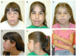
Newborns. All newborns with Cantú syndrome have hypertrichosis with thick scalp hair and excessive hair growth on the forehead, face, back, and extremities. Some have thick and/or curly eyelashes. The hypertrichosis usually persists over time.

Many newborns have macrosomia (large birth weight and birth length) and macrocephaly.

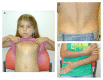
Generalized edema at birth (observed on occasion) usually resolves spontaneously.

Growth. Ultimate adult height is usually within the normal range; however, short stature has been seen in a few individuals.

Macrocephaly, often present at birth, typically persists throughout life. Some individuals who do not have macrocephaly at birth have developed progressive macrocephaly in childhood.

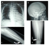
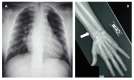
Skeletal abnormalities are usually asymptomatic and identified on radiographs. Characteristic skeletal abnormalities have included thickening of the calvaria, broad ribs, platyspondyly, ovoid vertebral bodies, scoliosis, narrow thorax and shoulders, pectus carinatum, hypoplastic ischium and pubic bones, Erlenmeyer-flask-like long bones with metaphyseal flaring, narrow obturator foramen, and coxa vara.

Gastroesophageal reflux is reported by just under half of individuals. A smaller percentage report intestinal dysfunction characterized by chronic constipation or slow intestinal motility.

Cardiac findings include the following:

  • Cardiac enlargement, with increased ventricular mass but normal chamber wall thickness and enlarged chambers of the heart, often present at birth. Despite the enlarged cardiac chambers, cardiac function is typically normal and ventricular contractility is increased on imaging studies [Grange et al 2006, Grange et al 2019]. Some patients note exercise intolerance, but others have been able to participate in organized sports without difficulty.
  • Patent ductus arteriosus (PDA) in 58% (and described as extremely large in some), often requiring surgical closure in infancy or early childhood
  • Bicuspid aortic valve with and without stenosis
  • Pericardial effusion in about 25% of affected individuals. Small pericardial effusions may be asymptomatic; large fluid accumulations result in symptoms such as exercise intolerance and require intervention.
  • Dilated aortic root and ascending aorta are present in about two thirds of individuals. The natural history is poorly understood. However, development of an aortic aneurysm is rare. Aortic aneurysm requiring surgical intervention was reported in one individual with an ABCC9 pathogenic variant [Hiraki et al 2014].

Tortuous vascularity including tortuous retinal vessels and multiple tortuous pulmonary arteriovenous communications have been reported [Scurr et al 2011, Grange et al 2019]. Abnormal tortuous vasculature in the brain is present in essentially all individuals who have been imaged specifically [Leon Guerrero et al 2016, Grange et al 2019].

Pulmonary hypertension has been reported in infants and young children, although the natural history is not well understood [Cantú et al 1982, Robertson et al 1999, Lazalde et al 2000, Kobayashi et al 2010, Scurr et al 2011]. In one child, pulmonary hypertension secondary to partial pulmonary venous obstruction was associated with severe mitral valve regurgitation that spontaneously resolved by age eight years [Kobayashi et al 2010]. In another individual, progressive (and ultimately fatal) pulmonary hypertension was reported [Park et al 2014]. In the majority of cases, pulmonary hypertension is mild and improves with age [Grange et al 2019].

Generalized edema, which may be present at birth, spontaneously resolves. Subsequently, edema involving the lower extremities and occasionally the arms and hands may develop over time, usually in adolescence or early adulthood. Puffiness of the eyelids is often observed. In one individual, lymphangiography demonstrated dilated lymphatic vessels in the legs with delayed lymphatic drainage [García-Cruz et al 2011]. In contrast, lymphatic studies were normal in another individual [Scurr et al 2011]. Therefore, it is unclear at this time whether the observed swelling is edema or lymphedema.

Intellect. Although the majority of affected individuals have normal intellect, mild learning disabilities and/or developmental delays have been observed, including delay in acquisition of early motor milestones (most likely related to decreased muscle tone) and delay in speech development. Ultimately, most affected individuals attend regular schools, and some are described as having a high IQ [Scurr et al 2011, Grange et al 2019].

Seizures are reported in about one quarter of individuals. Febrile, tonic-clonic, and absence seizure types have been observed as well as temporal lobe epilepsy.

Headaches are reported by many individuals, especially migraine-type headaches with associated aura, photophobia, and phonophobia, and occasionally with transient hemiparesis.

Behavioral problems have been reported in some individuals, including anxiety, mood swings, obsessive-compulsive disorder, and tics [Scurr et al 2011, Grange et al 2019]. Attention-deficit/hyperactivity disorder, autism spectrum disorder, and depression may also be present. Many individuals have self-reported these issues.

Features of a connective tissue abnormality are observed in many individuals with Cantú syndrome, including wrinkled or loose skin especially at birth, deep palmar and plantar creases, and joint hyperextensibility. Some have decreased subcutaneous fat with the appearance of a muscular build in childhood.

Less frequent features

The three individuals reported thus far with a pathogenic variant in KCNJ8 had typical clinical features seen in Cantú syndrome [Brownstein et al 2013, Cooper et al 2014, Chihara et al 2020]. The individual reported by Brownstein et al [2013] had the following additional abnormalities:

  • Brain MRI: cerebral atrophy and thin corpus callosum
  • Multiple tortuous venous collaterals and lack of flow in the inferior sagittal sinus
  • Systemic vasculature: dilated hepatic and celiac arteries, dilated and tortuous intrahepatic arteries and veins

Genotype-Phenotype Correlations

Current information about genotype-phenotype correlation in Cantú syndrome is limited.

No significant genotype-phenotype correlations for ABCC9 or KCNJ8 have been identified.

Penetrance

Penetrance for Cantú syndrome in familial cases reported thus far appears to be complete although with variable expression [Grange et al 2019]. In a few families, somatic mosaicism for an ABCC9 variant has been identified in one of the parents, resulting in much milder phenotypic manifestations [Grange et al 2019; DK Grange, unpublished].

Nomenclature

Cantú syndrome may also be referred to as hypertrichotic osteochondrodysplasia.

Prevalence

The prevalence of Cantú syndrome is unknown. To date, about 150 individuals have been reported with Cantú syndrome. Two previously reported conditions, acromegaloid facial appearance (AFA) syndrome and hypertrichosis with acromegaloid facial features (HAFF) syndrome, are now realized to be cases of Cantú syndrome with variable and sometimes milder phenotypic features. Cantú syndrome has been reported worldwide and in all ethnic groups.

Differential Diagnosis

Table 4.

Genes of Interest in the Differential Diagnosis of Cantú Syndrome

Gene(s) / Genetic MechanismDisorderMOIFeatures of This Disorder
Overlapping w/Cantú SyndromeDistinguishing from Cantú Syndrome
Abnormal methylation at 11p15.5
CDKN1C 1
Beckwith-Wiedemann syndrome Variable 2Neonatal macrosomia; coarse facial features w/macroglossia; umbilical herniaNeonatal hypoglycemia, & hyperinsulinism; ear pits & creases; omphalocele; hemihypertrophy; abdominal tumors in childhood (Wilms tumor, hepatoblastoma)
ATP6V1B2
KCNH1
KCNN3
Zimmermann-Laband syndrome (OMIM PS135500)ADHypertrichosis; coarse facial features; full lips; macrosomia at birth; PDA; aortic root dilatation; scoliosis; hypotoniaGingival hyperplasia or fibromatosis; bulbous nose; distal phalangeal hypoplasia; hypo/aplastic nails; hepatosplenomegaly; seizures; severe ID/DD in some persons
AGPAT2
BSCL2
Berardinelli-Seip congenital lipodystrophy ARMuscular build w/↓ subcutaneous fat in assoc w/cardiomegalyInsulin resistance; diabetes mellitus; hepatomegaly & hepatic steatosis; hypertrophy of skeletal muscles; hypertrophic cardiomyopathy different from cardiac involvement in Cantú syndrome
ANKRD1
BAG3
LMNA
MYBPC3
MYH6
MYH7
SCN5A
TNNT2
TTN
(~30 genes) 3
Dilated cardiomyopathy ADCardiomegalyAbsence of noncardiac findings
(Note: Persons w/Cantú syndrome have normal ventricular wall thickness & normal or enhanced myocardial function [despite enlargement of cardiac chambers] & high cardiac output.)
MYBPC3
MYH7
TNNI3
TNNT2
(~30 genes) 4
Hypertrophic cardiomyopathy AD
KCNK4 FHEIG syndrome (Bauer-Tartaglia syndrome) (OMIM 618381)ADHypertrichosis; coarse facial features; thick scalp hair; large mouth; hypotoniaOcular abnormalities (e.g. nystagmus & optic nerve hypoplasia); severe gingival hyperplasia; brachydactyly; ID/DD; epilepsy; lack of cardiac manifestations
GALNS MPS IVA (Morquio syndrome type A)ARCoarse facial features & hirsutism; some skeletal radiologic features (e.g., thickening of ribs)Flexion contractures; progressively worsening skeletal changes over time; progressive ID & neurologic deterioration in some persons; hepatomegaly & splenomegaly
GNPTAB Mucolipidosis III α/β (See GNPTAB Disorders.)AR
GNPTG Mucolipidosis III gamma AR
IDS MPS II (Hunter syndrome)XL
IDUA Severe MPS IAR
MAN2B1 Alpha-mannosidosis AR

AD = autosomal dominant; AR = autosomal recessive; DD = developmental delay; FHEIG syndrome = facial dysmorphism, hypertrichosis, epilepsy, intellectual/developmental delay, and gingival overgrowth syndrome; ID = intellectual disability; MOI = mode of inheritance; MPS = mucopolysaccharidosis; PDA = patent ductus arteriosus; XL = X-linked

1.

Beckwith-Wiedemann syndrome (BWS) is caused by an epigenetic or genomic alteration leading to abnormal methylation at 11p15.5 or a heterozygous BWS-causing pathogenic variant in CDKN1C.

2.

The risk to the sibs of a child with BWS depends on the genetic basis for BWS in the proband.

3.

Pathogenic variants in ANKRD1, BAG3, LMNA, MYBPC3, MYH6, MYH7, SCN5A, TNNT2, and TTN account for about one third of nonsyndromic dilated cardiomyopathy (DCM); about 30 genes are known to be associated with nonsyndromic DCM (see Dilated Cardiomyopathy Overview).

4.

Pathogenic variants in MYBPC3, MYH7, TNNI3, and TNNT2 account for more than 90% of nonsyndromic hypertrophic cardiomyopathy (HCM); about 30 genes are known to be associated with nonsyndromic HCM (see Hypertrophic Cardiomyopathy Overview).

Other Conditions

Congenital hypothyroidism. The macroglossia and hirsutism that can be seen in congenital hypothyroidism may overlap with features of Cantú syndrome.

Acromegaly. The macrocephaly, coarse facial features, and tall stature in some adults with Cantú syndrome have been confused with acromegaly due to excess human growth hormone.

Minoxidil treatment may lead to coarsening of facial features and hirsutism that has been called "pseudoacromegaly" [Ohko et al 2020]. Minoxidil has long been associated with hair growth and is used topically to treat scalp hair loss. When taken orally, it may cause generalized hirsutism, progressive coarsening of the facial features, and pericardial effusions, all of which can resemble the clinical features of Cantú syndrome.

Diazoxide treatment for hyperinsulinism may lead to hypertrichosis especially on the forehead, back, arms and legs, edema, and rarely pericardial effusion or pulmonary hypertension, which can resemble clinical features of Cantú syndrome [Herrera et al 2018].

Note: Both minoxidil and diazoxide can activate the same ATP-sensitive potassium (KATP) channels that are overactive in Cantú syndrome due to pathogenic variants in ABCC9 or KCNJ8.

Management

Evaluations Following Initial Diagnosis

To establish the extent of disease and needs in an individual diagnosed with Cantú syndrome, the evaluations summarized in Table 5 (if not performed as part of the evaluation that led to the diagnosis) are recommended.

Table 5.

Recommended Evaluations Following Initial Diagnosis in Individuals with Cantú Syndrome

System/ConcernEvaluationComment
Cardiac Cardiology assessment to incl echocardiogram & electrocardiogram
Brain imaging Brain MRI w/MRA & MRVShould be considered for all persons, but esp if history of headaches, migraine headaches, or hemiparesis
Skeletal
  • Radiographic skeletal survey to assess for bone abnormalities
  • Eval for scoliosis
Development Developmental eval for infants & young children
Neuropsychological eval for older personsEspecially for those w/concern for ASD, ADHD, OCD, or depression
Genetic
counseling
By genetics professionals 1To inform affected persons & their families re nature, MOI, & implications of Cantú syndrome to facilitate medical & personal decision making
Family support/
resources
Assess need for:

ADHD = attention-deficit/hyperactivity disorder; ASD = autism spectrum disorder; MOI = mode of inheritance; MRA = magnetic resonance angiogram; MRV = magnetic resonance venography; OCD = obsessive-compulsive disorder

1.

Medical geneticist, certified genetic counselor, or certified advanced genetic nurse

Treatment of Manifestations

Table 6.

Treatment of Manifestations in Individuals with Cantú Syndrome

Manifestation/ConcernTreatmentConsiderations/Other
Cardiac issues PDA, if present, often requires surgical or device closure in infancy or early childhood.
Pericardial effusion, if present, sometimes requires pericardiocentesis.Pericardial stripping may be required to prevent recurrent & hemodynamically significant pericardial effusion.
Peripheral edema Compression stockings & other standard mgmt
Hypertrichosis Referral to dermatologist for treatment optionsPossible treatments: shaving & (in teenagers & adults) use of depilatories or laser hair removal
Scoliosis Bracing or surgical correction
Migraines or other
types of headaches
Referral to neurologist for consideration of medication
Developmental delay See Developmental Delay Management Issues.

PDA = patent ductus arteriosus

Developmental Delay Management Issues

The following information represents typical management recommendations for individuals with developmental delay in the United States; standard recommendations may vary from country to country.

Ages 0-3 years. Referral to an early intervention program is recommended for access to occupational, physical, speech, and feeding therapy as well as infant mental health services, special educators, and sensory impairment specialists. In the US, early intervention is a federally funded program available in all states that provides in-home services to target individual therapy needs.

Ages 3-5 years. In the US, developmental preschool through the local public school district is recommended. Before placement, an evaluation is made to determine needed services and therapies and an individualized education plan is developed for those who qualify based on established motor, language, social, or cognitive delay. The early intervention program typically assists with this transition.

All ages. Consultation with a developmental pediatrician is recommended to ensure the involvement of appropriate community, state, and educational agencies (US) and to support parents in maximizing quality of life.

Motor Delay

Gross motor delay. Physical therapy may be recommended if there are gross motor delays in infancy or early childhood due to hypotonia.

Fine motor delay. Occupational therapy is recommended for difficulty with fine motor skills that affect adaptive function such as feeding, grooming, dressing, and writing.

Oral motor dysfunction should be assessed in infants and clinical feeding evaluations and/or radiographic swallowing studies should be obtained for choking/gagging during feeds, poor weight gain, frequent respiratory illnesses, or feeding refusal that is not otherwise explained. Assuming that the child is safe to eat by mouth, feeding therapy (typically from an occupational or speech therapist) may be needed to help improve coordination or sensory-related feeding issues. Feeds can be thickened or chilled for safety. When feeding dysfunction is severe, an NG-tube or G-tube may be necessary.

Social/Behavioral Concerns

Children may qualify for and benefit from interventions used in treatment of autism spectrum disorder, including applied behavior analysis (ABA). ABA therapy is targeted to the individual child's behavioral, social, and adaptive strengths and weaknesses and typically performed one on one with a board-certified behavior analyst.

Consultation with a developmental pediatrician may be helpful in guiding parents through appropriate behavior management strategies or providing prescription medications, such as medication used to treat attention-deficit/hyperactivity disorder, when necessary.

Surveillance

Table 7.

Recommended Surveillance for Individuals with Cantú Syndrome

System/ConcernEvaluationFrequency
Cardiac issues
  • Echocardiogram & EKG to monitor cardiac size & function & for evidence of pericardial effusion starting in infancy
  • Clinical eval & cardiac biomarkers such as BNP to monitor late development of high-output cardiac failure
Per cardiologist or annually
Peripheral
edema
Monitor w/history & examAnnually starting in adolescence
Cerebral
vasculature
abnormality
Brain MRI w/MRA & MRVIf persistent headaches or other neurologic symptoms develop
Headaches/
Migraines
Eval by neurologistPer neurologist
Scoliosis
  • Spine radiographs
  • Orthopedic eval if scoliosis is present
If concern based on physical exam

BNP = brain natriuretic peptide; EKG = electrocardiogram; MRA = magnetic resonance angiogram; MRV = magnetic resonance venography

Agents/Circumstances to Avoid

Avoid the following:

  • Minoxidil
  • Diazoxide
  • Angiotensin-converting enzyme inhibitors

Evaluation of Relatives at Risk

It is appropriate to clarify the genetic/clinical status of older and younger relatives of an affected individual in order to identify as early as possible those who should be evaluated and monitored for cardiac manifestations of Cantú syndrome, as well as peripheral edema and scoliosis (see Evaluations Following Initial Diagnosis and Surveillance).

Evaluations can include:

  • Molecular genetic testing if the causative pathogenic variant in the family is known;
  • Complete physical examination to assess for the characteristic clinical features, as well as an echocardiogram, electrocardiogram, and skeletal survey, should be performed if the pathogenic variant in the family is not known. Additional studies such as brain MRI with magnetic resonance angiogram and magnetic resonance venography may be indicated.

See Genetic Counseling for issues related to testing of at-risk relatives for genetic counseling purposes.

Pregnancy Management

Women affected by Cantú syndrome should be referred to a maternal-fetal medicine specialist for evaluation and management.

Therapies Under Investigation

Search ClinicalTrials.gov in the US and EU Clinical Trials Register in Europe for access to information on clinical studies for a wide range of diseases and conditions. Note: There may not be clinical trials for this disorder.

Genetic Counseling

Genetic counseling is the process of providing individuals and families with information on the nature, mode(s) of inheritance, and implications of genetic disorders to help them make informed medical and personal decisions. The following section deals with genetic risk assessment and the use of family history and genetic testing to clarify genetic status for family members; it is not meant to address all personal, cultural, or ethical issues that may arise or to substitute for consultation with a genetics professional. —ED.

Mode of Inheritance

Cantú syndrome is inherited in an autosomal dominant manner.

Risk to Family Members

Parents of a proband

  • In one study, 22% of individuals diagnosed with Cantú syndrome had an affected parent [Grange et al 2019].
  • Approximately 75% to 80% of individuals diagnosed with Cantú syndrome have the disorder as the result of a de novo pathogenic variant.
  • If a molecular diagnosis has been established in the proband and the proband appears to be the only affected family member (i.e., a simplex case), molecular genetic testing is recommended for the parents of the proband to confirm their genetic status and to allow reliable recurrence risk counseling.
  • If the causative ABCC9 or KCNJ8 pathogenic variant found in the proband is not identified in either parent, the following possibilities should be considered:
  • The family history of some individuals diagnosed with Cantú syndrome may appear to be negative because of failure to recognize the disorder in family members with a milder phenotype. Therefore, an apparently negative family history cannot be confirmed unless appropriate clinical evaluation and/or molecular genetic testing has been performed on the parents of the proband.

Sibs of a proband. The risk to the sibs of the proband depends on the clinical/genetic status of the proband's parents.

  • If a parent of the proband is affected and/or has the ABCC9 or KCNJ8 pathogenic variant identified in the proband, the risk to the sibs of inheriting the pathogenic variant is 50%. Because Cantú syndrome is associated with intrafamilial clinical variability, the phenotype, age of onset, and severity in sibs who inherit a pathogenic variant are not predictable [Grange et al 2019].
  • If the proband has a known Cantú syndrome-related pathogenic variant that cannot be detected in the leukocyte DNA of either parent, the recurrence risk to sibs is slightly greater than that of the general population because of the possibility of parental germline mosaicism [Grange et al 2019].
  • If the parents are clinically unaffected but their genetic status is unknown, risk to sibs of a proband is presumed to be low but increased over that of the general population because of the possibility of reduced penetrance in a heterozygous parent or parental germline mosaicism.

Offspring of a proband. Each child of an individual with Cantú syndrome has a 50% chance of inheriting the pathogenic variant; the phenotype, age of onset, and severity in offspring who inherit a causative pathogenic variant are not predictable.

Other family members. The risk to other family members depends on the status of the proband's parents: if a parent has a causative pathogenic variant, his or her family members may be at risk.

Related Genetic Counseling Issues

See Management, Evaluation of Relatives at Risk for information on evaluating at-risk relatives for the purpose of early diagnosis and treatment.

Family planning

  • The optimal time for determination of genetic risk and discussion of the availability of prenatal/preimplantation genetic testing is before pregnancy.
  • It is appropriate to offer genetic counseling (including discussion of potential risks to offspring and reproductive options) to young adults who are affected or at risk.

DNA banking. Because it is likely that testing methodology and our understanding of genes, pathogenic mechanisms, and diseases will improve in the future, consideration should be given to banking DNA from probands in whom a molecular diagnosis has not been confirmed (i.e., the causative pathogenic mechanism is unknown).

Prenatal Testing and Preimplantation Genetic Testing

If the Cantú syndrome-causing pathogenic variant has been identified in an affected family member, prenatal and preimplantation genetic testing are possible. The phenotype, age of onset, and severity are not predictable based on results of prenatal molecular genetic testing.

If the Cantú syndrome-causing pathogenic variant has not been identified in an affected family member, evaluation of a pregnancy at increased risk is recommended using ultrasound to look for fetal macrosomia and polyhydramnios and a fetal echocardiogram to evaluate for cardiovascular abnormalities.

In a fetus not known to be at increased risk for Cantú syndrome based on family history, the combination of fetal macrosomia and polyhydramnios should lead to consideration of Cantú syndrome. (Note: The etiology of polyhydramnios is diverse.)

Differences in perspective may exist among medical professionals and within families regarding the use of prenatal testing. While most centers would consider use of prenatal testing to be a personal decision, discussion of these issues may be helpful.

Resources

GeneReviews staff has selected the following disease-specific and/or umbrella support organizations and/or registries for the benefit of individuals with this disorder and their families. GeneReviews is not responsible for the information provided by other organizations. For information on selection criteria, click here.

Molecular Genetics

Information in the Molecular Genetics and OMIM tables may differ from that elsewhere in the GeneReview: tables may contain more recent information. —ED.

Table A.

Cantú syndrome: Genes and Databases

Data are compiled from the following standard references: gene from HGNC; chromosome locus from OMIM; protein from UniProt. For a description of databases (Locus Specific, HGMD, ClinVar) to which links are provided, click here.

Table B.

OMIM Entries for Cantú syndrome (View All in OMIM)

239850CANTU SYNDROME
600935POTASSIUM CHANNEL, INWARDLY RECTIFYING, SUBFAMILY J, MEMBER 8; KCNJ8
601439ATP-BINDING CASSETTE, SUBFAMILY C, MEMBER 9; ABCC9

Molecular Pathogenesis

ABCC9 and KCNJ8 encode the regulatory (SUR2) and pore-forming (Kir6.1) subunits of ATP-sensitive potassium (KATP) channels, respectively. KATP channels are prominent in cardiac, skeletal, and smooth muscle, as well as endothelial and other tissues. KATP channels are inhibited by nonhydrolytic binding of ATP, whereas MgADP has the opposite effect, activating channels through interaction with SUR subunits. Physiologic activity is thus effectively dependent on the [ADP]:[ATP] ratio. KATP activation results in membrane hyperpolarization and decrease in voltage-dependent Ca2+ entry, leading to inhibition of muscle contraction. All identified Cantú syndrome pathogenic variants result in increased SUR2/Kir6.2-dependent KATP channel activity. Primary reduction of smooth muscle activity is a major consequence, underlying at least the cardiovascular features.

Mechanism of disease causation. Cantú syndrome is caused by gain-of-function variants in either ABCC9 or KCNJ8.

Table 8.

Notable ABCC9 Pathogenic Variants

Reference SequencesDNA Nucleotide ChangePredicted Protein ChangeComment [Reference]
NM_005691​.4

NP_005682​.2

c.3461G>Ap.Arg1154GlnRecurrent variants [Grange et al 2019]
c.3460C>Tp.Arg1154Trp
c.3346C>Tp.Arg1116Cys
c.3347G>Ap.Arg1116His

Variants listed in the table have been provided by the authors. GeneReviews staff have not independently verified the classification of variants.

GeneReviews follows the standard naming conventions of the Human Genome Variation Society (varnomen​.hgvs.org). See Quick Reference for an explanation of nomenclature.

Chapter Notes

Author Notes

Dorothy K Grange, MD

Colin G Nichols, PhD

Dr Nichols's research is focused on the biology of ion channels, with emphasis on the molecular basis of potassium channel activity and the role of potassium channels in physiology and disease. Using various molecular biological and biophysical approaches, his laboratory is developing detailed understanding of the structural basis of channel activity, and animal models to understand the role of potassium channels in disease processes including diabetes, cardiovascular pathology and arrhythmias, and epilepsy.

Gautam K Singh, MD

Revision History

  • 1 October 2020 (ha) Comprehensive update posted live
  • 2 October 2014 (me) Review posted live
  • 2 October 2013 (dkg) Original submission

References

Literature Cited

  • Antzelevitch C, Yan GX. J wave syndromes. Heart Rhythm. 2010;7:549–58. [PMC free article: PMC2843811] [PubMed: 20153265]
  • Bienengraeber M, Olson TM, Selivanov VA, Kathmann EC, O'Cochlain F, Gao F, Karger AB, Ballew JD, Hodgson DM, Zingman LV, Pang YP, Alekseev AE, Terzic A. ABCC9 mutations identified in human dilated cardiomyopathy disrupt catalytic KATP channel gating. Nat Genet. 2004;36:382–7. [PMC free article: PMC1995438] [PubMed: 15034580]
  • Brownstein CA, Towne MC, Luquette LJ, Harris DJ, Marinakis NS, Meinecke P, Kutsche K, Campeau PM, Yu TW, Margulies DM, Agrawal PB, Beggs AH. Mutation of KCNJ8 in a patient with Cantú syndrome with unique vascular abnormalities - support for the role of K(ATP) channels in this condition. Eur J Med Genet. 2013;56:678–82. [PMC free article: PMC3902017] [PubMed: 24176758]
  • Cantú JM, García-Cruz D, Sánchez-Corona J, Hernández A, Nazará Z. A distinct osteochondrodysplasia with hypertrichosis-individualization of a probable autosomal recessive entity. Hum Genet. 1982;60:36–41. [PubMed: 7076246]
  • Chihara M, Asahina A, Itoh M. A novel mutation in the KCNJ8 gene encoding the Kir6.1 subunit of an ATP-sensitive potassium channel in a Japanese patient with Cantú syndrome. J Eur Acad Dermatol Venereol. 2020;34:e476–e478. [PubMed: 32215968]
  • Cooper PE, Reutter H, Woelfle J, Engels H, Grange DK, van Haaften G, van Bon BW, Hoischen A, Nichols CG. Cantú syndrome resulting from activating mutation in the KCNJ8 gene. Hum Mutat. 2014;35:809–13. [PMC free article: PMC4277879] [PubMed: 24700710]
  • García-Cruz D, Mampel A, Echeverria MI, Vargas AL, Castañeda-Cisneros G, Davalos-Rodriguez N, Patiño-Garcia B, Garcia-Cruz MO, Castañeda V, Cardona EG, Marin-Solis B, Cantu JM, Nuñez-Reveles N, Moran-Moguel C, Thavanati PK, Ramirez-Garcia S, Sanchez-Corona J. Cantu syndrome and lymphoedema. Clin Dysmorphol. 2011;20:32–7. [PubMed: 20890180]
  • Grange DK, Lorch SM, Cole PL, Singh GK. Cantú syndrome in a woman and her two daughters: further confirmation of autosomal dominant inheritance and review of the cardiac manifestations. Am J Med Genet. 2006;140:1673–80. [PubMed: 16835932]
  • Grange DK, Roessler HI, McClenaghan C, Duran K, Shields K, Remedi MS, Knoers NVAM, Lee J-M, Kirk EP, Scurr I, Smithson SF, Singh GK, van Haelst MM, Nichols CG, van Haaften G. Cantu syndrome: findings from 74 patients in the International Cantu Syndrome Registry. Am J Med Genet C Semin Med Genet. 2019;181:658–81. [PMC free article: PMC7654223] [PubMed: 31828977]
  • Herrera A, Vajravelu ME, Givler S, Mitteer L, Avitabile CM, Lord K, De León DD. Prevalence of adverse events in children with congenital hyperinsulinism treated with diazoxide. J Clin Endocrinol Metab. 2018;103:4365–72. [PMC free article: PMC6207144] [PubMed: 30247666]
  • Hiraki Y, Miyatake S, Hayashidani M, Nishimura Y, Matsuura H, Kamada M, Kawagoe T, Yunoki K, Okamoto N, Yofune H, Nakashima M, Tsurusaki Y, Satisu H, Murakami A, Miyake N, Nishimura G, Matsumoto N. Aortic aneurysm and craniosynostosis in a family with Cantú syndrome. Am J Med Genet A. 2014;164A:231–6. [PubMed: 24352916]
  • Hu D, Barajas-Martínez H, Terzic A, Park S, Pfeiffer R, Burashnikov E, Wu Y, Borggrefe M, Veltmann C, Schimpf R, Cai JJ, Nam GB, Deshmukh P, Scheinman M, Preminger M, Steinberg J, López-Izquierdo A, Ponce-Balbuena D, Wolpert C, Haïssaguerre M, Sánchez-Chapula JA, Antzelevitch C. ABCC9 is a novel Brugada and early repolarization syndrome susceptibility gene. Int J Cardiol. 2014;171:431–42. [PMC free article: PMC3947869] [PubMed: 24439875]
  • Kobayashi D, Cook AL, Williams DA. Pulmonary hypertension secondary to partial pulmonary venous obstruction in a child with Cantú syndrome. Pediatr Pulmonol. 2010;45:727–9. [PubMed: 20575102]
  • Lazalde B, Sanchez-Urbina R, Nuno-Arana I, Bitar WE, Ramirez-Duenas M. Autosomal dominant inheritance in Cantú syndrome (congenital hypertrichosis, osteochondrodysplasia, and cardiomegaly). Am J Med Genet. 2000;94:421–7. [PubMed: 11050630]
  • Leon Guerrero CR, Pathak S, Grange DK, Singh GK, Nichols CG, Lee JM, Vo KD. Neurologic and neuroimaging manifestations of Cantu syndrome: a case series. Neurology. 2016;87:270–6. [PMC free article: PMC4955281] [PubMed: 27316244]
  • Marques P, Spencer R, Morrison PJ, Carr IM, Dang MN, Bonthron DT, Hunter S, Korbonits M. Cantu syndrome with coexisting familial pituitary adenoma. Endocrine. 2018;59:677–84. [PMC free article: PMC5847123] [PubMed: 29327300]
  • Ohko K, Nakajima K, Nakajima H, Hiraki Y, Kubota K, Fukao T, Miyatake S, Matsumoto N, Sano S. Skin and hair abnormalities of Cantu syndrome: a congenital hypertrichosis due to a genetic alteration mimicking the pharmacological effect of minoxidil. J Dermatol. 2020;47:306–10. [PubMed: 31907964]
  • Olson TM, Alekseev AE, Moreau C, Liu XK, Zingman LV, Miki T, Seino S, Asirvatham SJ, Jahangir A, Terzic A. KATP channel mutation confers risk for vein of Marshall adrenergic atrial fibrillation. Nat Clin Pract Cardiovasc Med. 2007;4:110–6. [PMC free article: PMC2013306] [PubMed: 17245405]
  • Park JY, Koo SH, Jung YJ, Lim YJ, Chung ML. A patient with Cantú syndrome associated with fatal bronchopulmonary dysplasia and pulmonary hypertension. Am J Med Genet A. 2014;164A:2118–20. [PubMed: 24715715]
  • Richards S, Aziz N, Bale S, Bick D, Das S, Gastier-Foster J, Grody WW, Hegde M, Lyon E, Spector E, Voelkerding K, Rehm HL, et al. Standards and guidelines for the interpretation of sequence variants: a joint consensus recommendation of the American College of Medical Genetics and Genomics and the Association for Molecular Pathology. Genet Med. 2015;17:405–24. [PMC free article: PMC4544753] [PubMed: 25741868]
  • Robertson SP, Kirk E, Bernier F, Brereton J, Turner A, Bankier A. Congenital hypertrichosis. Osteochondrodysplasia and cardiomegaly: Cantú syndrome. Am J Med Genet. 1999;85:395–402. [PubMed: 10398267]
  • Scurr I, Wilson L, Lees M, Robertson S, Kirk E, Turner A, Morton J, Kidd A, Shashi V, Stanley C, Berry M, Irvine AD, Goudie D, Turner C, Brewer C, Smithson S. Cantú syndrome: report of nine new cases and expansion of the clinical phenotype. Am J Med Genet A. 2011;155A:508–18. [PubMed: 21344641]
  • Smeland MF, McClenaghan C, Roessler HI, Savelberg S, Hansen GÅM, Hjellnes H, Arntzen KA, Müller KI, Dybesland AR, Harter T, Sala-Rabanal M, Emfinger CH, Huang Y, Singareddy SS, Gunn J, Wozniak DF, Kovacs A, Massink M, Tessadori F, Kamel SM, Bakkers J, Remedi MS, Van Ghelue M, Nichols CG, van Haaften G. ABCC9-related Intellectual disability myopathy syndrome is a K(ATP) channelopathy with loss-of-function mutations in ABCC9. Nat Commun. 2019;10:4457. [PMC free article: PMC6773855] [PubMed: 31575858]
  • Theis NJ, Calvert T, McIntyre P, Robertson SP, Wheeler BJ. Cantu syndrome and hypopituitarism: implications for endocrine monitoring. Endocrinol Diabetes Metab Case Rep. 2019;2019:19–0103. [PMC free article: PMC6865860] [PubMed: 31743099]
  • Veeramah KR, Karafet TM, Wolf D, Samson RA, Hammer MF. The KCNJ8-S422L variant previously associated with J-wave syndromes is found at an increased frequency in Ashkenazi Jews. Eur J Hum Genet. 2014;22:94–8. [PMC free article: PMC3865407] [PubMed: 23632791]
Copyright © 1993-2024, University of Washington, Seattle. GeneReviews is a registered trademark of the University of Washington, Seattle. All rights reserved.

GeneReviews® chapters are owned by the University of Washington. Permission is hereby granted to reproduce, distribute, and translate copies of content materials for noncommercial research purposes only, provided that (i) credit for source (http://www.genereviews.org/) and copyright (© 1993-2024 University of Washington) are included with each copy; (ii) a link to the original material is provided whenever the material is published elsewhere on the Web; and (iii) reproducers, distributors, and/or translators comply with the GeneReviews® Copyright Notice and Usage Disclaimer. No further modifications are allowed. For clarity, excerpts of GeneReviews chapters for use in lab reports and clinic notes are a permitted use.

For more information, see the GeneReviews® Copyright Notice and Usage Disclaimer.

For questions regarding permissions or whether a specified use is allowed, contact: ude.wu@tssamda.

Bookshelf ID: NBK246980PMID: 25275207

Views

Tests in GTR by Gene

Related information

  • OMIM
    Related OMIM records
  • PMC
    PubMed Central citations
  • PubMed
    Links to PubMed
  • Gene
    Locus Links

Similar articles in PubMed

See reviews...See all...

Recent Activity

Your browsing activity is empty.

Activity recording is turned off.

Turn recording back on

See more...