U.S. flag

An official website of the United States government

NCBI Bookshelf. A service of the National Library of Medicine, National Institutes of Health.

Adam MP, Feldman J, Mirzaa GM, et al., editors. GeneReviews® [Internet]. Seattle (WA): University of Washington, Seattle; 1993-2024.

Cover of GeneReviews®

GeneReviews® [Internet].

Show details

21-Hydroxylase-Deficient Congenital Adrenal Hyperplasia

Synonyms: 21-OHD CAH, Virilizing Adrenal Hyperplasia

, MD, , MBBS, , MD, and , MD.

Author Information and Affiliations

Initial Posting: ; Last Update: February 4, 2016.

Estimated reading time: 36 minutes

Summary

Clinical characteristics.

21-hydroxylase deficiency (21-OHD) is the most common cause of congenital adrenal hyperplasia (CAH), a family of autosomal recessive disorders involving impaired synthesis of cortisol from cholesterol by the adrenal cortex. In 21-OHD CAH, excessive adrenal androgen biosynthesis results in virilization in all individuals and salt wasting in some individuals. A classic form with severe enzyme deficiency and prenatal onset of virilization is distinguished from a non-classic form with mild enzyme deficiency and postnatal onset. The classic form is further divided into the simple virilizing form (~25% of affected individuals) and the salt-wasting form, in which aldosterone production is inadequate (≥75% of individuals). Newborns with salt-wasting 21-OHD CAH are at risk for life-threatening salt-wasting crises. Individuals with the non-classic form of 21-OHD CAH present postnatally with signs of hyperandrogenism; females with the non-classic form are not virilized at birth.

Diagnosis/testing.

The diagnosis of classic 21-OHD CAH is established in newborns with characteristic clinical features, elevated serum 17-OHP, and elevated adrenal androgens. The diagnosis of non-classic 21-OHD is established by comparison of baseline serum 17-OHP and ACTH-stimulated serum 17-OHP or early morning elevated 17-OHP. Identification of biallelic pathogenic variants in CYP21A2 confirms the clinical diagnosis and allows for family studies.

Management.

Treatment of manifestations: Classic 21-OHD CAH: glucocorticoid replacement therapy, which needs to be increased during periods of stress. Salt-wasting form: mineralocorticoid 9α-fludrohydrocortisone therapy and often sodium chloride. Females who are virilized at birth may require feminizing genitoplasty and/or vaginal dilation. Symptomatic individuals with non-classic 21-OHD CAH may require treatment.

Prevention of primary manifestations: Newborn screening programs aim to identify infants with classic 21-OHD CAH in order to initiate glucocorticoid and mineralocorticoid treatment prior to a potentially life-threatening salt-wasting crisis.

Surveillance: Monitor:

  • Efficacy of glucocorticoid and mineralocorticoid replacement therapy every three to four months while children are actively growing, and less often thereafter;
  • For testicular adrenal rest tumors in males every three to five years after onset of puberty;
  • Weight, bone mineral density, fertility, cardiovascular and metabolic risks in adults.

Evaluation of relatives at risk: It is appropriate to measure 17-hydroxyprogesterone (17-OHP) of at-risk sibs to facilitate early diagnosis and treatment.

Genetic counseling.

21-OHD CAH is inherited in an autosomal recessive manner. Most parents are heterozygous for a pathogenic variant. Approximately 1% of pathogenic variants are de novo; thus, 1% of probands have only one parent who is heterozygous. In some instances during evaluation of a proband, a parent not previously known to be affected may be found to have biallelic pathogenic variants and the non-classic form of 21-OHD CAH. At conception, if the parents of a proband are both known to be heterozygotes, each sib has a 25% chance of being affected, a 50% chance of being an asymptomatic carrier, and a 25% chance of being unaffected and not a carrier. Carrier testing for at-risk relatives and prenatal testing for a pregnancy at increased risk are possible if the pathogenic variants in the family are known.

GeneReview Scope

21-Hydroxylase-Deficient Congenital Adrenal Hyperplasia: Included Phenotypes
  • Classic simple virilizing 21-OHD CAH
  • Classic salt-wasting 21-OHD CAH
  • Non-classic 21-OHD CAH

For synonyms and outdated names see Nomenclature.

Diagnosis

Suggestive Findings

21-hydroxylase-deficient congenital adrenal hyperplasia (21-OHD CAH) should be suspected in the following individuals:

  • Females who are virilized at birth, or who become virilized postnatally, or who have precocious puberty or adrenarche. Virilization affects maturation, growth (leading to tall stature), and sex hormone-sensitive areas (external genitalia, skin, and hair) (leading to secondary sexual characteristics).
  • Males with masculinization in childhood (i.e., premature adrenarche)
  • Any infant with a salt-losing crisis in the first four weeks of life. Individuals with untreated or poorly controlled salt wasting may have a decreased serum concentration of sodium, chloride, and total carbon dioxide (CO2), an increased serum concentration of potassium, and inappropriately increased urine concentration of sodium.
  • An infant with elevated 17-OHP concentration detected as positive newborn screening

Note: Females with 21-OHD CAH have a normal 46,XX karyotype; males with 21-OHD CAH have a normal 46,XY karyotype.

Newborn Screening

Newborn screening for 21-OHD CAH serves two purposes:

  • To identify infants, especially males, with the classic form of 21-OHD CAH who are at risk for life-threatening salt-wasting crises
  • To expedite the diagnosis of females with ambiguous genitalia

Note: Newborn screening rarely detects individuals with the non-classic form of 21-OHD CAH [Votava et al 2005].

As with newborn screening for other disorders, the concentration of 17-OHP is measured on a filter paper blood spot sample obtained by the heel-stick technique.

  • The majority of screening programs use a single screening test without retesting of samples with questionable 17-OHP concentrations. See Speiser et al [2010] (full text).
  • To improve efficacy of screening, some screening programs reevaluate samples with borderline first-tier test results with a second-tier test and some implement repeat screening in this situation [Sarafoglou et al 2012, Chan et al 2013]. Because of the high false-positive rate of immunoassay methods, liquid chromatography-tandem mass spectrometry was recommended as a second-tier test [Speiser et al 2010]. Some programs measure the concentration of different hormones (17-OHP, 21-deoxycortisol, and cortisol) as a second-tier test on samples with a positive first-tier test result [Janzen et al 2007]. Some US states mandate organic solvent extraction prior to immunoassay of dried blood spots in order to increase specificity.

Note: (1) Results on blood samples taken in the first 24 hours of life are elevated in all infants and may give false-positive results. (2) False-positive results may also be observed in low birth-weight infants or premature infants. Therefore, birth weight- or gestational age-adjusted normative data is used to determine if a test result is screen positive. (3) False-negative results may be observed in neonates receiving dexamethasone for management of unrelated problems.

Establishing the Diagnosis

21-OHD CAH. The diagnosis is established in a newborn with the following laboratory findings:

  • Serum 17-OHP is markedly elevated.
  • Adrenal androgens are elevated; Δ4-androstenedione, 21 deoxycortisol, and progesterone are increased in males and females with 21-OHD CAH; Testosterone and adrenal androgen precursors (Δ4-androstenedione, DHEA) are increased in affected females and prepubertal males.
  • Plasma renin activity is markedly elevated in individuals with the salt-wasting form of 21-OHD CAH.
    Note: In individuals with the salt-wasting form of 21-OHD CAH, the serum concentration of aldosterone is inappropriately low compared to the level of plasma renin activity (PRA) elevation. A reduced ratio of aldosterone to PRA indicates impaired aldosterone synthesis and can differentiate those individuals with the salt-wasting form of CAH from those with the simple virilizing form of CAH after the newborn period [Nimkarn et al 2007].
  • Identification of biallelic pathogenic variants in CYP21A2 (See Table 2.)

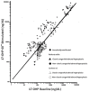
Non-classic 21-OHD CAH. The diagnosis is established in a proband based on the results of ONE of the two following laboratory tests (see Figure 1 and Table 1):

Figure 1. . 17-OHP nomogram for the diagnosis of steroid 21-hydroxylase deficiency (60-minute Cortrosyn™ stimulation test).

Figure 1.

17-OHP nomogram for the diagnosis of steroid 21-hydroxylase deficiency (60-minute Cortrosyn™ stimulation test). The data for this nomogram were collected between 1982 and 1991 at the Department of Pediatrics, the New York Hospital-Cornell Medical (more...)

  • 60-minute ACTH stimulation test. The serum concentration of 17-OHP measured at baseline and at 60 minutes after intravenous injection of a standard 250-µg bolus of synthetic ACTH (Cortrosyn™) are plotted on the nomogram in Figure 1.
  • 17-hydroxyprogesterone (17-OHP). A single early-morning (<8AM) measurement of plasma 17-OHP concentration (baseline values in affected individuals are not always elevated; see Table 1)
    Note: Normal ranges of 17-OHP for sex and pubertal status vary by laboratory, reflecting the methods used. In adult females, normal ranges depend on the phase of the menstrual cycle.

Table 1.

Diagnosis of 21-OHD CAH after Infancy Based on 17 OHP Levels

Classic FormNon-Classic FormUnaffected
Baseline 17-OHP level >10,000 ng/dL or 300 nmol/L200-10,000 ng/dL or 6-300 nmol/L 1<200 ng/dL or 6 nmol/L 1
17-OHP level after ACTH stimulation >10,000 ng/dL or 300 nmol/L1,000-10,000 ng/dL or 31-300 nmol/L<1,000 ng/dL or 50 nmol/L

Modified from Speiser et al [2010]

1.

Randomly measured 17-OHP can be normal in the non-classic form.

Molecular testing. Identification of biallelic pathogenic variants in CYP21A2 (see Table 2) confirms the diagnosis and allows for family studies. Molecular testing approaches can include single-gene testing, use of a multigene panel, and more comprehensive genomic testing:

  • Single-gene testing. Sequence analysis of CYP21A2 is performed first and followed by gene-targeted deletion/duplication analysis if only one or no pathogenic variant is found.
    Note: A large-scale gene conversion (see Molecular Genetics) can replace a large segment of functional CYP21A2 sequence with a segment of the CYP21A1P pseudogene that is nonfunctional as a result of more than one deleterious variant [Mao et al 2002]. Thus, when targeted analysis detects multiple pathogenic variants, it is possible that the pathogenic variants are either in trans configuration (i.e., are on separate chromosomes, one inherited from each parent) or in cis configuration (i.e., are on the same chromosome and thus represent only one mutated allele rather than two; most likely arising from gene conversion). To avoid diagnostic errors, studying both parents as well as the proband is recommended to confirm the pathogenic variants and to determine if they are in cis configuration or trans configuration.
  • A multigene panel that includes CYP21A2 and other genes of interest (see Differential Diagnosis) may also be considered. Note: (1) The genes included in the panel and the diagnostic sensitivity of the testing used for each gene vary by laboratory and are likely to change over time. (2) Some multigene panels may include genes not associated with the condition discussed in this GeneReview; thus, clinicians need to determine which multigene panel is most likely to identify the genetic cause of the condition while limiting identification of variants of uncertain significance and pathogenic variants in genes that do not explain the underlying phenotype. (3) In some laboratories, panel options may include a custom laboratory-designed panel and/or custom phenotype-focused exome analysis that includes genes specified by the clinician. (4) Methods used in a panel may include sequence analysis, deletion/duplication analysis, and/or other non-sequencing-based tests.
    For an introduction to multigene panels click here. More detailed information for clinicians ordering genetic tests can be found here.
  • More comprehensive genomic testing (when available) including exome sequencing, genome sequencing, and mitochondrial sequencing may be considered if serial single-gene testing (and/or use of a multigene panel) fails to confirm a diagnosis in an individual with features of 21-OHD CAH.
    For an introduction to comprehensive genomic testing click here. More detailed information for clinicians ordering genomic testing can be found here.

Table 2.

Molecular Genetic Testing Used in 21-Hydroxylase-Deficient Congenital Adrenal Hyperplasia

Gene 1MethodProportion of Probands with Pathogenic Variants 2 Detectable by Method
CYP21A2 Sequence analysis 3~70%-80% 4
Gene-targeted deletion/duplication analysis 5~20%-30% 6
1.
2.

See Molecular Genetics for information on allelic variants detected in this gene.

3.

Sequence analysis detects variants that are benign, likely benign, of uncertain significance, likely pathogenic, or pathogenic. Variants may include small intragenic deletions/insertions and missense, nonsense, and splice site variants; typically, exon or whole-gene deletions/duplications are not detected. For issues to consider in interpretation of sequence analysis results, click here.

4.

The majority of individuals from heterogeneous populations with 21-OHD CAH are compound heterozygotes [Krone et al 2000, New et al 2013].

5.

Gene-targeted deletion/duplication analysis detects intragenic deletions or duplications. Methods used may include quantitative PCR, long-range PCR, multiplex ligation-dependent probe amplification (MLPA), and a gene-targeted microarray designed to detect single-exon deletions or duplications.

6.

Approximately 20% of mutated alleles are deleted for a 30-kb gene segment that encompasses the 3' end of the CYP21A1P pseudogene, all of the adjacent C4B complement gene, and the 5' end of CYP21A2 (see Molecular Genetics).

Clinical Characteristics

Clinical Description

21-hydroxylase-deficient congenital adrenal hyperplasia (21-OHD CAH) occurs in a classic form and a non-classic form (Table 3).

In classic 21-OHD CAH prenatal exposure to potent androgens such as testosterone and Δ4-androstenedione at critical stages of sexual development virilizes the external genitalia of genetic females, often resulting in genital ambiguity at birth. The classic form is further divided into the simple virilizing form (~25% of individuals) and the salt-wasting form, in which aldosterone production is inadequate (≥75% of individuals). Newborns with salt-wasting CAH caused by 21-OHD CAH are at risk for life-threatening salt-wasting crises.

Individuals with the non-classic form of 21-OHD CAH have only moderate enzyme deficiency and present postnatally with signs of hyperandrogenism; females with the non-classic form are not virilized at birth.

Table 3.

Clinical Features in Individuals with Classic and Non-Classic 21-OHD CAH

Feature21-OHD CAH
ClassicNon-Classic
Prenatal virilization Present in femalesAbsent
Postnatal virilization Males and femalesVariable
Salt wasting ~75% of all individualsAbsent
Cortisol deficiency ~100%Rare

Classic Simple Virilizing 21-OHD CAH

Excess adrenal androgen production in utero results in genital virilization at birth in 46,XX females. In affected females, the excess androgens result in varying degrees of enlargement of the clitoris, fusion of the labioscrotal folds, and formation of a urogenital sinus. Because anti-müllerian hormone (AMH) is not secreted, the müllerian ducts develop normally into a uterus and fallopian tubes in affected females. It is not possible to distinguish between classic simple virilizing 21-OHD CAH and classic salt-wasting 21-OHD CAH based solely on the degree of virilization of an affected female at birth.

After birth, both females and males with classic simple virilizing 21-OHD CAH who do not receive glucocorticoid replacement therapy develop signs of androgen excess including precocious development of pubic and axillary hair, acne, rapid linear growth, and advanced bone age. Untreated males have progressive penile enlargement and small testes. Untreated females have clitoral enlargement, hirsutism, male pattern baldness, menstrual abnormalities, and reduced fertility.

The initial growth in the young child with untreated 21-OHD CAH is rapid; however, potential height is reduced and short adult stature results from premature epiphyseal fusion. Even if treatment with cortisol replacement therapy begins at an early age and secretion of excess adrenal androgens is controlled, individuals with 21-OHD CAH do not generally achieve the expected adult height. Bone age may be advanced compared to chronologic age.

Pubertal development. In boys and girls with proper glucocorticoid therapy and suppression of excessive adrenal androgen production, onset of puberty usually occurs at the appropriate chronologic age. However, exceptions occur even among individuals in whom the disease is well controlled [Trinh et al 2007].

It should be noted that in some previously untreated children, the start of glucocorticoid replacement therapy triggers true precocious puberty. This central precocious puberty may occur when glucocorticoid treatment releases the hypothalamic pituitary axis from inhibition by estrogens derived from excess adrenal androgen secretion.

Fertility. For most females who are adequately treated, menses are normal after menarche and pregnancy is possible [Lo et al 1999]. Overall fertility rates, however, are reported to be low. Reported reasons include inadequate vaginal introitus leading to unsatisfactory intercourse, pain with vaginal penetration [Gastaud et al 2007], elevated androgens leading to ovarian dysfunction, and psychosexual behaviors around gender identity and selection of sexual partner(s). Chronic anovulation, elevated progestin levels, and aberrant endometrial implantation have also been identified as reasons for subfertility [Witchel 2012].

In males, the main cause of subfertility is the presence of testicular adrenal rest tumors, which are thought to originate from aberrant adrenal tissue. In addition, hypogonadotropic hypogonadism may result from suppression of LH secretion by the pituitary by excessive adrenal androgens and their aromatization products [Ogilvie et al 2006a].

Adrenal medulla. In individuals with classic 21-OHD CAH, deficiency of cortisol also affects the development and functioning of the adrenal medulla, resulting in lower epinephrine and metanephrine concentrations than those found in unaffected individuals [Merke et al 2000].

Classic salt-wasting 21-OHD CAH. When the loss of 21-hydroxylase function is severe, adrenal aldosterone secretion is insufficient for sodium reabsorption by the distal renal tubules, resulting in salt wasting as well as cortisol deficiency and androgen excess. Infants with renal salt wasting have poor feeding, weight loss, failure to thrive, vomiting, dehydration, hypotension, hyponatremia, and hyperkalemic metabolic acidosis progressing to adrenal crisis (azotemia, vascular collapse, shock, and death). Adrenal crisis can occur as early as age one to four weeks.

Affected males who are not detected in a newborn screening program are at high risk for a salt-wasting adrenal crisis because their normal male genitalia do not alert medical professionals to their condition; they are often discharged from the hospital after birth without diagnosis and experience a salt-wasting crisis at home. Conversely, the ambiguous genitalia of females with the salt-wasting form usually prompts early diagnosis and treatment.

Although an overt salt-wasting crisis classifies the child as a salt waster, some degree of aldosterone deficiency, determined by the adrenal capacity to produce aldosterone in response to renin stimulation, was found in all forms of 21-OHD CAH [Nimkarn et al 2007].

Non-Classic 21-OHD CAH

Non-classic 21-OHD CAH may present at any time postnatally, with symptoms of androgen excess including acne, premature development of pubic hair, accelerated growth, advanced bone age, and as in classic 21-OHD CAH, reduced adult stature as a result of premature epiphyseal fusion [New 2006]. The mildly reduced synthesis of cortisol observed in individuals with non-classic 21-OHD CAH is not clinically significant.

Females with non-classic 21-OHD CAH. It is difficult to predict which affected women will show signs of virilization [Kashimada et al 2008]. Females with non-classic 21-OHD CAH are born with normal genitalia; postnatal symptoms may include hirsutism, frontal baldness, delayed menarche, menstrual irregularities, and infertility. Approximately 60% of adult women with non-classic 21-OHD CAH have hirsutism only; approximately 10% have hirsutism and a menstrual disorder; and approximately 10% have a menstrual disorder only. Many women with non-classic 21-OHD CAH develop polycystic ovaries. Non-classic 21-OHD CAH was identified in 2.2%-10% of women with hyper-androgenism [New 2006, Escobar-Morreale et al 2008, Fanta et al 2008]. The fertility rate among untreated women is reported to be 50% [Pang 1997].

Males with non-classic 21-OHD CAH. Little has been published about males with non-classic 21-OHD CAH. They may have early beard growth and an enlarged phallus with relatively small testes. Typically, they do not have impaired gonadal function; they tend to have normal sperm counts [New 2006]. Bilateral adrenocortical incidentoma was reported as the sole finding in an adult male with non-classic CAH [Nigawara et al 2008].

Gender role behavior. Prenatal androgen exposure in females with classic forms of 21-OHD CAH has a virilizing effect on the external genitalia and childhood behavior. Changes in childhood play behavior correlated with reduced female gender satisfaction and reduced heterosexual interest in adulthood. Affected adult females are more likely to have gender dysphoria, and experience less heterosexual interest and reduced satisfaction with the assignment to the female sex. Prenatal androgen exposure correlates with a decrease in self-reported femininity by adult females, but not an increase in self-reported masculinity by adult females [Long et al 2004].

The rates of bisexual and homosexual orientation, which were increased in women with all forms of 21-OHD CAH, were found to correlate with the degree of prenatal androgenization. Bisexual/homosexual orientation was correlated with global measures of masculinization of nonsexual behavior and predicted independently by the degree of both prenatal androgenization and masculinization of childhood behavior [Meyer-Bahlburg et al 2008].

In contrast, males with 21-OHD CAH do not show a general alteration in childhood play behavior, core gender identity, or sexual orientation [Hines et al 2004].

Pathogenesis. When the function of 21-hydroxylating cytochrome 450 is inadequate, the cortisol production pathway is blocked, leading to the accumulation of 17-hydroxyprogesterone (17-OHP). The excess 17-OHP is shunted into the intact androgen pathway where the 17,20-lyase enzyme converts the 17-OHP to Δ4-androstenedione, which is converted into androgens. Since the mineralocorticoid pathway requires minimal 21-hydroxylase activity, mineralocorticoid deficiency (salt wasting) is a feature of the most severe form of the disease.

The lack of steroid product impairs the negative feedback control of adrenocorticotropin (ACTH) secretion from the pituitary, leading to chronic stimulation of the adrenal cortex by ACTH, resulting in adrenal hyperplasia.

Genotype-Phenotype Correlations

A study by New et al [2013] that included the largest cohort of individuals with 21-OHD CAH demonstrated that the predictability of phenotype was less certain than previously thought. A direct genotype-phenotype correlation was found in approximately 50% of genotypes. The most unreliable predictions occurred in the simple virilizing form, where a wide phenotypic variety was observed with the same genotype. However, a strong correlation was noted for some genotypes that were exclusively found in salt-wasting and non-classic forms. For example, the Val281Leu pathogenic variant is exclusively associated with the non-classic form. In individuals with this form, the phenotype reflected the pathogenic variant with the less severe phenotypic effect of the two alleles.

Alleles can be grouped as severe or mild, based on residual enzyme activity (Table 4).

  • Salt-wasting 21-OHD CAH usually has the most severe pathogenic variants (e.g., homozygous deletions).
  • Non-classic 21-OHD CAH usually has one mild allele or both mild alleles.

In the context of prenatal diagnosis, it is important to distinguish classic and non-classic genotypes in order to determine the need to offer prenatal treatment.

  • In families in which the proband is a virilized female, predicting the risk of genital virilization in subsequent affected female fetuses is feasible.
  • In families in which the proband is a male, predicting the risk of genital virilization in subsequent affected female fetuses based on genotype is less reliable.

Classic 21-OHD CAH. The genotype for the classic form of 21-OHD CAH is predicted to be a severe pathogenic variant on both CYP21A2 alleles, with completely abolished enzyme activity determined by in vitro expression studies.

Note: The single-nucleotide variants c.293-13A>G or c.293-13C>G, among the most frequent pathogenic variants in classic 21-OHD CAH, cause premature splicing of the intron and a shift in the translational reading frame. Although most individuals (>90%) who are homozygous for one of these pathogenic variants have salt-wasting 21-OHD CAH, variation in severity of salt wasting is observed. This genotype-phenotype non-concordance can be explained by increased alternate splicing that can occur when the normal splicing is abolished by the splice site variant, allowing some protein production but with variable activity [Higashi et al 1988].

Among affected individuals who were compound heterozygotes for the pathogenic single-nucleotide variant p.Ile173Asn and a second severe variant, 76% had the simple virilizing phenotype while 23% had the salt wasting phenotype [New et al 2013]. It is postulated that subtle variations in transcription regulation or downstream protein translation may account for reduced 21-OH enzyme activity.

Non-classic 21-OHD CAH. Individuals with non-classic CAH are predicted to have two mild variants or one mild and one severe variant. Approximately two-thirds of individuals with non-classic 21-OHD CAH are compound heterozygotes. Pathogenic missense variants p.Pro31Leu in exon 1 and p.Val282Leu in exon 7 reduce enzyme activity and are generally associated with this form of the disease. However, variation in the phenotype associated with one mild variant can be observed:

Table 4.

Grouping of Common CYP21A2 Pathogenic Variants by Residual Enzyme Activity

Enzyme ActivityPhenotypeCYP21A2 Pathogenic Variant
0%Severe
(classic)
Whole-gene deletion (null variant)
Large-gene conversion
p.Gly111ValfsTer21
p.[Ile237Asn;Val238Glu;Met240Lys]
p.Leu308PhefsTer6
p.Gln319Ter
p.Arg357Trp
<1% 1c.293-13A>G
c.293C>G
2%-11%p.Ile173Asn
~20%-50%Mild (non-classic)p.Pro31Leu
p.Val282Leu
p.Pro454Ser
1.

Minimal residual activity

Contiguous gene deletion. A contiguous gene deletion involving CYP21A2 and TNX led to a combination of hypermobile Ehlers-Danlos syndrome and 21-OHD CAH [Burch et al 1997, Schalkwijk et al 2001].

Nomenclature

Terms used in the past for 21-OHD CAH include adrenogenital syndrome (AG syndrome) and congenital adrenocortical hyperplasia.

The non-classic form of 21-OHD CAH was previously referred to as the "attenuated" or "late-onset" form.

The salt-wasting form of 21-OHD CAH has also been called "salt-losing CAH."

Prevalence

Classic 21-OHD CAH. Analysis of data from almost 6.5 million newborns screened in different populations worldwide has demonstrated an overall incidence of 1:15,000 live births for the classic form of 21-OHD [van der Kamp & Wit 2004].

Prevalence in specific populations:

  • 1:300 in Yup'ik Eskimos of Alaska
  • 1:5,000 in Saudi Arabia
  • 1:10,000-1:16,000 in Europe and North America
  • 1:21,000 in Japan
  • 1:23,000 in New Zealand

Non-classic 21-OHD CAH. The prevalence of non-classic 21-OHD CAH in the general heterogeneous population of New York City was estimated at 1:100. The highest ethnic-specific non-classic disease prevalence (1:27) is found among Ashkenazi Jews. Other ethnic groups exhibiting high non-classic disease prevalence are: Hispanics (1:40), Slavs (1:50), and Italians (1:300) [Speiser et al 1985].

Differential Diagnosis

The production of cortisol in the zona fasciculata of the adrenal cortex occurs in five major enzyme-mediated steps. Congenital adrenal hyperplasia (CAH) results from deficiency in any one of these enzymes; impaired cortisol synthesis leads to chronic elevations of ACTH and overstimulation of the adrenal cortex resulting in hyperplasia. The five forms of CAH are summarized in Table 5. Impaired enzyme function at each step of adrenal cortisol biosynthesis leads to a unique combination of retained precursors and deficient products. The most common enzyme deficiency, accounting for more than 90% of all CAH, is 21-hydroxylase deficiency (21-OHD).

Table 5.

Enzyme Deficiencies Resulting in CAH

% of CAHDeficient EnzymeSubstrateProductAndrogenMineralo-corticoid
Unknown 1Steroidogenic acute regulatory protein (STAR)Mediates cholesterol transport across mitochondrial membraneDeficiency 2Deficiency 3
Unknown 13β-hydroxysteroid dehydrogenase (3β-HSD)Pregnenolone,
17-OH pregnenolone,
DHEA
Progesterone,
17-OHP,
Δ 4-androstenedione
Deficiency 2Deficiency 3
Unknown 117α-hydroxylasePregnenolone17-OH pregnenoloneDeficiency 2Excess 4
Progesterone17-OH (17-OHP)
>90%21-hydroxylaseProgesteroneDeoxycorticosterone (DOC)Excess 5Deficiency 3
17-hydroxy progesterone11-deoxycortisol
5%11β-hydroxylaseDeoxycorticosteroneCorticosteroneExcess 5Excess 4
1.

Unknown because of rarity of disease

2.

Males undervirilized at birth

3.

Associated with salt wasting

4.

Associated with hypertension

5.

Females virilized at birth or later

Non-classic 21-OHD CAH should be considered in females who present with any of the variable hyperandrogenic symptoms. A general occurrence rate of 1%-3% is reported in females with hyperandrogenism, but in certain populations the prevalence is much higher.

Cytochrome P450 oxidoreductase deficiency. A rare form of CAH not included in Table 5 is cytochrome P450 oxidoreductase deficiency, caused by mutation of POR. Urinary steroid excretion indicates an apparent combined partial deficiency of the two steroidogenic enzymes P450C17 (17-hydroxylase) and P450C21 (21-hydroxylase). Of note, cytochrome P450 oxidoreductase is important in the electron transfer from NADPH to both enzymes.

The phenotypic spectrum of cytochrome P450 oxidoreductase deficiency ranges from isolated steroid abnormalities to classic Antley-Bixler syndrome (ABS). Individuals with POR deficiency have cortisol deficiency, ranging from clinically insignificant to life threatening. Newborn males have ambiguous genitalia, including small penis and undescended testes; newborn females have vaginal atresia, fused labia minora, hypoplastic labia majora, and/or large clitoris. Craniofacial features of ABS, at the most severe end of the POR spectrum, can include craniosynostosis, choanal stenosis or atresia, stenotic external auditory canals, and hydrocephalus. Skeletal anomalies can include radiohumeral synostosis, neonatal fractures, congenital bowing of the long bones, camptodactyly, joint contractures, arachnodactyly, and clubfeet. Inheritance is autosomal recessive.

Management

Evaluations Following Initial Diagnosis

To establish the extent of disease and needs in an individual diagnosed with 21-hydroxylase-deficient congenital adrenal hyperplasia (21-OHD CAH), the following evaluations are recommended:

To assess for salt wasting

  • Plasma renin activity (PRA)
  • Serum electrolytes

To distinguish classic from non-classic forms of 21-OHD CAH

  • Baseline 17-OHP, Δ4-androstenedione, cortisol, and aldosterone
  • ACTH stimulation test to compare stimulated concentration of 17-OHP to the baseline level

To assess the degree of prenatal virilization in females

  • Careful physical examination of the external genitalia and its orifices
  • Vaginogram to assess the anatomy of urethra and vagina

To assess the degree of postnatal virilization in both males and females

  • Bone maturation assessment by bone age
  • Serum concentration of adrenal androgens (unconjugated dehydroepiandrosterone [DHEA], Δ4-androstenedione, and testosterone)

Consultation with a clinical geneticist and/or genetic counselor is recommended for those individuals with a new diagnosis of 21-OHD CAH.

Treatment of Manifestations

Clinical practice guidelines for the treatment of individuals with congenital adrenal hyperplasia due to 21-hydroxylase deficiency have been published [Speiser et al 2010] (full text).

It is imperative to make the diagnosis of 21-OHD CAH as quickly as possible in order to initiate therapy and arrest the effects of cortisol deficiency and mineralocorticoid deficiency, if present.

A multidisciplinary team of specialists in pediatric endocrinology, pediatric urology/surgery, clinical genetics, and psychology is essential for the diagnosis and management of the individual with ambiguous genitalia [Hughes et al 2006]. A pioneer project of CAH comprehensive care centers was implemented [Auchus et al 2010]. Two CAH comprehensive care centers which can provide multidisciplinary care from diagnosis through all stages of growth and development have been designated in the United States.

Classic 21-OHD CAH

Glucocorticoid replacement therapy. The goal of glucocorticoid replacement therapy is to replace deficient steroids, minimize adrenal sex hormone and glucocorticoid excess, prevent virilization, optimize growth, and promote fertility [Clayton et al 2002].

  • Hydrocortisone in tablet form is the treatment of choice in growing children. The use of oral hydrocortisone suspension is discouraged. Treatment for CAH principally involves glucocorticoid replacement therapy, usually in the form of hydrocortisone (10-15 mg/m2/24 hours) given orally in two or three daily divided doses [New et al 2013]. Glucocorticoid therapy for children involves balancing suppression of adrenal androgen secretion against iatrogenic Cushing's syndrome in order to maintain a normal linear growth rate and normal bone maturation.
    Note: Overtreatment with glucocorticosteroids can result in cushingoid features and should be avoided. It often occurs when serum concentration of 17-OHP is reduced to the physiologic range for age. An acceptable range for serum concentration of 17-OHP in the treated individual is higher (100-1,000 ng/dL) than normal, provided androgens are maintained in an appropriate range for sex and pubertal status.
  • During periods of stress (e.g., surgery, febrile illness, shock, major trauma), all individuals with classic 21-OHD CAH require increased amounts of glucocorticoids. Typically, two to three times the normal dose is administered orally or by intramuscular injection when oral intake is not tolerated.
  • Affected individuals should carry medical information regarding emergency steroid dosing.
  • Individuals with classic 21-OHD CAH require lifelong administration of glucocorticoids. After linear growth is complete, more potent glucocorticoids (e.g., prednisone and dexamethasone) that tend to suppress growth in childhood can be used.

Mineralocorticoid replacement therapy. Treatment with 9α-fludrohydrocortisone (Florinef®) (0.05-0.2 mg/day orally) and sodium chloride (1-2 g/day added to formula or foods) is necessary in individuals with the salt-wasting form of 21-OHD CAH.

  • All individuals with the classic form should be treated with both 9α-fludrohydrocortisone and sodium chloride supplement in the newborn period and early infancy [Speiser et al 2010].
  • Sodium chloride supplementation may not be necessary after infancy; the amount of mineralocorticoid required daily may likewise decrease with age.

Feminizing genitoplasty. Per the 2006 joint LWPES/ESPE (Lawson Wilkins Pediatric Endocrine Society/European Society for Paediatric Endocrinology) consensus statement [Lee et al 2006]:

"Surgery should only be considered in cases of severe virilization (Prader III-V) and be performed in conjunction, when appropriate, with repair of the common urogenital sinus. Because orgasmic function and erectile sensation may be disturbed by clitoral surgery, the surgical procedure should be anatomically based to preserve erectile function and the innervation of the clitoris. Emphasis is on functional outcome rather than a strictly cosmetic appearance. It is generally felt that surgery that is performed for cosmetic reasons in the first year of life relieves parental distress and improves attachment between the child and the parents; the systematic evidence for this belief is lacking."

The Endocrine Society clinical practice guidelines [Speiser et al 2010] (full text) state:

"[C]litoral and perineal reconstruction [should] be considered in infancy and performed by an experienced surgeon in a center with similarly experienced pediatric endocrinologists, mental health professionals, and social work services."

  • Although there are no randomized controlled studies of either the best age or the best methods for feminizing surgery, the recommended procedures are neurovascular-sparing clitoroplasty and vaginoplasty using total or partial urogenital mobilization.
  • When necessary, vaginoplasty is usually performed in late adolescence because routine vaginal dilation is required to maintain a patent vagina.

Precocious puberty. The true precocious puberty that may occur in 21-OHD CAH can be treated with analogs of luteinizing hormone-releasing hormone (LHRH).

Testicular adrenal rest tumors. Response of testicular adrenal rest tumors to intensified glucocorticoid treatment may decrease the tumor size and improve testicular function [Bachelot et al 2008]. Testis-sparing surgery is considered in males who fail medical treatment, but the outcome has not been favorable, perhaps because of long-standing obstruction of the tubules [Claahsen-van der Grinten et al 2008]. Assistive reproductive technologies (ART) may also be considered to achieve fertility [Sugino et al 2006].

Transition from adolescence to adulthood. Improved care for individuals with 21-OHD CAH has resulted in a good prognosis and normal life expectancy. However, a prospective cross-sectional study of adults with 21-OHD CAH in the UK showed the following [Arlt et al 2010]:

  • Affected individuals were significantly shorter and had a higher body mass index.
  • Women with classic CAH had increased diastolic blood pressure.
  • Metabolic abnormalities were common among studied individuals, and included obesity (41%), hypercholesterolemia (46%), insulin resistance (29%), osteopenia (40%), and osteoporosis (7%). Subjective health status was significantly impaired and fertility compromised.

Transition of pediatric individuals to medical care in the adult setting is an important step to ensure optimal lifelong treatment, aiming to achieve good health with a normal life expectancy and quality of life [Reisch et al 2011]. Care for adults with CAH requires a multidisciplinary approach, including psychological support by specialists [Ogilvie et al 2006a].

Adrenalectomy. Bilateral adrenalectomy has been reported as a treatment of individuals with severe 21-OHD CAH who are homozygous for a null variant and who have a history of poor control with hormone replacement therapy [Van Wyk et al 1996, Meyers & Grua 2000]. It is thought that these individuals may be more successfully treated as individuals with Addison disease; however, compliance with the medication regimen postoperatively is exceedingly important. Thus, bilateral adrenalectomy can be considered only in selected individuals who have failed medical therapy; the risk for non-compliance must be considered before surgery [Speiser et al 2010].

Only small series of adults undergoing adrenalectomy have been reported (see review in Bachelot et al [2008]), the largest of which included five persons [Ogilvie et al 2006b]. The three main indications for adrenalectomy were: infertility, virilization, and obesity. Improvements in all three areas were noted in all reported individuals. More long-term data are needed to determine the outcome of those undergoing adrenalectomy, since the potential increase in ACTH postoperatively can worsen adrenal rest tissues.

Non-Classic 21-OHD CAH

Individuals with non-classic 21-OHD CAH do not always require treatment. Many are asymptomatic throughout their lives, or symptoms may develop during puberty, after puberty, or post partum.

  • The hyperandrogenic symptoms that require treatment include advanced bone age, early pubic hair, precocious puberty, tall stature, and early arrest of growth in children; infertility, cystic acne, and short stature in both adult males and females; hirsutism, frontal balding, polycystic ovaries, and irregular menstrual periods in females; and testicular adrenal rest tissue in males [New 2006].
  • In previously treated individuals, an option of discontinuing therapy when symptoms resolve should be offered [Speiser et al 2010].

Traditionally, individuals with non-classic 21-OHD CAH have been treated with lower amounts of glucocorticoid than those required for individuals with classic 21-OHD CAH.

Prevention of Primary Manifestations

Salt-wasting crisis. Newborn screening programs aim to identify infants with classic 21-OHD CAH in order to initiate glucocorticoid and mineralocorticoid treatment prior to a potentially life-threatening salt-wasting crisis.

See Treatment of Manifestations, Glucocorticoid replacement therapy and Mineralocorticoid replacement therapy.

Prevention of Secondary Complications

Short stature. Short stature may result from glucocorticoid-induced growth suppression caused by over-treatment with glucocorticoids or from advanced skeletal maturation caused by inadequate glucocorticoid treatment. Evidence derived from observational studies suggests that the final height of individuals with CAH treated with glucocorticoids is lower than the population norm and lower than expected given parental height [Muthusamy et al 2010]. See Therapies Under Investigation for a discussion of treatment of short stature in individuals with CAH.

Surveillance

The following evaluations should be performed every three to four months when children are actively growing. Evaluation may be less often thereafter. The frequency of evaluation should vary depending on individual needs [Speiser et al 2010].

Efficacy of glucocorticoid replacement therapy is monitored by measurement of the following:

  • Early-morning serum concentrations of 17-OHP, Δ4-androstenedione, and testosterone approximately every three months during infancy and every three to six months thereafter. (In some instances, measurement of urinary pregnantriols and 17 ketosteroids in a 24-hour urine sample may help assess hormonal control. However, the process of urine collection makes it less practical than a simple blood draw.)
  • Linear growth, weight gain, pubertal development, and clinical signs of cortisol and androgen excess
  • Bone age to assess osseous maturation (at 6- to 12-month intervals)

Efficacy of mineralocorticoid replacement therapy is monitored by measurement of the following:

  • Blood pressure
  • Early morning plasma renin activity or direct renin assay in a controlled position (usually upright)

Monitoring for testicular abnormalities in males. Periodic imaging of the testes either by ultrasonography or MRI should begin after puberty and be repeated every three to five years.

Monitoring fertility and metabolic risks in adults. In affected adults, periodic measurements and/or monitoring of the following should be performed:

  • Fecundity and fertility
  • Weight
  • Lipid profile
  • Blood pressure
  • Bone mineral density

Imaging studies. No routine adrenal imaging or bone mineral density is recommended [Speiser et al 2010].

Agents/Circumstances to Avoid

Physical stress such as febrile illness, gastroenteritis with dehydration, surgery accompanied by general anesthesia, and major trauma can precipitate an adrenal crisis in individuals with classic CAH. Increased doses of glucocorticoids are recommended in these situations.

Evaluation of Relatives at Risk

If prenatal testing for 21-OHD CAH has not been performed, it is appropriate to evaluate newborn sibs of a proband in order to facilitate early diagnosis and treatment.

  • Serum 17-OHP concentration should be measured in addition to newborn screening.
  • Molecular genetic testing is indicated if the pathogenic variants in the family are known.

See Genetic Counseling for issues related to testing of at-risk relatives for genetic counseling purposes.

Pregnancy Management

Pregnant females with classic 21-OHD CAH. Pregnant females who have classic salt-wasting 21-OHD CAH need to be monitored closely by an endocrinologist during pregnancy. Maintenance doses of glucocorticoid and mineralocorticoid usually need to be increased because adrenal androgens tend to increase during pregnancy. Despite excess production of maternal adrenal androgens, the genitalia of their female fetuses are not virilized [Lo et al 1999].

Therapies Under Investigation

Affected female fetus with genital ambiguity. Through molecular genetic testing of fetal DNA, defects in 21-OHD CAH synthesis can be diagnosed in utero. Genital ambiguity in female fetuses may be reduced or eliminated by suppressing fetal androgen production through administration of dexamethasone to the mother beginning early in gestation and continuing until delivery. Prenatal treatment should continue to be considered experimental and should only be used within the context of a formal IRB-approved clinical trial. Noninvasive prenatal diagnostic methods for earlier diagnosis of affected female fetuses have been developed and may eliminate the unnecessary prenatal treatment of males and unaffected females [New et al 2014, Tardy-Guidollet et al 2014].

Treatment of short stature. Injections of human growth hormone alone or in combination with gonadotropin-releasing hormone (GnRH) may be used to improve height prognosis in individuals with 21-OHD CAH who have significant growth failure [Lin-Su et al 2011]. Aromatase inhibitors to slow bone age advancement have been used. However, these approaches are considered experimental treatment and should not be used outside of formally approved clinical trials [Speiser et al 2010].

Search ClinicalTrials.gov in the US and EU Clinical Trials Register in Europe for access to information on clinical studies for a wide range of diseases and conditions. Note: There may not be clinical trials for this disorder.

Genetic Counseling

Genetic counseling is the process of providing individuals and families with information on the nature, mode(s) of inheritance, and implications of genetic disorders to help them make informed medical and personal decisions. The following section deals with genetic risk assessment and the use of family history and genetic testing to clarify genetic status for family members; it is not meant to address all personal, cultural, or ethical issues that may arise or to substitute for consultation with a genetics professional. —ED.

Mode of Inheritance

21-hydroxylase-deficient congenital adrenal hyperplasia (21-OHD CAH) is inherited in an autosomal recessive manner.

Risk to Family Members

Parents of a proband

  • Most parents are heterozygotes (i.e., carriers of one CYP21A2 pathogenic variant).
  • Heterozygotes are asymptomatic but may have slightly elevated 17-OHP levels when stimulated with ACTH, as compared to individuals with two normal alleles (see Carrier Detection).
  • Approximately 1% of CYP21A2 pathogenic variants occur de novo and thus, 1% of probands have only one parent who is heterozygous [Krone et al 2000].
  • In some instances, a parent who was previously not known to be affected may be found to have the non-classic form of 21-OHD CAH. It is appropriate to evaluate both parents of a proband with molecular genetic testing and hormonal profiling to determine if either has non-classic 21-OHD CAH.

Sibs of a proband

  • At conception, if the parents of a proband are both heterozygotes, each sib has a 25% chance of being affected, a 50% chance of being an asymptomatic carrier, and a 25% chance of being unaffected and not a carrier.
  • If one parent of a proband is heterozygous and the other has 21-OHD CAH, each sib has a 50% chance of inheriting both pathogenic variants and being affected and a 50% chance of inheriting one pathogenic variant and being a carrier.
  • Heterozygotes are asymptomatic but may have slightly elevated 17-OHP levels when stimulated with ACTH, as compared to individuals with two normal alleles (see Carrier Detection).

Offspring of a proband

Other family members. Sibs of the proband's obligate heterozygous parents are at a 50% risk of also being carriers of a CYP21A2 pathogenic variant.

Carrier (Heterozygote) Detection

Molecular genetic testing. Carrier testing for at-risk relatives requires prior identification of the CYP21A2 pathogenic variants in the family.

Note: Another potential cause of misdiagnosis is CYP21A2 duplication [Koppens et al 2002]. In one study, 7% of CYP21A2 alleles in the population studied were duplications [Parajes et al 2008]. The common duplication haplotype consists of the p.Gln319Ter pathogenic allele coexisting with a normal CYP21A2 allele on the same chromosome [Kleinle et al 2009]. This could result in false positives during carrier screening of individuals who are not obligate carriers. A person carrying a functional gene and a duplicated copy with a pathogenic variant on the same chromosome may be incorrectly labeled a carrier, which may lead to an erroneous prenatal diagnosis [Lekarev et al 2013]. Such individuals may be identified by deletion/duplication analysis or haplotype analysis.

Hormonal testing. Although carriers may have slightly higher serum concentration of 17-OHP than non-carriers when stimulated with ACTH, overlap exists between heterozygotes and non-carriers. Thus, molecular genetic testing is the preferred method of carrier testing.

Related Genetic Counseling Issues

See Management, Evaluation of Relatives at Risk for information on evaluating at-risk relatives for the purpose of early diagnosis and treatment.

Family planning

  • The optimal time for determination of genetic risk, clarification of carrier status, and discussion of the availability of prenatal/preimplantation genetic testing is before pregnancy.
  • It is appropriate to offer genetic counseling (including discussion of potential risks to offspring and reproductive options) to young adults who are affected, are carriers, or are at risk of being carriers.

DNA banking. Because it is likely that testing methodology and our understanding of genes, pathogenic mechanisms, and diseases will improve in the future, consideration should be given to banking DNA from probands in whom a molecular diagnosis has not been confirmed (i.e., the causative pathogenic mechanism is unknown).

Prenatal Testing and Preimplantation Genetic Testing

High-risk pregnancies. Once the CYP21A2 pathogenic variants have been identified in an affected family member, molecular genetic prenatal testing for a pregnancy at increased risk and preimplantation genetic testing for 21-OHD CAH are possible.

Noninvasive prenatal diagnostic methods for earlier diagnosis of affected female fetuses have been developed and may eliminate the unnecessary prenatal treatment of males and unaffected females [New et al 2014, Tardy-Guidollet et al 2014].

Low-risk pregnancies. With the increase in use and improved resolution of prenatal ultrasonography, fetal genital and/or adrenal abnormalities may be detected more frequently than in the past [Saada et al 2004]. Pinhas-Hamiel et al [2002] detected genital ambiguity in 16 fetuses out of 10,000 who underwent prenatal ultrasound examination. Three of the 16 were ultimately diagnosed with 21-OHD CAH.

If ambiguous external genitalia are noted on routine ultrasound examination, a fetal karyotype, FISH for SRY, and ultrasound evaluation for müllerian structures should be obtained. A 46,XX karyotype in an SRY-negative fetus with a normal-appearing uterus should raise consideration of classic 21-OHD CAH. Amniocentesis or chorionic villus sampling for molecular genetic testing of CYP21A2 may be appropriate. Biochemical prenatal testing has been studied but is not appropriate for diagnosis of this disorder.

The prenatal diagnosis of 21-OHD CAH can be valuable in the medical management of the newborn and in preparation of the family for the related medical and social issues of 21-OHD CAH.

Resources

GeneReviews staff has selected the following disease-specific and/or umbrella support organizations and/or registries for the benefit of individuals with this disorder and their families. GeneReviews is not responsible for the information provided by other organizations. For information on selection criteria, click here.

Molecular Genetics

Information in the Molecular Genetics and OMIM tables may differ from that elsewhere in the GeneReview: tables may contain more recent information. —ED.

Table A.

21-Hydroxylase-Deficient Congenital Adrenal Hyperplasia: Genes and Databases

GeneChromosome LocusProteinLocus-Specific DatabasesHGMDClinVar
CYP21A2 6p21​.33 Steroid 21-hydroxylase CYP21A2 database
CYP21A2 @ PharmVar
CYP21A2 CYP21A2

Data are compiled from the following standard references: gene from HGNC; chromosome locus from OMIM; protein from UniProt. For a description of databases (Locus Specific, HGMD, ClinVar) to which links are provided, click here.

Table B.

OMIM Entries for 21-Hydroxylase-Deficient Congenital Adrenal Hyperplasia (View All in OMIM)

201910ADRENAL HYPERPLASIA, CONGENITAL, DUE TO 21-HYDROXYLASE DEFICIENCY
613815CYTOCHROME P450, FAMILY 21, SUBFAMILY A, POLYPEPTIDE 2; CYP21A2

Gene structure. The functional gene for adrenal 21-hydroxylase, CYP21A2, is located approximately 30 kb from a nonfunctional pseudogene, CYP21A1P, on chromosome 6p in the human leukocyte antigen (HLA) gene cluster. CYP21A2 and CYP21A1P, the latter of which is inactive because of the presence of multiple deleterious variants, share a high level of nucleotide sequence identity (98% between exons and 96% between introns). Both the functional gene and the pseudogene comprise ten exons. For a detailed summary of gene and protein information, see Table A, Gene.

Benign variants. Five benign variants of the functional gene CYP21A2 are given in Table 6.

Pathogenic variants. CYP21A2 and CYP21A1P occur in a region of other repeated (duplicated) genes arranged in tandem. This arrangement facilitates recombination events between repeated sequences. Such recombination events are a major cause of CYP21A2 pathogenic variants that result in 21-OHD CAH. Recombination resulting from unequal crossing over during meiosis between the functional CYP21A2 homologs can result in gross CYP21A2 deletion or duplication. The high degree of sequence similarity between CYP21A2 and CYP21A1P facilitates gene conversion [Higashi et al 1988, Tusié-Luna & White 1995, Wedell 1998], a phenomenon whereby a segment of functional CYP21A2 is replaced by a segment copied from the CYP21A1P pseudogene. Therefore, the segment of the converted CYP21A2 has sequence variants typical of the pseudogene. These variants are pathogenic and inactivate normal CYP21A2 expression and/or translation of normal protein.

  • Small-scale gene conversions account for some of the common pathogenic variants, such as a combination of p.Pro31Leu, c.293-13A or C>G, and p.Gly111ValfsTer21 on the same allele, detected by allele-specific PCR method.
  • Large-scale gene conversions also occur, some of which may require additional testing (see Establishing the Diagnosis, Molecular testing).
  • Approximately 20%-30% of mutated alleles are the result of meiotic recombination between repeated sequences that result in a 30-kb deletion that encompasses the 3' end of the CYP21A1P pseudogene, all of the adjacent C4B complement gene, and the 5' end of CYP21A2, thereby producing a nonfunctional chimeric pseudogene [White et al 1988].
  • Another common pathogenic variant is c.293-13A>G or c.293-13C>G, occurring with a frequency of 20%-30%, leading to aberrant splicing and truncated small or unusual protein.

Nine pathogenic variants in the nonfunctional pseudogene inactivate the functional gene when transferred from CYP21A1P to CYP21A2 by gene conversion [Wedell 1998]. These nine pathogenic variants, together with CYP21A2 deletion and apparent large gene conversions, account for approximately 95% of all disease-causing CYP21 alleles [Wedell 1998].

More than 100 pathogenic variants, including single-nucleotide variants, small deletions, small insertions, and complex rearrangements of the gene, have been described to date. (For more information, see Table A.)

Table 6.

Selected CYP21A2 Variants

Variant ClassificationDNA Nucleotide Change
(Alias 1)
Predicted Protein Change
(Alias 1)
Reference Sequences
Benign c.25_27dupCTGp.Leu9dup 2 NM_000500​.5
NP_000491​.2
c.308G>Ap.Arg103Lys
(Lys102Arg)
c.552C>Gp.Asp184Glu
(Asp183Glu)
c.806G>Cp.Ser269Thr
(Ser268Thr)
c.1482C>Tp.Asn494Ser
(Asn493Ser)
Pathogenic c.92C>Tp.Pro31Leu
(Pro30Leu)
c.293-13A>G
(659A>G)
--
c.293-13C>G
(659C>G)
--
c.332_339del
(8-bp deletion in exon 3 or 707_714del)
p.Gly111ValfsTer21
(G110_Y112delfs)
c.518T>Ap.Ile173Asn
(Ile172Asn)
c.[701T>A;713T>A;719T>A]p.[Ile237Asn;Val238Glu;Met240Lys]
(I236N, V237E, M239K)
(exon 6 mutation cluster)
c.844G>Tp.Val282Leu
(Val281Leu)
c.844G>Cp.Val282Leu
(Val281Leu)
c.923dupT
(Leu307insT)
p.Leu308PhefsTer6
(F306+T)
c.955C>Tp.Gln319Ter
(Gln318Ter)
c.1069C>Tp.Arg357Trp
(Arg356Trp)
c.1360C>Tp.Pro454Ser
(Pro453Ser)
Whole-gene deletion--
Whole-gene duplication--

Variants listed in the table have been provided by the authors. GeneReviews staff have not independently verified the classification of variants.

GeneReviews follows the standard naming conventions of the Human Genome Variation Society (varnomen​.hgvs.org). See Quick Reference for an explanation of nomenclature.

1.

Variant designation that does not conform to current naming conventions

2.

Normal gene product. The encoded protein is predicted to contain 494 amino acids with a molecular weight of 55 kd. The enzyme is at most 28% homologous to other cytochrome P450 enzymes.

Abnormal gene product. Aberration of the gene product depends on the specific pathogenic variant. Approximately 20% of the pathogenic variants are meiotic recombinations deleting a 30-kb gene segment that encompasses the 3' end of the CYP21A1P pseudogene, all of the adjacent C4B complement gene, and the 5' end of CYP21A2, producing a nonfunctional chimeric pseudogene.

References

Published Guidelines / Consensus Statements

  • Joint LWPES/ESPE CAH Working Group. Consensus statement on 21-hydroxylase deficiency from the Lawson Wilkins Pediatric Endocrine Society and the European Society for Paediatric Endocrinology. Available online. 2002. Accessed 2-14-22. [PubMed: 12213842]
  • Lee PA, Houk CP, Ahmed SF, Hughes IA. Consensus statement on management of intersex disorders. International Consensus Conference on Intersex organized by the Lawson Wilkins Pediatric Endocrine Society and the European Society for Paediatric Endocrinology. Pediatrics. 2006;118:e488–500. [PubMed: 16882788]
  • Speiser PW, Azziz R, Baskin LS, Ghizzoni L, Hensle TW, Merke DP, Meyer-Bahlburg HF, Miller WL, Montori VM, Oberfield SE, Ritzen M, White PC. Endocrine Society. Congenital adrenal hyperplasia due to steroid 21-hydroxylase deficiency: an Endocrine Society clinical practice guideline. J Clin Endocrinol Metab. 2010;95:4133–60. [PMC free article: PMC2936060] [PubMed: 20823466]

Literature Cited

  • Arlt W, Willis DS, Wild SH, Krone N, Doherty EJ, Hahner S, Han TS, Carroll PV, Conway GS, Rees DA, Stimson RH, Walker BR, Connell JM, Ross RJ., United Kingdom Congenital Adrenal Hyperplasia Adult Study Executive. (CaHASE). Health status of adults with congenital adrenal hyperplasia: a cohort study of 203 patients. J Clin Endocrinol Metab. 2010;95:5110–21.
  • Auchus RJ, Witchel SF, Leight KR, Aisenberg J, Azziz R, Bachega TA, Baker LA, Baratz AB, Baskin LS, Berenbaum SA, Breault DT, Cerame BI, Conway GS, Eugster EA, Fracassa S, Gearhart JP, Geffner ME, Harris KB, Hurwitz RS, Katz AL, Kalro BN, Lee PA, Alger Lin G, Loechner KJ, Marshall I, Merke DP, Migeon CJ, Miller WL, Nenadovich TL, Oberfield SE, Pass KA, Poppas DP, Lloyd-Puryear MA, Quigley CA, Riepe FG, Rink RC, Rivkees SA, Sandberg DE, Schaeffer TL, Schlussel RN, Schneck FX, Seely EW, Snyder D, Speiser PW, Therrell BL, Vanryzin C, Vogiatzi MG, Wajnrajch MP, White PC, Zuckerman AE. Guidelines for the development of comprehensive care centers for congenital adrenal hyperplasia: guidance from the CARES Foundation Initiative. Int J Pediatr Endocrinol. 2010;2010:275213. [PMC free article: PMC3025377] [PubMed: 21274448]
  • Bachelot A, Chakthoura Z, Rouxel A, Dulon J, Touraine P. Classical forms of congenital adrenal hyperplasia due to 21-hydroxylase deficiency in adults. Horm Res. 2008;69:203–11. [PubMed: 18204267]
  • Burch GH, Gong Y, Liu W, Dettman RW, Curry CJ, Smith L, Miller WL, Bristow J. Tenascin-X deficiency is associated with Ehlers-Danlos syndrome. Nat Genet. 1997;17:104–8. [PubMed: 9288108]
  • Chan CL, McFann K, Taylor L, Wright D, Zeitler PS, Barker JM. Congenital adrenal hyperplasia and the second newborn screen. J Pediatr. 2013;163:109–13.e1. [PubMed: 23414665]
  • Claahsen-van der Grinten HL, Otten BJ, Hermus AR, Sweep FC, Hulsbergen-van de Kaa CA. Testicular adrenal rest tumors in patients with congenital adrenal hyperplasia can cause severe testicular damage. Fertil Steril. 2008;89:597–601. [PubMed: 17543962]
  • Clayton PE, Miller WL, Oberfield SE, Ritzén EM, Sippell WG, Speiser PW., ESPE/ LWPES CAH Working Group. Consensus statement on 21-hydroxylase deficiency from the European Society for Paediatric Endocrinology and the Lawson Wilkins Pediatric Endocrine Society. Horm Res. 2002;58:188–95. [PubMed: 12324718]
  • Escobar-Morreale HF, Sanchón R, San Millán JL. A prospective study of the prevalence of nonclassical congenital adrenal hyperplasia among women presenting with hyperandrogenic symptoms and signs. J Clin Endocrinol Metab. 2008;93:527–33. [PubMed: 18000084]
  • Fanta M, Cibula D, Vrbíková J. Prevalence of nonclassic adrenal hyperplasia (NCAH) in hyperandrogenic women. Gynecol Endocrinol. 2008;24:154–7. [PubMed: 18335331]
  • Gastaud F, Bouvattier C, Duranteau L, Brauner R, Thibaud E, Kutten F, Bougnères P. Impaired sexual and reproductive outcomes in women with classical forms of congenital adrenal hyperplasia. J Clin Endocrinol Metab. 2007;92:1391–6. [PubMed: 17284631]
  • Higashi Y, Tanae A, Inoue H, Hiromasa T, Fujii-Kuriyama Y. Aberrant splicing and missense mutations cause steroid 21-hydroxylase [P-450(C21)] deficiency in humans: possible gene conversion products. Proc Natl Acad Sci U S A. 1988;85:7486–90. [PMC free article: PMC282216] [PubMed: 2845408]
  • Higashi Y, Yoshioka H, Yamane M, Gotoh O, Fujii-Kuriyama Y. Complete nucleotide sequence of two steroid 21-hydroxylase genes tandemly arranged in human chromosome: a pseudogene and a genuine gene. Proc Natl Acad Sci USA. 1986;83:2841–5. [PMC free article: PMC323402] [PubMed: 3486422]
  • Hines M, Brook C, Conway GS. Androgen and psychosexual development: core gender identity, sexual orientation and recalled childhood gender role behavior in women and men with congenital adrenal hyperplasia (CAH). J Sex Res. 2004;41:75–81. [PubMed: 15216426]
  • Hughes IA, Houk C, Ahmed SF, Lee PA., LWPES Consensus Group. ESPE Consensus Group. Consensus statement on management of intersex disorders. Arch Dis Child. 2006;91:554–63. [PMC free article: PMC2082839] [PubMed: 16624884]
  • Janzen N, Peter M, Sander S, Steuerwald U, Terhardt M, Holtkamp U, Sander J. Newborn screening for congenital adrenal hyperplasia: additional steroid profile using liquid chromatography-tandem mass spectrometry. J Clin Endocrinol Metab. 2007;92:2581–9. [PubMed: 17456574]
  • Kashimada K, Ono M, Onishi T, Koyama S, Toyoura T, Imai K, Saisho S, Mizutani S. Clinical course of patients with nonclassical 21-hydroxylase deficiency (21-OHD) diagnosed in infancy and childhood. Endocr J. 2008;55:397–404. [PubMed: 18385531]
  • Kleinle S, Lang R, Fischer GF, Vierhapper H, Waldhauser F, Födinger M, Baumgartner-Parzer SM. Duplications of the functional CYP21A2 gene are primarily restricted to Q318X alleles: evidence for a founder effect. J Clin Endocrinol Metab. 2009;94:3954–8. [PubMed: 19773403]
  • Koppens PF, Hoogenboezem T, Degenhart HJ. Duplication of the CYP21A2 gene complicates mutation analysis of steroid 21-hydroxylase deficiency: characteristics of three unusual haplotypes. Hum Genet. 2002;111:405–10. [PubMed: 12384784]
  • Krone N, Braun A, Roscher AA, Knorr D, Schwarz HP. Predicting phenotype in steroid 21-hydroxylase deficiency? Comprehensive genotyping in 155 unrelated, well defined patients from southern Germany. J Clin Endocrinol Metab. 2000;85:1059–65. [PubMed: 10720040]
  • Lee PA, Houk CP, Ahmed SF, Hughes IA. Consensus statement on management of intersex disorders. International Consensus Conference on Intersex organized by the Lawson Wilkins Pediatric Endocrine Society and the European Society for Paediatric Endocrinology. Pediatrics. 2006;118:e488–500. [PubMed: 16882788]
  • Lekarev O, Tafuri K, Lane AH, Zhu G, Nakamoto JM, Buller-Burckle AM, Wilson TA, New MI. Erroneous prenatal diagnosis of congenital adrenal hyperplasia owing to a duplication of the CYP21A2 gene. J Perinatol. 2013;33:76–8. [PubMed: 23269230]
  • Lin-Su K, Harbison MD, Lekarev O, Vogiatzi MG, New MI. Final adult height in children with congenital adrenal hyperplasia treated with growth hormone. J Clin Endocrinol Metab. 2011;96:1710–7. [PMC free article: PMC3206397] [PubMed: 21450983]
  • Lo JC, Schwitzgebel VM, Tyrrell JB, Fitzgerald PA, Kaplan SL, Conte FA, Grumbach MM. Normal female infants born of mothers with classic congenital adrenal hyperplasia due to 21-hydroxylase deficiency. J Clin Endocrinol Metab. 1999;84:930–6. [PubMed: 10084573]
  • Long DN, Wisniewski AB, Migeon CJ. Gender role across development in adult women with congenital adrenal hyperplasia due to 21-hydroxylase deficiency. J Pediatr Endocrinol Metab. 2004;17:1367–73. [PubMed: 15526714]
  • Mao R, Nelson L, Kates R, Miller CE, Donaldson DL, Tang W, Ward K. Prenatal diagnosis of 21-hydroxylase deficiency caused by gene conversion and rearrangements: pitfalls and molecular diagnostic solutions. Prenat Diagn. 2002;22:1171–6. [PubMed: 12478627]
  • Merke DP, Chrousos GP, Eisenhofer G, Weise M, Keil MF, Rogol AD, Van Wyk JJ, Bornstein SR. Adrenomedullary dysplasia and hypofunction in patients with classic 21-hydroxylase deficiency. N Engl J Med. 2000;343:1362–8. [PubMed: 11070100]
  • Meyer-Bahlburg HF, Dolezal C, Baker SW, New MI. Sexual orientation in women with classical or non-classical congenital adrenal hyperplasia as a function of degree of prenatal androgen excess. Arch Sex Behav. 2008;37:85–99. [PubMed: 18157628]
  • Meyers RL, Grua JR. Bilateral laparoscopic adrenalectomy: a new treatment for difficult cases of congenital adrenal hyperplasia. J Pediatr Surg. 2000;35:1586–90. [PubMed: 11083429]
  • Muthusamy K, Elamin MB, Smushkin G, Murad MH, Lampropulos JF, Elamin KB, Abu Elnour NO, Gallegos-Orozco JF, Fatourechi MM, Agrwal N, Lane MA, Albuquerque FN, Erwin PJ, Montori VM. Clinical review: Adult height in patients with congenital adrenal hyperplasia: a systematic review andmetaanalysis. J Clin Endocrinol Metab. 2010;95:4161–72. [PubMed: 20823467]
  • New MI. Extensive clinical experience: nonclassical 21-hydroxylase deficiency. J Clin Endocrinol Metab. 2006;91:4205–14. [PubMed: 16912124]
  • New MI, Abraham M, Gonzalez B, Dumic M, Razzaghy-Azar M, Chitayat D, Sun L, Zaidi M, Wilson RC, Yuen T. Genotype-phenotype correlation in 1,507 families with congenital adrenal hyperplasia owing to 21-hydroxylase deficiency. Proc Natl Acad Sci U S A. 2013;110:2611–6. [PMC free article: PMC3574953] [PubMed: 23359698]
  • New MI, Tong YK, Yuen T, Jiang P, Pina C, Chan KC, Khattab A, Liao GJ, Yau M, Kim SM, Chiu RW, Sun L, Zaidi M, Lo YM. Noninvasive prenatal diagnosis of congenital adrenal hyperplasia using cell-free fetal DNA in maternal plasma. J Clin Endocrinol Metab. 2014;99:E1022–30. [PMC free article: PMC4037720] [PubMed: 24606108]
  • Nigawara T, Kageyama K, Sakihara S, Takayasu S, Kawahara M, Imai A, Ohyama C, Usui T, Sasano H, Suda T. A male case of nonclassical 21-hydroxylase deficiency first manifested in his sixties with adrenocortical incidentaloma. Endocr J. 2008;55:291–7. [PubMed: 18323673]
  • Nimkarn S, Lin-Su K, Berglind N, Wilson RC, New MI. Aldosterone-to-renin ratio as a marker for disease severity in 21-hydroxylase deficiency congenital adrenal hyperplasia. J Clin Endocrinol Metab. 2007;92:137–42. [PubMed: 17032723]
  • Ogilvie CM, Crouch NS, Rumsby G, Creighton SM, Liao LM, Conway GS. Congenital adrenal hyperplasia in adults: a review of medical, surgical and psychological issues. Clin Endocrinol (Oxf). 2006a;64:2–11. [PubMed: 16402922]
  • Ogilvie CM, Rumsby G, Kurzawinski T, Conway GS. Outcome of bilateral adrenalectomy in congenital adrenal hyperplasia: one unit's experience. Eur J Endocrinol. 2006b;154:405–8. [PubMed: 16498053]
  • Pang S. Congenital adrenal hyperplasia. Endocrinol Metab Clin North Am. 1997;26:853–91. [PubMed: 9429863]
  • Parajes S, Quinteiro C, Domínguez F, Loidi L. High frequency of copy number variations and sequence variants at CYP21A2 locus: implication for the genetic diagnosis of 21-hydroxylase deficiency. PLoS ONE. 2008;3:e2138. [PMC free article: PMC2364643] [PubMed: 18478071]
  • Pinhas-Hamiel O, Zalel Y, Smith E, Mazkereth R, Aviram A, Lipitz S, Achiron R. Prenatal diagnosis of sex differentiation disorders: the role of fetal ultrasound. J Clin Endocrinol Metab. 2002;87:4547–53. [PubMed: 12364433]
  • Reisch N, Arlt W, Krone N. Health problems in congenital adrenal hyperplasia due to 21-hydroxylase deficiency. Horm Res Paediatr. 2011;76:73–85. [PubMed: 21597280]
  • Saada J, Grebille AG, Aubry MC, Rafii A, Dumez Y, Benachi A. Sonography in prenatal diagnosis of congenital adrenal hyperplasia. Prenat Diagn. 2004;24:627–30. [PubMed: 15305351]
  • Sarafoglou K, Banks K, Gaviglio A, Hietala A, McCann M, Thomas W. Comparison of one-tier and two-tier newborn screening metrics for congenital adrenal hyperplasia. Pediatrics. 2012;130:e1261–8. [PubMed: 23071209]
  • Schalkwijk J, Zweers MC, Steijlen PM, Dean WB, Taylor G, van Vlijmen IM, van Haren B, Miller WL, Bristow J. A recessive form of the Ehlers-Danlos syndrome caused by tenascin-X deficiency. N Engl J Med. 2001;345:1167–75. [PubMed: 11642233]
  • Speiser PW, Azziz R, Baskin LS, Ghizzoni L, Hensle TW, Merke DP, Meyer-Bahlburg HF, Miller WL, Montori VM, Oberfield SE, Ritzen M, White PC. Endocrine Society. Congenital adrenal hyperplasia due to steroid 21-hydroxylase deficiency: an Endocrine Society clinical practice guideline. J Clin Endocrinol Metab. 2010;95:4133–60. [PMC free article: PMC2936060] [PubMed: 20823466]
  • Speiser PW, Dupont B, Rubinstein P, Piazza A, Kastelan A, New MI. High frequency of nonclassical steroid 21-hydroxylase deficiency. Am J Hum Genet. 1985;37:650–67. [PMC free article: PMC1684620] [PubMed: 9556656]
  • Stikkelbroeck NM, Hoefsloot LH, de Wijs IJ, Otten BJ, Hermus AR, Sistermans EA. CYP21 gene mutation analysis in 198 patients with 21-hydroxylase deficiency in The Netherlands: six novel mutations and a specific cluster of four mutations. J Clin Endocrinol Metab. 2003;88:3852–9. [PubMed: 12915679]
  • Sugino Y, Usui T, Okubo K, Nagahama K, Takahashi T, Okuno H, Hatayama H, Ogawa O, Shimatsu A, Nishiyama H. Genotyping of congenital adrenal hyperplasia due to 21-hydroxylase deficiency presenting as male infertility: case report and literature review. J Assist Reprod Genet. 2006;23:377–80. [PMC free article: PMC3455103] [PubMed: 17033937]
  • Tardy-Guidollet V, Menassa R, Costa JM, David M, Bouvattier-Morel C, Baumann C, Houang M, Lorenzini F, Philip N, Odent S, Guichet A, Morel Y. New management strategy of pregnancies at risk of congenital adrenal hyperplasia using fetal sex determination in maternal serum: French cohort of 258 cases (2002-2011). J Clin Endocrinol Metab. 2014;99:1180–8. [PubMed: 24471566]
  • Trinh L, Nimkarn S, New MI, Lin-Su K. Growth and pubertal characteristics in patients with congenital adrenal hyperplasia due to 21-hydroxylase deficiency. J Pediatr Endocrinol Metab. 2007;20:883–91. [PubMed: 17937061]
  • Tusié-Luna MT, White PC. Gene conversions and unequal crossovers between CYP21 (steroid 21-hydroxylase gene) and CYP21P involve different mechanisms. Proc Natl Acad Sci U S A. 1995;92:10796–800. [PMC free article: PMC40699] [PubMed: 7479886]
  • van der Kamp HJ, Wit JM. Neonatal screening for congenital adrenal hyperplasia. Eur J Endocrinol. 2004;151 Suppl 3:U71–5. [PubMed: 15554889]
  • Van Wyk JJ, Gunther DF, Ritzén EM, Wedell A, Cutler GB Jr, Migeon CJ, New MI. The use of adrenalectomy as a treatment for congenital adrenal hyperplasia. J Clin Endocrinol Metab. 1996;81:3180–90. [PubMed: 8784066]
  • Votava F, Török D, Kovács J, Möslinger D, Baumgartner-Parzer SM, Sólyom J, Pribilincová Z, Battelino T, Lebl J, Frisch H, Waldhauser F. Middle European Society for Paediatric Endocrinology -- Congenital Adrenal Hyperplasia (MESPE-CAH) Study Group. Estimation of the false-negative rate in newborn screening for congenital adrenal hyperplasia. Eur J Endocrinol. 2005;152:869–74. [PubMed: 15941926]
  • Wedell A. Molecular genetics of congenital adrenal hyperplasia (21-hydroxylase deficiency): implications for diagnosis, prognosis and treatment. Acta Paediatr. 1998;87:159–64. [PubMed: 9512201]
  • White PC, New MI, Dupont B. Structure of human steroid 21-hydroxylase genes. Proc Natl Acad Sci U S A. 1986;83:5111–5. [PMC free article: PMC323900] [PubMed: 3487786]
  • White PC, Vitek A, Dupont B, New MI. Characterization of frequent deletions causing steroid 21-hydroxylase deficiency. Proc Natl Acad Sci U S A. 1988;85:4436–40. [PMC free article: PMC280444] [PubMed: 3260033]
  • Witchel SF. Management of CAH during pregnancy: optimizing outcomes. Curr Opin Endocrinol Diabetes Obes. 2012;19:489–96. [PubMed: 23108200]

Chapter Notes

Author History

Brian Betensky, Weill Medical College of Cornell University (2004-2006)
Prasanna K Gangishetti, MBBS (2016-present)
Maria I New, MD (2001-present)
Saroj Nimkarn, MD (2006-present)
Andrea Putnam, MS; New York Weill Cornell Center (2001-2004)
Mabel Yau, MD (2016-present)

Revision History

  • 4 February 2016 (sw) Comprehensive update posted live
  • 29 August 2013 (me) Comprehensive update posted live
  • 24 August 2010 (cd) Revision: edits to Management and Genetic Counseling
  • 15 September 2009 (me) Comprehensive update posted live
  • 7 September 2006 (me) Comprehensive update posted live
  • 15 April 2004 (me) Comprehensive update posted live
  • 26 February 2002 (me) Review posted live
  • 15 June 2001 (mn) Original submission
Copyright © 1993-2024, University of Washington, Seattle. GeneReviews is a registered trademark of the University of Washington, Seattle. All rights reserved.

GeneReviews® chapters are owned by the University of Washington. Permission is hereby granted to reproduce, distribute, and translate copies of content materials for noncommercial research purposes only, provided that (i) credit for source (http://www.genereviews.org/) and copyright (© 1993-2024 University of Washington) are included with each copy; (ii) a link to the original material is provided whenever the material is published elsewhere on the Web; and (iii) reproducers, distributors, and/or translators comply with the GeneReviews® Copyright Notice and Usage Disclaimer. No further modifications are allowed. For clarity, excerpts of GeneReviews chapters for use in lab reports and clinic notes are a permitted use.

For more information, see the GeneReviews® Copyright Notice and Usage Disclaimer.

For questions regarding permissions or whether a specified use is allowed, contact: ude.wu@tssamda.

Bookshelf ID: NBK1171PMID: 20301350

Views

Tests in GTR by Gene

Related information

  • OMIM
    Related OMIM records
  • PMC
    PubMed Central citations
  • PubMed
    Links to PubMed
  • Gene
    Locus Links

Similar articles in PubMed

See reviews...See all...

Recent Activity

Your browsing activity is empty.

Activity recording is turned off.

Turn recording back on

See more...