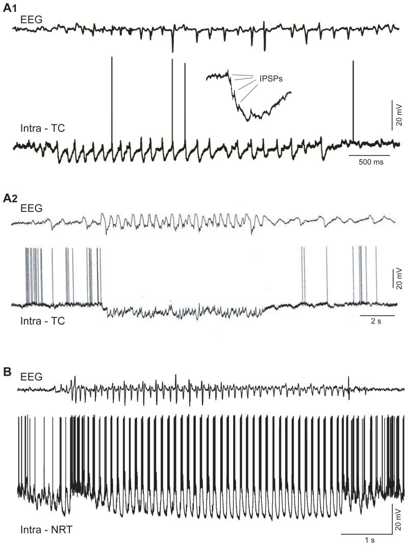
Figure 4. Intracellular correlates of TC and NRT neuron activity during spontaneous SWDs in vivo.

Figure 4Intracellular correlates of TC and NRT neuron activity during spontaneous SWDs in vivo

A1. Intracellular recording (bottom trace) from a TC neurons in GAERS in vivo during a spontaneous SWD (top trace) shows the presence of bursts of IPSPs occurring in synchrony with each spike and wave complex. Note the individual IPSPs (arrows) in the enlarged burst in the inset. B. Intracellular recording (bottom trace) from a cat TC neurons in vivo during a SWDs (top trace) shows the presence of IPSPs superimposed on a long-lasting hyperpolarization that lasts for the entire duration of the SWD. B. Intracellular recording (bottom trace) from an NRT neurons in GAERS in vivo during a spontaneous SWD (top trace) shows the presence of large low-threshold Ca2+ potential in synchrony with each spike and wave complex. (A1 and B: reproduced with permission from Crunelli and Leresche 2001, © Nature Publishing Group; A2: reproduced with permission from Steriade and Contreras 1995, © Society for Neuroscience).

From: GABA-A Receptor Function in Typical Absence Seizures
Cover of Jasper's Basic Mechanisms of the Epilepsies
Jasper's Basic Mechanisms of the Epilepsies [Internet]. 4th edition.
Noebels JL, Avoli M, Rogawski MA, et al., editors.
Copyright © 2012, Michael A Rogawski, Antonio V Delgado-Escueta, Jeffrey L Noebels, Massimo Avoli and Richard W Olsen.

All Jasper's Basic Mechanisms of the Epilepsies content, except where otherwise noted, is licensed under a Creative Commons Attribution-NonCommercial-NoDerivs 3.0 Unported license, which permits copying, distribution and transmission of the work, provided the original work is properly cited, not used for commercial purposes, nor is altered or transformed.

NCBI Bookshelf. A service of the National Library of Medicine, National Institutes of Health.