U.S. flag

An official website of the United States government

NCBI Bookshelf. A service of the National Library of Medicine, National Institutes of Health.

Adam MP, Feldman J, Mirzaa GM, et al., editors. GeneReviews® [Internet]. Seattle (WA): University of Washington, Seattle; 1993-2024.

Cover of GeneReviews®

GeneReviews® [Internet].

Show details

Von Willebrand Disease

Synonym: Von Willebrand Factor Deficiency

, MD.

Author Information and Affiliations

Initial Posting: ; Last Update: November 14, 2024.

Estimated reading time: 47 minutes

Summary

Clinical characteristics.

Von Willebrand disease (VWD) is characterized by mucocutaneous bleeding and excessive bleeding with trauma and procedures. Individuals with more severe forms of VWD are also at-risk for musculoskeletal bleeding. Mucocutaneous bleeding can include easy bruising, prolonged bleeding from minor wounds, epistaxis, oral cavity bleeding, heavy menstrual bleeding, gastrointestinal bleeding, and bleeding with hemostatic challenges such as dental work, childbirth, and surgery. Bleeding severity can vary widely in VWD, even between affected individuals within the same family. For some with VWD the bleeding phenotype may only become apparent upon hemostatic challenge, while others may have frequent spontaneous bleeding.

Diagnosis.

The diagnosis of VWD is established in a proband with excessive bleeding by identification of a quantitative or qualitative deficiency in von Willebrand factor (VWF) and/or identification of a VWD-causative variant(s) in VWF by molecular genetic testing.

Management.

Targeted therapy: VWF replacement; desmopressin.

Supportive care: People with VWD benefit from care in a comprehensive bleeding disorders program. Treatment is often given on demand for active bleeding episodes or for prevention of bleeding with hemostatic challenges. Prophylaxis should be considered in those with more severe bleeding. Useful adjunctive hemostatic treatments include antifibrinolytics, hormonal therapies for female reproductive tract bleeding, and local anatomic measures. Antifibrinolytic treatment (e.g., tranexamic acid) can be used alone for some hemostatic challenges or in combination with VWF-increasing treatment. Hormonal treatments can be useful in managing female reproductive tract bleeding either alone or in combination with antifibrinolytics and/or VWD-specific treatment.

Surveillance: Assessment in a center experienced in the comprehensive management of bleeding disorders to determine bleeding frequency and severity, treatment efficacy and tolerability, CBC, iron levels, VWF levels, and VWF inhibitors in those at risk (type 3 VWD) annually in those receiving treatment or every two to three years in those without bleeding and not requiring treatment. Evaluation by a physiotherapist, including consideration of musculoskeletal ultrasound evaluation for those with more severe VWD and/or low factor VIII levels, to monitor joint health (as in individuals with hemophilia A). Gynecology evaluation as needed for females with heavy bleeding. Gastroenterology evaluation with any suspected gastrointestinal bleeding or per gastroenterologist. Infectious disease assessment as needed in those exposed to blood products prior to 1985 or with other risk factors.

Agents/circumstances to avoid: Activities with a high risk of trauma; high contact sports with risk of head injury; medications with effects on platelet function (e.g., aspirin, clopidogrel); nutritional supplements that may impair hemostasis (e.g., fish oil, turmeric); invasive procedures without prior consultation with a hematologist (e.g., circumcision, dental, dermatologic, piercings). NSAIDs can increase bleeding risk and should be used cautiously and only for brief periods.

Evaluation of relatives at risk: It is appropriate to evaluate apparently asymptomatic at-risk relatives of an affected individual to allow early diagnosis and treatment as needed. In newborns, assess cord blood for low VWF and factor VIII levels to inform neonatal treatment. Molecular genetic testing of cord blood can be informative, particularly if the familial VWD-causing variant(s) are known.

Pregnancy management: Monitor VWF and factor VIII levels throughout pregnancy; postpartum hemorrhage prophylaxis with antifibrinolytics unless contraindicated; VWF replacement can be used to increase VWF levels.

Genetic counseling.

Type 1, type 2A, and type 2M VWD are typically caused by a heterozygous VWF variant and inherited in an autosomal dominant manner; rarely, these VWD types are associated with biallelic VWF variants. Type 2B VWD is typically caused by a heterozygous VWF variant and inherited in an autosomal dominant manner. Type 2N and type 3 VWD are caused by biallelic VWF variants and inherited in an autosomal recessive manner.

Autosomal dominant inheritance: Many individuals diagnosed with autosomal dominant VWD have an affected parent. Each child of an individual with autosomal dominant VWD has a 50% chance of inheriting the VWD-causing variant. VWD often exhibits variable penetrance and expressivity.

Autosomal recessive inheritance: If both parents are known to be heterozygous for a VWD-causing variant, each sib of an affected individual has at conception a 25% chance of being affected, a 50% chance of being heterozygous, and a 25% chance of inheriting neither of the familial VWD-causing variants. Heterozygous sibs of an individual with type 2N VWD are often asymptomatic; however, a small proportion of these heterozygous individuals may have some mild bleeding symptoms and lower factor VIII levels and may be diagnosed with VWD. Heterozygous sibs of an individual with type 3 VWD may be asymptomatic or may have bleeding symptoms and low VWF levels and may be diagnosed with type 1 VWD. Molecular genetic testing for at-risk relatives is most informative if the VWD-causing variant(s) have been identified in an affected family member.

Once the familial VWD-causing variant(s) have been identified, prenatal and preimplantation genetic testing for VWD are possible.

GeneReview Scope

Von Willebrand Disease: Phenotypic Spectrum

PhenotypeVon Willebrand Disease Subtype
Partial quantitative deficiency of functionally normal VWFType 1 VWD
Type 1C VWD
Qualitative defect in one or more VWF functions; divided into four subtypes depending on which VWF function is perturbedType 2A VWD
Type 2B VWD
Type 2M VWD
Type 2N VWD
Undetectable VWF levelsType 3 VWD

VWD = von Willebrand disease; VWF = von Willebrand factor

Diagnosis

International guidelines on the diagnosis of von Willebrand disease (VWD) have been published [James et al 2021].

Suggestive Findings

VWD should be suspected in probands with the following clinical and laboratory findings.

Clinical findings

  • Excessive bruising, particularly without recognized trauma
  • Prolonged bleeding from cutaneous wounds
  • Prolonged, recurrent, and/or severe nosebleeds
  • Bleeding from the gums after brushing or flossing teeth, particularly lifelong or in the absence of periodontal disease, and/or prolonged bleeding following dental cleaning or dental extractions
  • Excessive female reproductive tract bleeding including uterine bleeding (e.g., heavy menstrual bleeding, postpartum hemorrhage) and ovarian hemorrhage
  • Prolonged bleeding following invasive procedures, surgery, or trauma
  • Excessive gastrointestinal bleeding, or gastrointestinal bleeding with angiodysplasia
  • More severe forms may develop hemarthrosis and/or hemarthropathy

Routine laboratory findings

  • Complete blood count (CBC) is often normal but may show microcytic anemia (in those with iron deficiency) or thrombocytopenia in those with type 2B VWD. In type 2B VWD, thrombocytopenia is accompanied by platelet clumping that can be seen on blood smear.
  • Activated partial thromboplastin time (aPTT) is often normal but may be prolonged when the factor VIII clotting activity level (FVIII:C) is low (i.e., <20-30 IU/dL), as can be seen in some individuals with type 1 and type 2 VWD, and in all individuals with type 2N and type 3 VWD.
  • Prothrombin time is normal.

Establishing the Diagnosis

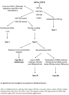
The diagnosis of VWD can be established in a proband with excessive bleeding by identification of one of the following (see Figure 1):

Figure 1. . An approach to the diagnosis of von Willebrand disease (VWD) [James et al 2021].

Figure 1.

An approach to the diagnosis of von Willebrand disease (VWD) [James et al 2021]. VWF levels refer to VWF antigen (VWF:Ag) and/or platelet-dependent VWF activity of 30-50 IU/dL, with the caveat that the laboratory’s lower limit of the normal range (more...)

  • Decreased quantity of von Willebrand factor (VWF)
  • Qualitative defect in VWF identified on a VWF activity assay or VWF multimer gel
  • VWD-causative variant(s) in VWF by molecular genetic testing

Note: Additional testing can inform VWD type (see Table 1 and Figure 2).

Figure 2. . Algorithms for additional testing in suspected type 2B von Willebrand disease (VWD) (left) and suspected type 2N VWD (right).

Figure 2.

Algorithms for additional testing in suspected type 2B von Willebrand disease (VWD) (left) and suspected type 2N VWD (right). GPIb = glycoprotein Ib; RIPA = ristocetin-induced platelet agglutination; VWF:FVIII = von Willebrand factor/Factor VIII ratio. (more...)

VWD laboratory assays (see Table 1) [Bodó et al 2015, Kalot et al 2022a]

  • VWF:Ag. Decreased quantity of VWF protein (antigen) measured using ELISA or latex immunoassay can be identified in individuals with type 1 VWD and in some individuals with type 2 VWD. VWF:Ag is undetectable in individuals with type 3 VWD (see Table 1) [Castaman et al 2010].
  • Platelet-dependent VWF activity assays
    • VWF:GPIbR. Assay that tests ristocetin-induced binding of VWF to glycoprotein Ib (GPIb). Decreased binding activity can be identified in individuals with most types of VWD. The results of this assay can be artifactually low in individuals with benign VWF variants that interfere with the ristocetin-VWF interaction.
    • VWF:GPIbM. Assay that tests the binding of VWF to a GPIb fragment with a gain-of-function variant. Decreased binding activity can be identified in individuals with most types of VWD. This assay does not use ristocetin.
    • VWF:RCo. Ristocetin cofactor activity assay that uses reagent platelets and ristocetin to test the ability of VWF to agglutinate platelets. This assay can have a high coefficient of variation. Individuals with a benign VWF variant that interferes with ristocetin can have artifactually low results. Decreased VWF:RCo activity can be identified in individuals with most types of VWD. Current guidance recommends using newer platelet-dependent VWF activity assays instead of ristocetin-based assays [James et al 2021].
  • Factor VIII (FVIII) activity assays used in individuals with VWD include either the one-stage FVIII activity assay or the chromogenic FVIII activity assay. FVIII activity is always low in type 2N and type 3 VWD and is often low in most other forms of VWD.

Assays to inform the type of VWD

  • VWF multimer analysis. SDS-agarose gel electrophoresis is used to determine the distribution of sizes of VWF multimers. Normal plasma contains VWF complexes ranging from VWF dimers to very large VWF multimers (500 kD to >10,000 kD). Multimers are classified as low (1-5 bands), intermediate (6-9 bands), and high (≥10 bands) molecular weight. High-molecular-weight multimers are decreased or absent in all individuals with type 2A VWD and many with type 2B VWD; intermediate- and low-molecular-weight multimers may also be decreased or absent in individuals with type 2A or type 2B VWD. Abnormalities detected in VWF multimer satellite ("triplet") band patterns can give clues as to the underlying pathogenesis of the VWD [Budde et al 2008, Stockschlaeder et al 2014]. However, protocols that can detect VWF triplet band multimer patterns are not available in all clinical laboratories that do multimer assays.
  • Ristocetin-induced platelet agglutination (RIPA) can assess the ability of VWF to agglutinate platelets in platelet-rich plasma in the presence of specific concentrations of ristocetin. Agglutination at a low ristocetin concentration (~0.5-0.7 mg/mL) or spontaneous agglutination in the absence of ristocetin indicates enhanced VWF platelet binding due to either type 2B VWD or platelet-type VWD (see Differential Diagnosis).
  • VWF:FVIIIB. A functional assay that measures the ability of VWF to bind reagent FVIII. Binding is decreased in individuals with type 2N VWD.
  • VWF:CB. Collagen-binding assays measure the ability of VWF to bind to specific collagens. A reduced VWF:CB-to-VWF:Ag ratio can be identified in individuals with type 2A, 2B, or 2M VWD. This assay is necessary to identify isolated VWF collagen-binding defects in some forms of type 2M VWD. Most clinical VWF:CB assays use a mixture of collagen type I and type III or collagen type III alone. Assays to assess for isolated deficient binding to collagen types IV and VI, as is found in some individuals with type 2M VWD, are available in specialty reference laboratories.

Table 1.

Classification of VWD Based on Specific Laboratory Tests

VWD TypeVWF:Act 1, 2VWF:Ag 2VWF:Act-to-VWF:Ag RatioVWF:CB 2FVIII IU/dL 2Multimer PatternOther
1LowLow≥0.7LowLow or normalEssentially normal 3Normal VWF half-life after desmopressin
1CLowLow≥0.7LowLow or normalEssentially normal 3↓ VWF half-life after desmopressin
2ALow; rarely, can be normalLow; can be normal<0.7Low; rarely, can be normalLow or normalAbnormal; ↓ HMW
2BLow; rarely, can be normalLow; can be normalOften <0.7; can be ≥0.7Low; can be normalLow or normalOften abnormal; ↓ HMWClassification requires identification of VWD-causing VWF variant or ↑ RIPA; 4 often ↓ platelet count 5
2MLow; rarely, can be normalLow; can be normal<0.7Low; can be normalLow or normalEssentially normal 3Classification requires ↓ VWF:Act-to-VWF:Ag ratio or ↓ VWF:CB-to-VWF:Ag ratio (<0.7) 6
2NNormal; can be lowNormal; can be low≥0.7Normal; can be low<40NormalClassification requires FVIII activity < VWF:Ag & ↓ VWF:FVIIIB or identification of VWD-causing VWF variants 7
3AbsentAbsentNAAbsent<10AbsentNo detectable VWF

FVIII = factor VIII; HMW = high-molecular-weight VWF multimers; NA = not applicable; RIPA = ristocetin-induced platelet agglutination; VWD = von Willebrand disease; VWF = von Willebrand factor; VWF:Act = platelet-dependent VWF activity; VWF:Ag = VWF antigen; VWF:CB = VWF collagen binding; VWF:FVIIIB = binding of FVIII by VWF

1.

VWF:Act assays include VWF:RCo, VWF:GPIbR, and VWF:GPIbM. VWF activity assays that do not use ristocetin are preferred when available [James et al 2021].

2.

"Low" refers to VWF:Act <50 IU/dL, VWF:Ag <50 IU/dL, VWF:CB <50 IU/dL, or FVIII activity below the laboratory reference range (usually <50 IU/dL).

3.

All sizes of multimers are present, but there may be subtle abnormalities in multimer pattern.

4.

Clinical presentation of type 2B VWD is identical to platelet-type VWD (PT-VWD). Type 2B VWD is distinguished from PT-VWD using molecular genetic testing or specialized RIPA mixing studies.

5.

Thrombocytopenia and decrease in high-molecular-weight VWF multimers usually occur together. In those with a normal platelet count and normal VWF multimer distribution, the predisposition to thrombocytopenia and abnormal VWF multimer distribution is often unmasked by physical stressors.

6.

Reduction in the ability of VWF to bind to collagen. Collagen types I/III are bound by the VWF A3 domain, while collagen types IV and VI are bound by the VWF A1 domain. Most clinical laboratories do not have assays to test VWF binding to all relevant collagen types.

7.

Clinical presentation of type 2N VWD is similar to hemophilia A except that inheritance of type 2N VWD is autosomal recessive and hemophilia A is X-linked. Type 2N VWD can be distinguished from hemophilia A through molecular genetic testing or decreased VWF:FVIIIB.

Molecular genetic testing can be used to establish the diagnosis of VWD in a proband with ambiguous or conflicting quantitative and qualitative VWF testing and/or classify VWD subtype (see Table 2).

  • Identification of a heterozygous (or, rarely, biallelic) VWD-causing variant in VWF can establish the diagnosis in individuals with types 1C, 2A, 2B, and 2M VWD. Note: The proportion of individuals with type 1 VWD who have a VWD-causing variant identified is lower than other forms of VWD; however, molecular genetic testing can still inform the diagnosis.
  • Identification of biallelic VWD-causing variants in VWF can establish the diagnosis in individuals with types 2N and 3 VWD.

Note: (1) Molecular genetic testing should be done in a laboratory experienced in testing for VWD. (2) Laboratories may not report the common benign VWF variant p.Asp1472His, which causes artificially decreased results on VWF:GPIbR and VWF:RCo assays.

  • Single-gene testing. Sequence analysis of VWF is performed first to detect missense, nonsense, and splice site variants and small deletions/insertions. Depending on the sequencing method used, large deletions or duplications and VWF-VWFP1 pseudogene conversions may not be detected (see Molecular Genetics, VWF-specific laboratory technical considerations). If no VWD-causing variant is detected by the sequencing method used in an individual with clinical and laboratory features of VWD, the next step is to perform testing that can identify large VWF deletions, duplications, and other structural variants.
  • A multigene panel that includes VWF, F8, and GPIBA (see Differential Diagnosis) may be considered to identify the genetic cause of the condition and/or distinguish VWD from hemophilia A and PT-VWD. Note: (1) The genes included in the panel and the diagnostic sensitivity of the testing used for each gene vary by laboratory and are likely to change over time. (2) Some multigene panels may include genes not associated with the condition discussed in this GeneReview. (3) In some laboratories, panel options may include a custom laboratory-designed panel and/or custom phenotype-focused exome analysis that includes genes specified by the clinician. (4) Methods used in a panel may include sequence analysis, deletion/duplication analysis, and/or other non-sequencing-based tests. (5) Multigene panels usually cannot detect the common recurring F8 intron 1 and intron 22 inversions.
    For an introduction to multigene panels click here. More detailed information for clinicians ordering genetic tests can be found here.

Table 2.

Molecular Genetic Testing Used in von Willebrand Disease

Gene 1VWD subtypeProportion of VWD-Causative Variants 2 Identified by Method
Sequence analysis 3Gene-targeted deletion/duplication analysis 4
VWF Type 1 VWD w/VWF 30-50 IU/dL40%-60%Rare 5
Type 1 VWD w/VWF ≤30 IU/dL80%-90% 6, 7≤6% 6, 7
Type 2 VWD
Type 3 VWD

VWD = von Willebrand disease

1.

See Table A. Genes and Databases for chromosome locus and protein.

2.

See Molecular Genetics for information on variants detected in this gene.

3.

Sequence analysis detects missense, nonsense, splice site variants, and small insertions or deletions. Sequence analysis should include regulatory regions and the VWF promotor. Larger deletions, duplications, structural variants including pseudogene conversions, and non-coding deep intronic variants are not often detected. For issues to consider in interpretation of sequence analysis results, click here.

4.

Gene-targeted deletion/duplication methods can detect deletions or duplications. Methods used may include a range of techniques such as quantitative PCR, long-range PCR, custom analyses of next generation sequencing data, multiplex ligation-dependent probe amplification (MLPA), comparative genomic hybridization (CGH), and a gene-targeted microarray designed to detect single-exon deletions or duplications.

5.
6.

Cumming et al [2006], Goodeve et al [2007], James et al [2007a], Yadegari et al [2012], Veyradier et al [2016], Laffan et al [2021], Kalot et al [2022b], and data derived from the subscription-based professional view of Human Gene Mutation Database [Stenson et al 2020]

7.

Sequencing and analysis of VWF exons 23 to 34 is complicated by the presence of a partial pseudogene, VWFP1, on chromosome 22 (see Molecular Genetics).

Clinical Characteristics

Clinical Description

Von Willebrand disease (VWD) is a congenital bleeding disorder. Symptoms may only become apparent on hemostatic challenge, and increased or prolonged bleeding may only be identified after recurrent exposure to hemostatic challenges. Thus, it may take some time before a bleeding history becomes apparent. Individuals with VWD primarily manifest excessive mucocutaneous bleeding (e.g., bruising, epistaxis, ear, nose, and throat bleeding, oral bleeding, heavy menstrual bleeding) and do not tend to experience musculoskeletal bleeding in the absence of trauma unless the factor VIII clotting (FVIII:C) level is low, as can be seen in individuals with type 2N or type 3 VWD.

Type 1 VWD typically manifests as mucocutaneous bleeding. However, the severity can range from mild to severe. Epistaxis and bruising are common. Heavy menstrual bleeding is the most common finding in females who menstruate [Ragni et al 2016].

Type 1C VWD. Individuals have accelerated clearance of VWF [James et al 2021]. Bleeding occurs in the same pattern as in type 1 VWD, but symptoms tend to be milder compared to similarly low VWF levels in type 1 VWD.

Type 2A VWD. Individuals with type 2A VWD can present with mild-to-severe mucocutaneous bleeding [Veyradier et al 2016].

Type 2B VWD. Individuals with type 2B VWD can present with mild-to-severe mucocutaneous bleeding. Thrombocytopenia may be present. A hallmark of type 2B VWD is a worsening of thrombocytopenia during stressful situations (e.g., severe infection, surgery, pregnancy) or in those treated with desmopressin [Federici et al 2009].

Type 2M VWD. Individuals with type 2M VWD can present with mild-to-severe mucocutaneous bleeding, including bleeding after surgeries and postpartum hemorrhage [Castaman et al 2012, Larsen et al 2013, Maas et al 2022].

Type 2N VWD. The clinical manifestations of type 2N VWD include joint, muscle, and mucocutaneous bleeding and excessive bleeding at the time of surgery or procedures [van Meegeren et al 2015, Seidizadeh et al 2021].

Type 3 VWD manifests with moderate-to-severe bleeding including excessive mucocutaneous bleeding, musculoskeletal bleeding, and risk for intracranial hemorrhage [Metjian et al 2009, Ahmad et al 2013, Kasatkar et al 2014, Tosetto et al 2020].

Associated complications

  • Gastrointestinal (GI) angiodysplasia occurs in all types of VWD, most commonly in adults with type 2A VWD, followed by type 3 and type 1 VWD [Chornenki et al 2022]. It occurs throughout the GI tract, particularly the colon, small intestine, and stomach [Franchini & Mannucci 2014]. GI angiodysplasia presents with GI bleeding that may begin insidiously, sometimes only recognizable through identification of iron deficiency or anemia or on endoscopy. As angiodysplasia progresses, GI bleeding generally worsens, and bleeding episodes may become life-threatening.
  • Alloantibodies against VWF. The development of alloantibodies against VWF (anti-VWF antibodies) is an uncommon but serious complication of VWD treatment. An estimated 5%-10% of individuals with type 3 VWD develop anti-VWF antibodies, although these antibodies are exceedingly rare in other VWD types [Franchini & Mannucci 2018]. Affected individuals present with reduced or absent response to treatment with VWF replacement or, in rare instances, with anaphylactic reaction. Individuals with large VWF deletions and those who have had multiple episodes of VWF replacement (i.e., multiple exposure days) are at highest risk for this complication.

Genotype-Phenotype Correlations

The three phenotypes reflect a partial quantitative deficiency of VWF (type 1 VWD), complete quantitative deficiency of VWF (type 3 VWD), or qualitative defects in VWF (type 2 VWD). See Molecular Genetics for details regarding the genotypes associated with each subtype of VWD.

Individuals with large deletions of VWF are at highest risk for alloantibody development, although null alleles have also been associated with this complication [Franchini & Mannucci 2018].

Penetrance

Type 1 VWD (autosomal dominant). VWD-causing variants resulting in plasma VWF levels lower than 30 IU/dL tend to be more penetrant.

Other autosomal dominant types (2A, 2B, and 2M). VWD-causing variants are often fully penetrant, particularly for type 2A and type 2B VWD.

Nomenclature

Clarifications and changes in VWD nomenclature are listed in Table 3.

Table 3.

Clarifications and Changes in von Willebrand Disease Nomenclature

TermCurrent Term &/or Definition
Platelet-dependent VWF activityRefers to VWF activity w/regard to platelets, incl VWF:RCo, VWF:GPIbR, and VWF:GPIbM 1
Type 1C VWDRefers to type 1 VWD w/accelerated VWF clearance
VWF:CBRefers to VWF collagen binding
FVIII RAg (FVIII-related antigen)Now called VWF:Ag 1
RiCof (ristocetin cofactor activity)Now called VWF:RCo 1
Von Willebrand's diseaseVon Willebrand disease
vWDVWD
vWFVWF

VWD = von Willebrand disease; VWF = von Willebrand factor

1.

See Establishing the Diagnosis, VWD laboratory assays

Platelet-type von Willebrand disease (PT-VWD), previously called pseudo-VWD, is a platelet disorder caused by pathogenic variants in GP1BA and, thus, is not a form of VWD (see Differential Diagnosis) [Fu et al 2024].

Acquired von Willebrand syndrome (AVWS), previously known as acquired VWD, is the preferred terminology for defects in VWF concentration, structure, or function that are neither inherited nor due to disease-causing variants in VWF, and instead arise as consequences of other medical conditions (see Differential Diagnosis) [Franchini & Mannucci 2020].

Prevalence

VWD affects 0.1% of the population. Prior estimates that VWD affects up to 1% of the population likely included people with low VWF without excessive bleeding. One in 10,000 individuals seek tertiary care referral for VWD.

Type 3 VWD affects 0.5:1,000,000 to 6:1,000,000.

Founder variants in VWF that account for a significant proportion of disease-causing variants in a specific population have been reported in the Romani and Amish populations (see Table 9).

Differential Diagnosis

Mild-to-moderate hemophilia A and platelet-type von Willebrand disease (PT-VWD) can be difficult to distinguish phenotypically from von Willebrand disease (VWD) (see Table 4).

Table 4.

Genetic Disorders in the Differential Diagnosis of von Willebrand Disease

GeneDisorderMOIPhenotypeDiagnosis
F8 Mild-to-moderate hemophilia AXLPersons w/low FVIII due to VWD may be misdiagnosed w/hemophilia A.
↓ levels of FVIII:C (~2-40 IU/dL) & normal-to-low levels of VWF can be seen in both type 2N VWD & mild hemophilia A.
The frequency of bleeding episodes in hemophilia A varies widely, from multiple times per yr to once every 10 yrs.
Molecular genetic testing can distinguish VWD & hemophilia A. Molecular testing identifies a causative F8 variant in >98% of persons w/hemophilia A. 1
VWF:FVIIIB assay can also be used to discriminate between type 2N VWD & hemophilia A. 2
Both molecular & phenotypic testing have fallibilities in interpretation. 3
GP1BA Platelet-type VWD 4 (PT-VWD) (OMIM 177820)ADPT-VWD is a platelet disorder; platelet transfusion is required for major bleeding challenges.
The PT-VWD phenotype is identical to type 2B VWD.
In PT-VWD the half-life of VWF is ↓ as a result of ↑ binding to the abnormal GpIbα on platelets, which can often also necessitate VWF replacement treatment.
VWD & PT-VWD can be distinguished by molecular genetic testing.
Alternately, RIPA mixing assays using plasma & platelets of affected persons & controls to determine which component is defective 5 can be done, but this method is becoming less available.

AD = autosomal dominant; FVIII = factor VIII; FVIII:C = factor VIII clotting; MOI = mode of inheritance; RIPA = ristocetin-induced platelet agglutination; VWD = von Willebrand disease; VWF = von Willebrand factor; VWF:FVIIIB = VWF factor VIII binding; XL = X-linked

1.
2.

VWF:FVIIIB assay determines ability of VWF to bind FVIII; it is available although on a limited basis.

3.
4.

One study identified pathogenic variants in GP1BA in up to 15% of persons diagnosed with type 2B VWD [Hamilton et al 2011].

5.

Acquired von Willebrand syndrome (AVWS) is a bleeding disorder of qualitative or quantitative deficiency of von Willebrand factor (VWF) that can occur in a variety of conditions and is neither inherited nor due to disease-causing variants in VWF [Sucker et al 2009, Federici et al 2013, Mital 2016, Franchini & Mannucci 2020]. AVWS is most often seen in persons older than age 40 years with no prior bleeding history. AVWS has diverse pathology and causes including the following:

  • Lymphoproliferative or plasma cell proliferative disorders, paraproteinemias (monoclonal gammopathy of unknown significance), chronic lymphocytic leukemia, multiple myeloma, and Waldenstrom macroglobulinemia. Antibodies against VWF have been detected in some individuals with these disorders.
  • Autoimmune disorders including systemic lupus erythematosus, scleroderma, and antiphospholipid antibody syndrome
  • Shear-induced VWF conformational changes leading to relative loss of high-molecular-weight VWF multimers (e.g., aortic valve stenosis, ventricular septal defect, left ventricular assist devices)
  • Markedly increased blood platelet count (e.g., essential thrombocythemia or other myeloproliferative disorders)
  • Removal of VWF from circulation by aberrant binding to tumor cells (e.g., Wilms tumor or certain lymphoproliferative disorders) or amyloidosis
  • Decreased VWF synthesis (e.g., hypothyroidism)
  • Certain drugs (e.g., valproic acid, ciprofloxacin, griseofulvin, hydroxyethyl starch, N-acetylcysteine)

Management

Clinical practice guidelines for von Willebrand disease (VWD) have been published [Connell et al 2021].

Evaluations Following Initial Diagnosis

To establish the extent of disease and needs in an individual diagnosed with VWD, the evaluations summarized in Table 5 (if not performed as part of the evaluation that led to the diagnosis) are recommended.

Table 5.

Von Willebrand Disease: Recommended Evaluations Following Initial Diagnosis

System/ConcernEvaluationComment
Hematology
  • Ensure complete & modern VWD eval has been performed whenever possible prior to invasive procedures or other hemostatic risks.
  • Assess history of bleeding.
  • Assess VWF levels.
  • Assess FVIII levels.
Very old or historical diagnoses of VWD w/o recent laboratory confirmation should not be relied upon in treatment planning.
VWF inhibitor testing (when available) in those w/type 3 VWD or suspected presence of inhibitors
CBC; iron studies incl ferritinTo assess for anemia & iron deficiency to identify persons who should have iron replacement
Musculoskeletal Joint & muscle eval (as would be done for persons w/hemophilia A)In those w/low FVIII levels or clinical suspicion
Gynecology Gynecologic eval for those w/heavy menstrual bleeding &/or ovarian hemorrhage (See also Pregnancy Management.)To assess for other anatomic contributors to bleeding & to coordinate mgmt w/hormonal therapies, IUD, & procedures if needed
Gastrointestinal Endoscopic eval for anatomic causes of bleeding, incl angiodysplasia in those w/suspected GI bleeding
Infectious disease Screening for hepatitis B, hepatitis C, & HIVIn persons who received blood products or plasma-derived clotting factor concentrates prior to 1985 or w/other risk factors
Genetic counseling By genetics professionals, 1 incl assessment of family history of bleeding to help identify relatives at risk for VWDTo obtain a pedigree & inform affected persons & their families re nature, MOI, & implications of VWD to facilitate medical & personal decision making

CBC = complete blood count; FVIII = factor VIII; IUD = intrauterine device; MOI = mode of inheritance; VWD = von Willebrand disease; VWF = von Willebrand factor

1.

Medical geneticist, certified genetic counselor, certified advanced genetic nurse

Treatment of Manifestations

Targeted Therapy

In GeneReviews, a targeted therapy is one that addresses the specific underlying mechanism of disease causation (regardless of whether the therapy is significantly efficacious for one or more manifestation of the genetic condition); would otherwise not be considered without knowledge of the underlying genetic cause of the condition; or could lead to a cure. —ED

Von Willebrand factor (VWF) factor replacement (plasma derived or recombinant) is the mainstay of VWD treatment. Desmopressin can be used in individuals who are responsive to desmopressin for non-major, time-limited hemostatic challenges. Guidance on the treatment of VWD has been published [Connell et al 2021].

Intravenous infusion of VWF replacement

  • VWF replacement options include pathogen-inactivated plasma-derived products containing both VWF and factor VIII (FVIII), plasma-derived products depleted of FVIII, and recombinant VWF that does not provide FVIII [Weyand & Flood 2021].
  • Bleeding episodes can be prevented or controlled with intravenous infusion of VWF-containing replacement treatments.
  • Plasma-derived concentrates are prepared from pooled blood donations from many donors. Procedures such as pasteurization and detergents inactivate potential pathogens.
  • Recombinant VWF is manufactured in vitro [Franchini & Mannucci 2016].

Desmopressin

  • Many individuals with type 1 VWD respond to intranasal, intravenous, or subcutaneous treatment with desmopressin [Leissinger et al 2014]. Desmopressin promotes release of stored VWF and raises VWF and FVIII levels three- to fourfold.
  • Following the diagnosis of type 1 VWD, a desmopressin challenge is recommended to assess VWF response [Connell et al 2021]. Desmopressin challenges are no longer recommended routinely in individuals with other types of VWD, although it may be occasionally useful in some individuals with type 2 VWD.
  • Individuals with type 1C VWD will have accelerated VWF clearance after diagnostic desmopressin challenge [James et al 2021]. Desmopressin is generally contraindicated in type 1C VWD for surgery [Connell et al 2021].
  • Desmopressin should be used with caution, particularly in those younger than age two years, because of the potential difficulty in restricting fluids in this age group.
  • Repeat dosing of desmopressin over a short period of time results in tachyphylaxis: a decrease of released VWF after repeated administration.
  • Desmopressin is contraindicated in individuals with arteriovascular disease and in those older than age 70 years.
  • In persons who do not tolerate desmopressin, who have a poor VWF response, who face a moderate or major hemostatic challenge, who cannot sufficiently fluid restrict, or who have a duration of hemostatic need longer than a few days, VWF replacement treatment is required.
  • Note: Because desmopressin can cause hyponatremia through the retention of free water (which can lead to seizures, coma, or death), fluid intake should be restricted for 24 hours following its administration to minimize this risk.

Table 6.

Targeted Therapy by von Willebrand Disease Type

VWD TypeTreatmentComment
1Treatment w/VWF replacement is often required for moderate or severe hemostatic challenges & may be required for minor challenges.Use of VWF replacement depends upon VWF levels & bleeding risk.
Desmopressin can be used for minor & some moderate, time-limited (2-3 days) hemostatic challenges in responsive persons.Responsiveness to desmopressin is variable & should be confirmed prior to therapeutic use.
2ATreatment w/VWF replacement is usually required for moderate or severe hemostatic challenges & may be required for minor challenges.Desmopressin response is usually poor.
2BTreatment w/VWF replacement is usually required for moderate or severe hemostatic challenges & may be required for minor challenges.Desmopressin response is usually poor. Treatment w/desmopressin can precipitate or worsen thrombocytopenia in many but not all persons w/type 2B VWD. 1
Platelet counts should be monitored, as platelet transfusions may need to be added to VWF replacement.
2MTreatment w/VWF replacement is usually required for moderate or severe hemostatic challenges & may be required for minor challenges.Desmopressin response is generally poor.
2NTreatment w/VWF replacement is usually required for moderate or severe hemostatic challenges & may be required for minor challenges.
  • Because the FVIII levels are low, a VWF replacement product containing VWF as well as FVIII is preferred in initial treatment over a product containing VWF alone.
  • Desmopressin response is generally poor.
3
  • Treatment w/VWF replacement is usually required for moderate or severe hemostatic challenges & is often required for minor challenges.
  • Many individuals require prophylaxis w/VWF replacement to prevent musculoskeletal bleeding & subsequent joint damage.
  • Because the FVIII levels are low, a VWF replacement product containing VWF as well as FVIII is preferred in initial treatment over a product containing VWF alone.
  • Desmopressin is not effective.
  • Some persons w/type 3 VWD can develop alloantibodies (inhibitors) against VWF.

FVIII = factor VIII; VWD = von Willebrand disease; VWF = von Willebrand factor

1.

Supportive Care

Supportive care to improve quality of life, maximize function, and reduce complications is recommended. This ideally involves multidisciplinary care by a comprehensive bleeding disorders program (see Table 7). The goal is to develop treatment plans that enable people with VWD to live full and active lives.

Table 7.

Von Willebrand Disease: Treatment of Manifestations

Manifestation/ConcernTreatmentConsiderations/Other
Hematologic
  • Fibrinolytic inhibitors (i.e., tranexamic acid, aminocaproic acid) for treatment or prevention of bleeding episodes & as adjunctive therapy for invasive procedures
  • Tranexamic acid can be used alone for minor bleeding 1
Fibrinolytic inhibitors are relatively contraindicated if there is gross hematuria.
Epistaxis
  • Nasal care (humidity, water-based gels, saline sprays, treatment of allergies or infections), nasal clamps, nasal packing, nasal cautery, VWF replacement
  • VWF prophylaxis for refractive or severe recurrent bleeding
  • Treatment w/tranexamic acid (or aminocaproic acid) ± VWF replacement should be considered for frequent bleeding.
  • Treatment should be given prior to cautery.
Heavy menstrual bleeding Treatment options incl:
  • Hormonal treatments (e.g., combined oral contraceptive pill, progestin-only pills, progestin-only injections, hormonal IUDs)
  • Tranexamic acid (or aminocaproic acid)
  • Desmopressin
  • VWF replacement
  • Combination treatment w/antifibrinolytic &/or hormonal treatment &/or VWD treatment
  • Risks & benefits of hormonal therapy should be discussed w/affected person prior to starting treatment.
  • Use of tranexamic acid w/hormonal therapy (in those not desiring pregnancy) or tranexamic acid w/o hormonal therapy (in those desiring conception or w/contraindication) are suggested over desmopressin. Responsiveness to desmopressin should be confirmed prior to therapeutic use. 1
Infectious disease Encourage vaccination for hepatitis B
GI angiodysplasia
  • Endoscopic intervention for active lesions (cautery, injection), VWF replacement, tranexamic acid
  • VWF prophylaxis for more severe GI bleeding
For significant persistent or periodic bleeding from angiodysplasia, also consider adjunctive agents. 2

GI = gastrointestinal; IUD = intrauterine device; VWF = von Willebrand factor

1.
2.

Surveillance

To monitor existing manifestations, the individual's response to supportive care, and the emergence of new manifestations, the evaluations summarized in Table 8 are recommended.

Table 8.

Von Willebrand Disease: Recommended Surveillance

System/ConcernEvaluationFrequency
Hematology
  • Assessment at hematology treatment center w/experience w/bleeding disorders
  • Determination of frequency of bleeding, factors causing incr bleeding, & treatment efficacy & tolerability
  • CBC & iron levels
  • VWF levels
  • Assess for VWD inhibitors in those at risk (type 3 VWD)
  • Annually, particularly in those receiving treatment
  • Every 2-3 years in those w/o bleeding & not on treatment
Musculoskeletal
  • Assessment of joint scores & mobility by PT
  • Musculoskeletal ultrasound when applicable
In those w/more severe VWD &/or low FVIII levels, or w/evidence of musculoskeletal bleeding, assess on same schedule as hemophilia A.
Gynecology Gynecologic evalAs needed for females w/heavy menstrual bleeding or other reproductive tract bleeding
Gastrointestinal Gastroenterology eval for iron deficiency, anemia, or signs of GI bleedingAt time of active bleeding & per gastroenterologist recommendation
Infectious disease Assessment by specialist in hepatitis B &/or hepatitis C (usually hepatologist) & HIV infection (usually infectious disease specialist); surveillance is disease specific & may incl monitoring viral load, antiviral titer, liver imaging or biopsy, liver function tests, hepatocellular carcinoma surveillance, blood counts, & eval of other comorbidities.As recommended by hepatologist &/or infectious disease specialist in those exposed to blood products prior to 1985 or w/other risk factors

CBC = complete blood count; FVIII = factor VIII; GI = gastrointestinal; PT = physical therapist; VWD = von Willebrand disease; VWF = von Willebrand factor

Agents/Circumstances to Avoid

Activities with a high risk of trauma should be approached with caution and full protective gear should be used in sports and recreational activities; high contact sports with risk of head injury should be avoided.

Medications that affect platelet function (e.g., aspirin, clopidogrel) should only be used for clear medical indications and with counseling and monitoring for bleeding, as these medications can worsen bleeding symptoms.

Nonsteroidal anti-inflammatory drugs (NSAIDs) can increase bleeding risk and should be used cautiously and only for brief periods.

Nutritional supplements that may impair hemostasis (e.g., fish oil, turmeric) should be avoided.

All invasive procedures require prior consultation with a hematologist regardless of how minor they are perceived to be by the proceduralist, including common outpatient medical procedures (e.g., circumcision, dental, dermatologic) and non-medical procedures (e.g., piercings).

Evaluation of Relatives at Risk

Newborn at risk for VWD. The mother should be counseled during pregnancy regarding the increased risk for bleeding complications (see Pregnancy Management) and a high-risk obstetrician, neonatologist, and pediatric hematologist should be notified of the risk of VWD in the infant before birth. Evaluation of newborns at risk for VWD can include:

  • VWD laboratory assays. For newborns at-risk for more severe forms of type 1, type 2, or type 3 VWD, cord blood should be obtained to assess for low VWF and FVIII levels to inform neonatal treatment. Assess platelet count on cord blood to evaluate newborns at risk for type 2B VWD. The stress of birth, sample handling, and the immature neonatal hemostasis system can all affect clotting factor levels in cord blood.
    In newborns with early life complications and anticipated hemostatic challenges (e.g., invasive procedures and/or NICU hospitalization), it is important to assess VWF and FVIII levels as early as possible. More definitive VWD testing (see Establishing the Diagnosis) should be done at an older age to confirm cord blood findings or for diagnosis in otherwise healthy infants without anticipated hemostatic challenges.
  • VWF molecular genetic testing of cord blood. Molecular genetic testing is most informative when the familial VWF variant(s) are known. (If the familial VWD-causing variant[s] are not known, the absence of an identified VWD-causing variant in a cord blood sample cannot be used to rule out the possibility of VWD.)

Other at-risk relatives. It is appropriate to evaluate even apparently asymptomatic at-risk relatives of an affected individual to allow early diagnosis and treatment as needed [Goodeve 2016]. Evaluations can include:

  • VWD hemostasis factor assays sensitive and specific for VWD (see Table 1) regardless of if the type of VWD and/or VWD-causing variant(s) in the family are known;
  • Molecular genetic testing if the VWD-causing variant(s) in the family are known.

See Genetic Counseling for issues related to testing of at-risk relatives for genetic counseling purposes.

Pregnancy Management

VWF levels increase throughout pregnancy, with the peak occurring four hours after delivery [James et al 2015, Johnsen & MacKinnon 2022]. Nonetheless, pregnant people with VWD are at increased risk for bleeding complications, and care should be provided in centers with experience in perinatal management of bleeding disorders [Johnsen & MacKinnon 2022].

The 2021 VWD guidelines do not address target VWF levels for delivery [Connell et al 2021]. Some providers will treat to keep VWF levels >100 IU/dL for delivery, while nearly all providers agree that a VWF level <50 IU/dL should be increased prior to delivery [Punt et al 2020, Johnsen & MacKinnon 2022, Lavin et al 2022, Lim et al 2024]. VWD 2021 guidelines suggest a target VWF level of ≥50 IU/dL for those undergoing neuraxial anesthesia [Connell et al 2021].

Although deliveries should occur based on obstetric indications, use of instrumentation to assist delivery (e.g., forceps, vacuum) should be minimized whenever possible [Johnsen & MacKinnon 2022]. Episiotomies should be avoided whenever possible. Fetal scalp electrodes and scalp blood sampling should be avoided in individuals with most forms of VWD due to the risk that the fetus has VWD.

Delayed, secondary postpartum bleeding rates are high in individuals with VWD. VWD 2021 guidelines suggest the use of tranexamic acid for postpartum hemorrhage prophylaxis. The guidelines do not address duration of postpartum treatment [Connell et al 2021]. VWF level rapidly returns to pre-pregnancy levels following delivery [James et al 2015].

Therapies Under Investigation

Search ClinicalTrials.gov in the US and EU Clinical Trials Register in Europe for access to information on clinical studies for a wide range of diseases and conditions. Note: There may not always be clinical trials for this disorder.

Genetic Counseling

Genetic counseling is the process of providing individuals and families with information on the nature, mode(s) of inheritance, and implications of genetic disorders to help them make informed medical and personal decisions. The following section deals with genetic risk assessment and the use of family history and genetic testing to clarify genetic status for family members; it is not meant to address all personal, cultural, or ethical issues that may arise or to substitute for consultation with a genetics professional. —ED.

Mode of Inheritance

Type 1, type 2A, and type 2M von Willebrand disease (VWD) are typically caused by a heterozygous VWF variant and inherited in an autosomal dominant manner; rarely, these VWD types are associated with biallelic VWF variants.

Type 2B VWD is typically caused by a heterozygous VWF variant and inherited in an autosomal dominant manner.

Type 2N and type 3 VWD are caused by biallelic VWF variants and inherited in an autosomal recessive manner.

Autosomal Dominant Inheritance – Risk to Family Members

Parents of a proband

  • Many individuals diagnosed with autosomal dominant VWD have an affected parent.
  • A proband with autosomal dominant VWD may have the disorder as the result of a de novo VWD-causing variant. De novo VWD-causing variants have been reported rarely in type 1 VWD [James et al 2007a, Ahmad et al 2014] and type 2 VWD [Shen et al 2016, Chen et al 2022].
  • If the proband appears to be the only affected family member (i.e., a simplex case), hemostasis laboratory assays and (if a molecular diagnosis has been established in the proband) VWF molecular genetic testing are recommended for the parents of the proband to confirm their status, allow reliable recurrence counseling, and determine their need for VWD treatment.
  • If the VWD-causing variant identified in the proband is not identified in either parent and parental identity testing has confirmed biological maternity and paternity, the following possibilities should be considered:
    • The proband has a de novo VWD-causing variant.
    • The proband inherited a VWD-causing variant from a parent with gonadal (or somatic and gonadal) mosaicism. Note: Testing of parental leukocyte DNA may not detect all instances of somatic mosaicism and will not detect a VWD-causing variant that is present in the germ (gonadal) cells only.
  • The family history of some individuals diagnosed with autosomal dominant VWD may appear to be negative because of failure to recognize the disorder in family members, reduced penetrance, variable expressivity, early death of an informative relative (such as a parent) before the recognition of symptoms, or delay of significant hemostatic challenges in affected relatives. Therefore, an apparently negative family history cannot be confirmed without appropriate evaluations (e.g., VWD laboratory assays) and/or molecular genetic testing (to establish that neither parent has VWD-causing variant[s]).

Sibs of a proband. The risk to the sibs of the proband depends on the clinical/genetic status of the proband's parents:

  • If a parent of the proband is affected and/or is known to have the VWD-causing variant identified in the proband, the risk to the sibs is 50%. VWD often exhibits variable penetrance and expressivity, meaning that not all sibs who inherit a VWF variant will have symptoms of VWD, and that those who have VWD-related manifestations can bleed with variable severity.
  • If the proband has a known VWD-causing variant that cannot be detected in the leukocyte DNA of either parent, the recurrence risk to sibs is slightly greater than that of the general population because of the possibility of parental gonadal mosaicism [Chen et al 2022].
  • If the parents are clinically unaffected but their genetic status is unknown, the risk to the sibs of a proband is increased over that of the general population because of the possibility of reduced penetrance in a heterozygous parent (see Penetrance) or parental gonadal mosaicism.

Offspring of a proband

  • Each child of an individual with autosomal dominant VWD has a 50% chance of inheriting the VWD-causing variant.
  • If the proband's reproductive partner also has VWD-causing variant(s), offspring are at risk of inheriting biallelic VWD-causing variants and potentially having a more severe phenotype or different VWD type than the proband.

Other family members. The risk to other family members depends on the status of the proband's parents: if a parent is affected, the parent's family members may be at risk.

Autosomal Recessive Inheritance – Risk to Family Members

Parents of a proband

  • The parents of an individual with autosomal recessive VWD are typically heterozygous for one VWD-causing variant. Alternatively, it is possible that one or both parents have biallelic VWD-causing variants.
  • If a molecular diagnosis has been established in the proband, molecular genetic testing is recommended for the parents of the proband to confirm their genetic status and allow reliable recurrence risk assessment. If a VWD-causing variant is detected in only one parent and parental identity testing has confirmed biological maternity and paternity, the following possibilities should be considered:
    • One of the VWD-causing variants identified in the proband occurred as a de novo event in the proband or as a postzygotic de novo event in a mosaic parent [Jónsson et al 2017].
    • Uniparental isodisomy for the parental chromosome with the VWD-causing variant resulted in homozygosity for the VWD-causing variant in the proband. Maternal uniparental isodisomy has been reported in type 3 VWD [Boisseau et al 2011].
  • Heterozygous parents of an individual with type 2N VWD are often asymptomatic. However, a small proportion of these heterozygous individuals may show some mild bleeding symptoms and lower factor VIII (FVIII) levels and may be diagnosed with VWD.
  • Heterozygous parents of an individual with type 3 VWD can have normal VWF levels and be asymptomatic. However, between 15% and 50% may show some mild bleeding symptoms and may be diagnosed with type 1 VWD [Nichols et al 2008, Bowman et al 2013, Christopherson et al 2022].

Sibs of a proband

  • If both parents are known to be heterozygous for a VWD-causing variant, each sib of an affected individual has at conception a 25% chance of being affected, a 50% chance of being heterozygous, and a 25% chance of inheriting neither of the familial VWD-causing variants.
  • Heterozygous sibs of an individual with type 2N VWD are often asymptomatic. However, a small proportion of these heterozygous individuals may show some mild bleeding symptoms and lower FVIII levels and may be diagnosed with VWD.
  • Heterozygous sibs of an individual with type 3 VWD can have normal VWF levels and be asymptomatic. However, between 15% and 50% may have some bleeding symptoms and low VWF levels and be diagnosed with type 1 VWD [Nichols et al 2008, Bowman et al 2013, Christopherson et al 2022].

Offspring of a proband. Unless an affected individual's reproductive partner also has a VWD-causing variant(s), offspring will be obligate heterozygotes for a VWD-causing variant.

Other family members. Each sib of the proband's parents is at a 50% risk of being heterozygous for a VWD-causing variant.

Heterozygote detection. Molecular genetic heterozygote testing for at-risk relatives is most informative if the VWD-causing variants have been identified in an affected family member.

Related Genetic Counseling Issues

See Management, Evaluation of Relatives at Risk for information on evaluating at-risk relatives for the purpose of early diagnosis and treatment.

Family planning

  • The optimal time for determination of genetic risk and discussion of the availability of prenatal/preimplantation genetic testing is before pregnancy.
  • It is appropriate to offer genetic counseling (including discussion of potential risks to offspring and reproductive options) to young adults who have VWD-causing variant(s).
  • Genetic testing should be considered for the reproductive partners of individuals with VWD-causing variant(s), particularly if consanguinity is likely and/or if both partners have the same ancestral background. VWF founder variants have been identified in individuals of Romani heritage and in the Amish population (see Table 9).

DNA banking. Because it is likely that testing methodology and our understanding of genes, pathogenic mechanisms, and diseases will improve in the future, consideration should be given to retaining DNA from probands in whom a molecular diagnosis has not been confirmed (i.e., the causative pathogenic mechanism is unknown) or if the genetic findings incompletely explain the VWD phenotype. For more information, see Huang et al [2022].

Prenatal Testing and Preimplantation Genetic Testing

Once the VWD-causing variant(s) have been identified in an affected family member, prenatal and preimplantation genetic testing for VWD are possible.

Differences in perspective may exist among medical professionals and within families regarding the use of prenatal and preimplantation genetic testing. While most health care professionals would consider use of prenatal and preimplantation genetic testing to be a personal decision, discussion of these issues may be helpful.

Resources

GeneReviews staff has selected the following disease-specific and/or umbrella support organizations and/or registries for the benefit of individuals with this disorder and their families. GeneReviews is not responsible for the information provided by other organizations. For information on selection criteria, click here.

Molecular Genetics

Information in the Molecular Genetics and OMIM tables may differ from that elsewhere in the GeneReview: tables may contain more recent information. —ED.

Table A.

Von Willebrand Disease: Genes and Databases

Data are compiled from the following standard references: gene from HGNC; chromosome locus from OMIM; protein from UniProt. For a description of databases (Locus Specific, HGMD, ClinVar) to which links are provided, click here.

Table B.

OMIM Entries for Von Willebrand Disease (View All in OMIM)

193400VON WILLEBRAND DISEASE, TYPE 1; VWD1
277480VON WILLEBRAND DISEASE, TYPE 3; VWD3
613160VON WILLEBRAND FACTOR; VWF
613554VON WILLEBRAND DISEASE, TYPE 2; VWD2

Molecular Pathogenesis

The von Willebrand facto (VWF) protein is comprised of a 22-amino acid signal peptide, a 741-amino acid propeptide, and a 2,050-amino acid mature protein (see Figure 3). VWF has two key functions:

Figure 3. . VWF protein structure and location of VWD-causing VWF variants by VWD type.

Figure 3.

VWF protein structure and location of VWD-causing VWF variants by VWD type. Bold horizontal lines indicate the approximate position of exons where VWD-causing variants are most prevalent; thinner lines indicate exons with variants of lower frequency. (more...)

  • Binding to the subendothelium at sites of vascular damage and recruiting platelets to sites of clotting; and
  • Protecting factor VIII (FVIII) from proteolytic degradation in circulation and transporting it to sites of clot generation.

VWF has two sites of synthesis: endothelial cells and megakaryocytes, the precursors of platelets. During synthesis, tail-to-tail disulfide-linked dimers are formed through the C-terminal cystine knot (CK) domains, followed by head-to-head disulfide-linked VWF oligomers of up to 40 dimers in length.

During intracellular processing, the VWF propeptide (VWFpp) is cleaved by furin in the Golgi, plays a critical role in VWF multimerization, remains non-covalently associated with VWF through intracellular trafficking and storage, and is secreted into the plasma along with VWF [Haberichter 2015]. The ratio of VWFpp to mature VWF (von Willebrand factor antigen or VWF:Ag) can be used to estimate relative half-life of mature VWF [Haberichter et al 2008] and provide information about the mechanism of pathogenicity [Eikenboom et al 2013]. The VWFpp-to-VWF:Ag ratio correlates poorly with rapid VWF clearance in individuals with VWF levels of 30-50 IU/dL [Doherty et al 2023].

In circulation, high-molecular-wight VWF is cleaved to smaller VWF multimer forms by ADAMTS13 (a disintegrin and metalloprotease with thrombospondin type 1 motif) between amino acids 1605 and 1606 following secretion. The pattern of multimer proteolysis products by agarose-SDS gel analysis can help inform the underlying mechanism [Saadalla et al 2023].

Type 1 von Willebrand disease (VWD). Partial quantitative deficiency in type 1 VWD is mostly associated with missense variants [Seidizadeh et al 2024].

Type 1C VWD. VWD-causing variants associated with type 1C VWD are predominantly missense [Haberichter et al 2008, Millar et al 2008, Eikenboom et al 2013] or other structural variants [Sadler et al 2023] located in the D3 and A1 domains and reduce the residence time of VWF in plasma. The "Vicenza" variant, p.Arg1205His, is the most common of these VWD-causing variants.

Type 2 VWD. Qualitative deficiency (type 2 VWD) almost always results from VWD-causing missense variants in functionally important areas of VWF. Most VWD-causing variants seen in types 2A, 2B, and 2M VWD are located in exon 28.

  • Type 2A VWD. Missense variants result in a loss of high- or intermediate-molecular-weight multimers by (1) impaired dimer assembly, (2) impaired multimer assembly, (3) enhanced susceptibility to VWF cleaving protease encoded by ADAMTS13 [Hassenpflug et al 2006], and (4) intracellular retention [Schneppenheim et al 2010]. All result in the loss of high-molecular-weight forms of VWF and less effective clot formation.
  • Type 2B VWD. Missense variants that allow spontaneous, inappropriate binding of VWF to platelet glycoprotein GpIbα, without the conformational change in VWF precipitated by its engagement in clotting, are associated with type 2B VWD. The platelet-VWF complex is removed from circulation and can result in mild-to-moderate thrombocytopenia in up to 50% of individuals [Castaman & Federici 2016]. Higher-molecular-weight multimers bind platelets preferentially, favoring their loss. VWF platelet binding can also enhance susceptibility to the VWF cleaving protease (encoded by ADAMTS13), contributing to the loss of high-molecular-weight multimers.
  • Type 2M VWD. Missense variants (often in the A1 domain) that result in poor binding of VWF to GpIbα (see Figure 3) alter protein confirmation and render the domain incapable of binding GpIbα, but without the loss of high-molecular-weight multimers associated with type 2A VWD [James et al 2007b]. Missense variants in the A1 domain, and less commonly the A3 domain, may also reduce affinity for subendothelial collagen types I/III (A3 domain) and IV/VI (A1 domain) [Flood et al 2012, Flood et al 2015].
  • Type 2N VWD. Affinity of VWF for FVIII is reduced as a result of alteration of key amino acids in the FVIII binding site or of conformational change having an indirect effect on FVIII binding by VWF (such as through lack of cleavage of the propeptide from mature VWF).

Type 3 VWD. Both alleles are affected by VWD-causing variants (null or missense) that result in lack of VWF secretion from the cell. Most individuals with type 3 VWD have two null alleles and therefore produce no significant quantity of VWF. Approximately 25% of alleles have missense variants (see Figure 3) [Seidizadeh et al 2024]. Some may impair VWF multimerization, resulting in intracellular retention and lack of secretion into plasma.

Mechanism of disease causation. Loss of function for types 1, 2A, 2M, 2N, and 3 VWD; gain of function for type 2B VWD

VWF-specific laboratory technical considerations. VWF has a partial pseudogene, VWFP1 (exons 23-34), which complicates analysis of these exons [Laffan et al 2021]. Domain structure and exons encoding each VWF domain are shown in Figure 3. Gene conversion events with VWFP1 may impact exons 23-34 [Goodeve 2010, Laffan et al 2021]. There can be sufficient differences between VWF and VWFP1 to recognize a conversion event, usually through two or more sequential sequence variants from the pseudogene sequence (VWFP1) [Ahmad et al 2019]. Conversions of 6 bp to 335 bp are most commonly seen and have been reported in types 1, 2B, 2M, and 3 VWD.

Table 9.

VWF Pathogenic Variants Referenced in This GeneReview

Reference SequencesDNA Nucleotide ChangePredicted Protein ChangeComment [Reference]
NM_000552​.5
NP_000543​.3
c.3614G>Ap.Arg1205HisAssoc w/type 1C VWD (See Molecular Pathogenesis.)
c.3931C>Tp.Gln1311TerFounder variant in Romani population in Spain; homozygotes present w/type 3 VWD [Casaña et al 2000]
c.4120C>Tp.Arg1374CysFounder variant in Amish population from Indiana & Ohio, US [Gupta et al 2016]
c.4414G>Cp.Asp1472HisCommon benign variant that causes artificially ↓ results on VWF:GPIbR & VWF:RCo assays

VWD = von Willebrand disease; VWF = von Willebrand factor; VWF:GPIbR = ristocetin-induced binding of VWF to glycoprotein Ib (GPIb); VWF:RCo = ristocetin cofactor activity testing ability of VWF to agglutinate platelets

Variants listed in the table have been provided by the author. GeneReviews staff have not independently verified the classification of variants.

GeneReviews follows the standard naming conventions of the Human Genome Variation Society (varnomen​.hgvs.org). See Quick Reference for an explanation of nomenclature.

Chapter Notes

Author Notes

Dr Jill Johnsen is a Professor of Medicine at the University of Washington and a physician scientist with expertise in classic hematology. Dr Johnsen sees people with bleeding disorders at the Washington Center for Bleeding Disorders and studies the genetics and biology of variation in clotting factors and blood groups (blood types). A major focus of her work is on the genetics of von Willebrand factor. Dr Johnsen's academic program is dedicated to improving the diagnosis and care of patients through research. More information and links can be found at hemonc.uw.edu/people/jill-johnsen.

Acknowledgments

Dr Johnsen would like to acknowledge funding that has supported research in VWD including the National Heart Lung and Blood Institute (NIH), Octapharma, Takeda, and the Washington Center for Bleeding Disorders; funding from Hemophilia Alliance that supports genotyping for eligible people with VWD at member HTCs in the US; colleagues that have collaborated with in VWD care and research including Kerry Lannert, Shelley Fletcher, Gayle Teramura, Barbara Konkle, Rebecca Kruse-Jarres, Livia Hegerova, Dan Sabath, Tracy Tun, David Lillicrap, Dan Hampshire, Pam Christopherson, Veronica Flood, Andrew Yee, and David Ginsburg; and the many people living with and impacted by von Willebrand disease who have been critical to advancing this field and improving care.

Author History

Anne Goodeve, PhD; University of Sheffield (2009-2024)
Paula James, MD, FRCPC; Queen's University (2009-2024)
Jill Johnsen, MD (2024-present)

Revision History

  • 14 November 2024 (sw) Comprehensive update posted live
  • 5 October 2017 (ha) Comprehensive update posted live
  • 24 July 2014 (me) Comprehensive update posted live
  • 13 October 2011 (me) Comprehensive update posted live
  • 4 June 2009 (et) Review posted live
  • 4 December 2008 (ag) Original submission

References

Published Guidelines / Consensus Statements

  • Castaman G, Goodeve A, Eikenboom J; European Group on von Willebrand Disease. Principles of care for the diagnosis and treatment of von Willebrand disease. Available online. 2013.
  • Connell NT, Flood VH, Brignardello-Petersen R, Abdul-Kadir R, Arapshian A, Couper S, Grow JM, Kouides P, Laffan M, Lavin M, Leebeek FWG, O'Brien SH, Ozelo MC, Tosetto A, Weyand AC, James PD, Kalot MA, Husainat N, Mustafa RA. ASH ISTH NHF WFH 2021 guidelines on the management of von Willebrand disease. Blood Adv. 2021;5:301-25. [PubMed]
  • James PD, Connell NT, Ameer B, Di Paola J, Eikenboom J, Giraud N, Haberichter S, Jacobs-Pratt V, Konkle B, McLintock C, McRae S, R Montgomery R, O'Donnell JS, Scappe N, Sidonio R, Flood VH, Husainat N, Kalot MA, Mustafa RA. ASH ISTH NHF WFH 2021 guidelines on the diagnosis of von Willebrand disease. Blood Adv. 2021;5:280-300. [PubMed]
  • Johnsen J, Ginsburg D. von Willebrand Disease. In: Kaushansky K, Levi M, eds. Williams Hematology Hemostasis and Thrombosis. New York, NY: McGraw-Hill Education; 2017.
  • Laffan M, Benson G, Farrelly C, Gomez K, Jones A, Maclean R, O'Donnell J, Lavin M. An expert consensus to define how higher standards of equitable care for von Willebrand disease can be achieved in the UK and Republic of Ireland. Haemophilia. 2023;29:819-26. [PubMed]
  • Laffan MA, Lester W, O'Donnell JS, Will A, Tait RC, Goodeve A, Millar CM, Keeling DM. The diagnosis and management of von Willebrand disease: a United Kingdom Haemophilia Centre Doctors Organization guideline approved by the British Committee for Standards in Haematology. Br J Haematol. 2014;167:453-65. [PubMed]
  • Platton S, Baker P, Bowyer A, Keenan C, Lawrence C, Lester W, Riddell A, Sutherland M. Guideline for laboratory diagnosis and monitoring of von Willebrand disease: A joint guideline from the United Kingdom Haemophilia Centre Doctors' Organisation and the British Society for Haematology. Br J Haematol. 2024;204:1714-31. [PubMed]

Literature Cited

  • Ahmad F, Budde U, Jan R, Oyen F, Kannan M, Saxena R, Schneppenheim R. Phenotypic and molecular characterisation of type 3 von Willebrand disease in a cohort of Indian patients. Thromb Haemost. 2013;109:652-60. [PubMed: 23407766]
  • Ahmad F, Kannan M, Obser T, Budde U, Schneppenheim S, Saxena R, Schneppenheim R. Characterization of VWF gene conversions causing von Willebrand disease. Br J Haematol. 2019;184:817-25. [PubMed: 30488424]
  • Ahmad F, Oyen F, Jan R, Budde U, Schneppenheim R, Saxena R. Germline de novo mutations and linkage markers vs. DNA sequencing for carrier detection in von Willebrand disease. Haemophilia. 2014;20:e311-7. [PubMed: 24712919]
  • Bodó I, Eikenboom J, Montgomery R, Patzke J, Schneppenheim R, Di Paola J, et al. Platelet-dependent von Willebrand factor activity. Nomenclature and methodology: communication from the SSC of the ISTH. J Thromb Haemost. 2015;13:1345-50. [PMC free article: PMC5576173] [PubMed: 25858564]
  • Boisseau P, Giraud M, Ternisien C, Veyradier A, Fressinaud E, Lefrancois A, Bezieau S, Fouassier M. An unexpected transmission of von Willebrand disease type 3: the first case of maternal uniparental disomy 12. Haematologica. 2011;96:1567-8. [PMC free article: PMC3186323] [PubMed: 21750090]
  • Bowman M, Tuttle A, Notley C, Brown C, Tinlin S, Deforest M, Leggo J, Blanchette VS, Lillicrap D, James P, et al. The genetics of Canadian type 3 von Willebrand disease: further evidence for co-dominant inheritance of mutant alleles. J Thromb Haemost. 2013;11:512-20. [PMC free article: PMC3904644] [PubMed: 23311757]
  • Budde U, Schneppenheim R, Eikenboom J, Goodeve A, Will K, Drewke E, Castaman G, Rodeghiero F, Federici A, Batlle J, Perez A, Meyer D, Mazurier C, Goudemand J, Ingerslev J, Habart D, Vorlova Z, Holmberg L, Lethagen S, Pasi J, Hill F, Peake I. Detailed von Willebrand factor multimer analysis in patients with von Willebrand disease in the European study, molecular and clinical markers for the diagnosis and management of type 1 von Willebrand disease (MCMDM-1VWD). J Thromb Haemost. 2008;6:762-71. [PubMed: 18315556]
  • Cartwright A, Webster SJ, de Jong A, Dirven RJ, Bloomer LDS, Al-Buhairan AM, Budde U, Halldén C, Habart D, Goudemand J, Peake IR, Eikenboom JCJ, Goodeve AC, Hampshire DJ. Characterization of large in-frame von Willebrand factor deletions highlights differing pathogenic mechanisms. Blood Adv. 2020;4:2979-90. [PMC free article: PMC7362359] [PubMed: 32609846]
  • Casaña P, Martínez F, Haya S, Lorenzo JI, Espinós C, Aznar JA. Q1311X: a novel nonsense mutation of putative ancient origin in the von Willebrand factor gene. Br J Haematol. 2000;111:552–5. [PubMed: 11122100]
  • Castaman G, Federici AB. Type 2B von Willebrand disease: a matter of plasma plus platelet abnormality. Semin Thromb Hemost. 2016;42:478-82. [PubMed: 27148840]
  • Castaman G, Federici AB, Tosetto A, La Marca S, Stufano F, Mannucci PM, Rodeghiero F. Different bleeding risk in type 2A and 2M von Willebrand disease: a 2-year prospective study in 107 patients. J Thromb Haemost. 2012;10:632-8. [PubMed: 22329792]
  • Castaman G, Tosetto A, Cappelletti A, Goodeve A, Federici AB, Batlle J, Meyer D, Goudemand J, Eikenboom JC, Schneppenheim R, Budde U, Ingerslev J, Lethagen S, Hill F, Peake IR, Rodeghiero F. Validation of a rapid test (VWF-LIA) for the quantitative determination of von Willebrand factor antigen in type 1 von Willebrand disease diagnosis within the European multicenter study MCMDM-1VWD. Thromb Res. 2010;126:227-31. [PubMed: 20650506]
  • Chen M, Shen MC, Chang SP, Ma GC, Huang YC, Lin CY. Origin and timing of de novo variants implicated in type 2 von Willebrand disease. J Cell Mol Med. 2022;26:5403-13. [PMC free article: PMC9639050] [PubMed: 36226571]
  • Chornenki NLJ, Ocran E, James PD. Special considerations in GI bleeding in VWD patients. Hematology Am Soc Hematol Educ Program. 2022;2022:624-30. [PMC free article: PMC9820382] [PubMed: 36485078]
  • Christopherson PA, Haberichter SL, Flood VH, Perry CL, Sadler BE, Bellissimo DB, Di Paola J, Montgomery RR; Zimmerman Program Investigators. Molecular pathogenesis and heterogeneity in type 3 VWD families in U.S. Zimmerman program. J Thromb Haemost. 2022;20:1576-88. [PubMed: 35343054]
  • Connell NT, Flood VH, Brignardello-Petersen R, Abdul-Kadir R, Arapshian A, Couper S, Grow JM, Kouides P, Laffan M, Lavin M, Leebeek FWG, O'Brien SH, Ozelo MC, Tosetto A, Weyand AC, James PD, Kalot MA, Husainat N, Mustafa RA. ASH ISTH NHF WFH 2021 guidelines on the management of von Willebrand disease. Blood Adv. 2021;5:301-25. [PMC free article: PMC7805326] [PubMed: 33570647]
  • Cumming A, Grundy P, Keeney S, Lester W, Enayat S, Guilliatt A, Bowen D, Pasi J, Keeling D, Hill F, Bolton-Maggs PH, Hay C, Collins P. An investigation of the von Willebrand factor genotype in UK patients diagnosed to have type 1 von Willebrand disease. Thromb Haemost. 2006;96:630-41. [PubMed: 17080221]
  • Doherty D, Lavin M, Byrne M, Nolan M, O'Sullivan JM, Ryan K, O'Connell NM, Haberichter SL, Christopherson PA, Di Paola J, James PD, O'Donnell JS. Enhanced VWF clearance in low VWF pathogenesis: limitations of the VWFpp/VWF:Ag ratio and clinical significance. Blood Adv. 2023;7:302-8. [PMC free article: PMC9898599] [PubMed: 35523118]
  • Eikenboom J, Federici AB, Dirven RJ, Castaman G, Rodeghiero F, Budde U, Schneppenheim R, Batlle J, Canciani MT, Goudemand J, Peake I, Goodeve A, et al. VWF propeptide and ratios between VWF, VWF propeptide, and FVIII in the characterization of type 1 von Willebrand disease. Blood. 2013;121:2336-9. [PMC free article: PMC4191434] [PubMed: 23349392]
  • Federici AB, Budde U, Castaman G, Rand JH, Tiede A. Current diagnostic and therapeutic approaches to patients with acquired von Willebrand syndrome: a 2013 update. Semin Thromb Hemost. 2013;39:191-201. [PubMed: 23397553]
  • Federici AB, Mannucci PM, Castaman G, Baronciani L, Bucciarelli P, Canciani MT, Pecci A, Lenting PJ, De Groot PG. Clinical and molecular predictors of thrombocytopenia and risk of bleeding in patients with von Willebrand disease type 2B: a cohort study of 67 patients. Blood 2009;113:526-34. [PubMed: 18805962]
  • Flood VH, Gill JC, Christopherson PA, Bellissimo DB, Friedman KD, Haberichter SL, Lentz SR, Montgomery RR. Critical von Willebrand factor A1 domain residues influence type VI collagen binding. J Thromb Haemost. 2012;10:1417-24. [PMC free article: PMC3809952] [PubMed: 22507569]
  • Flood VH, Schlauderaff AC, Haberichter SL, Slobodianuk TL, Jacobi PM, Bellissimo DB, Christopherson PA, Friedman KD, Gill JC, Hoffmann RG, Montgomery RR, et al. Crucial role for the VWF A1 domain in binding to type IV collagen. Blood. 2015;125:2297-304. [PMC free article: PMC4383803] [PubMed: 25662333]
  • Franchini M, Mannucci PM. Acquired von Willebrand syndrome: focused for hematologists. Haematologica. 2020;105:2032-7. [PMC free article: PMC7395262] [PubMed: 32554559]
  • Franchini M, Mannucci PM. Alloantibodies in von Willebrand Disease. Semin Thromb Hemost. 2018;44:590-4. [PubMed: 29165738]
  • Franchini M, Mannucci PM. Gastrointestinal angiodysplasia and bleeding in von Willebrand disease. Thromb Haemost. 2014;112:427-31. [PubMed: 24898873]
  • Franchini M, Mannucci PM. Von Willebrand factor (Vonvendi(R)): the first recombinant product licensed for the treatment of von Willebrand disease. Expert Rev Hematol. 2016;9:825-30. [PubMed: 27427955]
  • Fu A, Kazmirchuk TDD, Bradbury-Jost C, Golshani A, Othman M. Platelet-type von Willebrand disease: complex pathophysiology and insights on novel therapeutic and diagnostic strategies. Semin Thromb Hemost. 2024. Epub ahead of print. [PubMed: 39191406]
  • Goodeve A. Diagnosing von Willebrand disease: genetic analysis. Hematology Am Soc Hematol Educ Program. 2016;2016:678-82. [PMC free article: PMC6065508] [PubMed: 27913546]
  • Goodeve A, Eikenboom J, Castaman G, Rodeghiero F, Federici AB, Batlle J, Meyer D, Mazurier C, Goudemand J, Schneppenheim R, Budde U, Ingerslev J, Habart D, Vorlova Z, Holmberg L, Lethagen S, Pasi J, Hill F, Hashemi Soteh M, Baronciani L, Hallden C, Guilliatt A, Lester W, Peake I. Phenotype and genotype of a cohort of families historically diagnosed with type 1 von Willebrand disease in the European study, Molecular and Clinical Markers for the Diagnosis and Management of Type 1 von Willebrand Disease (MCMDM-1VWD). Blood. 2007;109:112-21. [PubMed: 16985174]
  • Goodeve AC. The genetic basis of von Willebrand disease. Blood Rev. 2010;24:123-34. [PubMed: 20409624]
  • Gupta S, Heiman M, Duncan N, Hinckley J, Di Paola J, Shapiro AD. Variable bleeding phenotype in an Amish pedigree with von Willebrand disease. Am J Hematol. 2016;91:E431–5. [PMC free article: PMC5031525] [PubMed: 27414491]
  • Haberichter SL. Von Willebrand factor propeptide: biology and clinical utility. Blood. 2015;126:1753-61. [PMC free article: PMC4600015] [PubMed: 26215113]
  • Haberichter SL, Castaman G, Budde U, Peake I, Goodeve A, Rodeghiero F, Federici AB, Batlle J, Meyer D, Mazurier C, Goudemand J, Eikenboom J, Schneppenheim R, Ingerslev J, Vorlova Z, Habart D, Holmberg L, Lethagen S, Pasi J, Hill FG, Montgomery RR. Identification of type 1 von Willebrand disease patients with reduced von Willebrand factor survival by assay of the VWF propeptide in the European study: molecular and clinical markers for the diagnosis and management of type 1 VWD (MCMDM-1VWD). Blood. 2008;111:4979-85. [PMC free article: PMC2384129] [PubMed: 18344424]
  • Hamilton A, Ozelo M, Leggo J, Notley C, Brown H, Frontroth JP, Angelillo-Scherrer A, Baghaei F, Enayat SM, Favaloro E, Lillicrap D, Othman M. Frequency of platelet type versus type 2B von Willebrand disease. An international registry-based study. Thromb Haemost. 2011;105:501-8. [PubMed: 21301777]
  • Hassenpflug WA, Budde U, Obser T, Angerhaus D, Drewke E, Schneppenheim S, Schneppenheim R. Impact of mutations in the von Willebrand factor A2 domain on ADAMTS13-dependent proteolysis. Blood. 2006;107:2339-45. [PubMed: 16322474]
  • Huang SJ, Amendola LM, Sternen DL. Variation among DNA banking consent forms: points for clinicians to bank on. J Community Genet. 2022;13:389-97. [PMC free article: PMC9314484] [PubMed: 35834113]
  • James AH, Konkle BA, Kouides P, Ragni MV, Thames B, Gupta S, Sood S, Fletcher SK, Philipp CS. Postpartum von Willebrand factor levels in women with and without von Willebrand disease and implications for prophylaxis. Haemophilia. 2015;21:81-7. [PubMed: 25333737]
  • James PD, Connell NT, Ameer B, Di Paola J, Eikenboom J, Giraud N, Haberichter S, Jacobs-Pratt V, Konkle B, McLintock C, McRae S, R Montgomery R, O'Donnell JS, Scappe N, Sidonio R, Flood VH, Husainat N, Kalot MA, Mustafa RA. ASH ISTH NHF WFH 2021 guidelines on the diagnosis of von Willebrand disease. Blood Adv. 2021;5:280-300. [PMC free article: PMC7805340] [PubMed: 33570651]
  • James PD, Notley C, Hegadorn C, Leggo J, Tuttle A, Tinlin S, Brown C, Andrews C, Labelle A, Chirinian Y, O'Brien L, Othman M, Rivard G, Rapson D, Hough C, Lillicrap D. The mutational spectrum of type 1 von Willebrand disease: results from a Canadian cohort study. Blood. 2007a;109:145-54. [PubMed: 17190853]
  • James PD, Notley C, Hegadorn C, Poon MC, Walker I, Rapson D, Lillicrap D. Challenges in defining type 2M von Willebrand disease: results from a Canadian cohort study. J Thromb Haemost. 2007b;5:1914-22. [PubMed: 17596142]
  • Johnsen JM, Fletcher SN, Dove A, McCracken H, Martin BK, Kircher M, Josephson NC, Shendure J, Ruuska SE, Valentino LA, Pierce GF, Watson C, Cheng D, Recht M, Konkle BA. Results of genetic analysis of 11 341 participants enrolled in the My Life, Our Future hemophilia genotyping initiative in the United States. J Thromb Haemost. 2022;20:2022-34. [PubMed: 35770352]
  • Johnsen JM, MacKinnon HJ. JTH in clinic - obstetric bleeding: VWD and other inherited bleeding disorders. J Thromb Haemost. 2022;20:1568-75. [PubMed: 35621921]
  • Jónsson H, Sulem P, Kehr B, Kristmundsdottir S, Zink F, Hjartarson E, Hardarson MT, Hjorleifsson KE, Eggertsson HP, Gudjonsson SA, Ward LD, Arnadottir GA, Helgason EA, Helgason H, Gylfason A, Jonasdottir A, Jonasdottir A, Rafnar T, Frigge M, Stacey SN, Th Magnusson O, Thorsteinsdottir U, Masson G, Kong A, Halldorsson BV, Helgason A, Gudbjartsson DF, Stefansson K. Parental influence on human germline de novo mutations in 1,548 trios from Iceland. Nature. 2017;549:519-22. [PubMed: 28959963]
  • Kalot MA, Husainat N, Abughanimeh O, Diab O, El Alayli A, Tayiem S, Madoukh B, Dimassi A, Qureini A, Ameer B, Eikenboom J, Giraud N, Haberichter S, Jacobs-Pratt V, Konkle BA, McRae S, Montgomery R, O'Donnell JS, Brignardello-Petersen R, Flood V, Connell NT, James P, Mustafa RA. Laboratory assays of VWF activity and use of desmopressin trials in the diagnosis of VWD: a systematic review and meta-analysis. Blood Adv. 2022a;6:3735-45. [PMC free article: PMC9631556] [PubMed: 35192687]
  • Kalot MA, Husainat N, El Alayli A, Abughanimeh O, Diab O, Tayiem S, Madoukh B, Dimassi AB, Qureini A, Ameer B, Eikenboom JCJ, Giraud N, McLintock C, McRae S, Montgomery RR, O'Donnell JS, Scappe N, Sidonio RF, Brignardello-Petersen R, Flood VH, Connell NT, James PD, Mustafa RA. Von Willebrand factor levels in the diagnosis of von Willebrand disease: a systematic review and meta-analysis. Blood Adv. 2022b;6:62-71. [PMC free article: PMC8753202] [PubMed: 34610118]
  • Kasatkar P, Shetty S, Ghosh K. Genetic heterogeneity in a large cohort of Indian type 3 von Willebrand disease patients. PLoS One. 2014;9:e92575. [PMC free article: PMC3967998] [PubMed: 24675615]
  • Laffan M, Sathar J, Johnsen JM. Von Willebrand disease: diagnosis and treatment, treatment of women, and genomic approach to diagnosis. Haemophilia. 2021;27 Suppl 3:66-74. [PubMed: 32578345]
  • Larsen DM, Haberichter SL, Gill JC, Shapiro AD, Flood VH. Variability in platelet- and collagen-binding defects in type 2M von Willebrand disease. Haemophilia. 2013;19:590-4. [PMC free article: PMC3688679] [PubMed: 23496210]
  • Lavin M, Sánchez Luceros A, Kouides P, Abdul-Kadir R, O'Donnell JS, Baker RI, Othman M, Haberichter SL; ISTH Von Willebrand Factor and Women’s Health Scientific Subcommittees. Examining international practices in the management of pregnant women with von Willebrand disease. J Thromb Haemost. 2022;20:82-91. [PubMed: 34661341]
  • Leissinger C, Carcao M, Gill JC, Journeycake J, Singleton T, Valentino L. Desmopressin (DDAVP) in the management of patients with congenital bleeding disorders. Haemophilia. 2014;20:158-67. [PubMed: 23937614]
  • Lim MY, Rodgers GM, Branch DW, Simonsen SE. Targeting a higher plasma VWF level at time of delivery in pregnant individuals with von Willebrand disease: outcomes at a single-institution cohort study. Haemophilia. 2024;30:470-7. [PubMed: 38343098]
  • Maas DPMSM, Atiq F, Blijlevens NMA, Brons PPT, Krouwel S, Laros-van Gorkom BAP, Leebeek FWG, Nieuwenhuizen L, Schoormans SCM, Simons A, Meijer D, van Heerde WL, Schols SEM. Von Willebrand disease type 2M: correlation between genotype and phenotype. J Thromb Haemost. 2022;20:316-27. [PMC free article: PMC9299039] [PubMed: 34758185]
  • Metjian AD, Wang C, Sood SL, Cuker A, Peterson SM, Soucie JM, Konkle BA. Bleeding symptoms and laboratory correlation in patients with severe von Willebrand disease. Haemophilia. 2009;15:918-25. [PubMed: 19473418]
  • Millar CM, Riddell AF, Brown SA, Starke R, Mackie I, Bowen DJ, Jenkins PV, van Mourik JA. Survival of von Willebrand factor released following DDAVP in a type 1 von Willebrand disease cohort: influence of glycosylation, proteolysis and gene mutations. Thromb Haemost. 2008;99:916-24. [PubMed: 18449422]
  • Mital A. Acquired von Willebrand syndrome. Adv Clin Exp Med. 2016;25:1337-44. [PubMed: 28028990]
  • Nichols WL, Hultin MB, James AH, Manco-Johnson MJ, Montgomery RR, Ortel TL, Rick ME, Sadler JE, Weinstein M, Yawn BP. Von Willebrand disease (VWD): evidence-based diagnosis and management guidelines, the National Heart, Lung, and Blood Institute (NHLBI) Expert Panel report (USA). Haemophilia. 2008;14:171-232. [PubMed: 18315614]
  • Othman M, Kaur H, Favaloro EJ, Lillicrap D, Di Paola J, Harrison P, Gresele P, Willebrand D, Platelet P, et al. Platelet type von Willebrand disease and registry report: communication from the SSC of the ISTH. J Thromb Haemost. 2016;14:411-4. [PubMed: 26882161]
  • Punt MC, Waning ML, Mauser-Bunschoten EP, Kruip MJHA, Eikenboom J, Nieuwenhuizen L, Makelburg ABU, Driessens MHE, Duvekot JJ, Peters M, Middeldorp JM, Bloemenkamp KWM, Schutgens REG, Lely AT, Van Galen KPM. Maternal and neonatal bleeding complications in relation to peripartum management in women with Von Willebrand disease: a systematic review. Blood Rev. 2020;39:100633. [PubMed: 31718817]
  • Ragni MV, Machin N, Malec LM, James AH, Kessler CM, Konkle BA, Kouides PA, Neff AT, Philipp CS, Brambilla DJ. Von Willebrand factor for menorrhagia: a survey and literature review. Haemophilia. 2016;22:397-402. [PMC free article: PMC4874860] [PubMed: 26843404]
  • Saadalla A, Seheult J, Pruthi RK, Chen D. Von Willebrand factor multimer analysis and classification: a comprehensive review and updates. Semin Thromb Hemost. 2023;49:580-91. [PubMed: 36174612]
  • Sadler B, Christopherson PA, Perry CL, Bellissimo DB, Haberichter SL, Haller G, Antunes L, Flood VH, Di Paola J, Montgomery RR; Zimmerman Program Investigators. Characterization of copy-number variants in a large cohort of patients with von Willebrand disease reveals a relationship between disrupted regions and disease type. Res Pract Thromb Haemost. 2023;7:102232. [PMC free article: PMC10704516] [PubMed: 38077814]
  • Schneppenheim R, Michiels JJ, Obser T, Oyen F, Pieconka A, Schneppenheim S, Will K, Zieger B, Budde U. A cluster of mutations in the D3 domain of von Willebrand factor correlates with a distinct subgroup of von Willebrand disease: type 2A/IIE. Blood. 2010;115:4894-901. [PubMed: 20351307]
  • Seidizadeh O, Baronciani L, Lillicrap D, Peyvandi F. Application of genetic testing for the diagnosis of von Willebrand disease. J Thromb Haemost. 2024;22:2115-28. [PMC free article: PMC11548015] [PubMed: 38762018]
  • Seidizadeh O, Peyvandi F, Mannucci PM. Von Willebrand disease type 2N: an update. J Thromb Haemost. 2021;19:909-16. [PubMed: 33497541]
  • Shen MC, Chen M, Ma GC, Chang SP, Lin CY, Lin BD, Hsieh HN. De novo mutation and somatic mosaicism of gene mutation in type 2A, 2B and 2M VWD. Thromb J. 2016;14(Suppl 1):36. [PMC free article: PMC5056463] [PubMed: 27766062]
  • Stenson PD, Mort M, Ball EV, Chapman M, Evans K, Azevedo L, Hayden M, Heywood S, Millar DS, Phillips AD, Cooper DN. The Human Gene Mutation Database (HGMD®): optimizing its use in a clinical diagnostic or research setting. Hum Genet. 2020;139:1197-207. [PMC free article: PMC7497289] [PubMed: 32596782]
  • Stockschlaeder M, Schneppenheim R, Budde U. Update on von Willebrand factor multimers: focus on high-molecular-weight multimers and their role in hemostasis. Blood Coagul Fibrinolysis. 2014;25:206-16. [PMC free article: PMC3969155] [PubMed: 24448155]
  • Sucker C, Michiels JJ, Zotz RB. Causes, etiology and diagnosis of acquired von Willebrand disease: a prospective diagnostic workup to establish the most effective therapeutic strategies. Acta Haematol. 2009;121:177-82. [PubMed: 19506364]
  • Tosetto A, Badiee Z, Baghaipour MR, Baronciani L, Battle J, Berntorp E, Bodó I, Budde U, Castaman G, Eikenboom JCJ, Eshghi P, Ettorre C, Goodeve A, Goudemand J, Hay CRM, Hoorfar H, Karimi M, Keikhaei B, Lassila R, Leebeek FWG, Lopez Fernandez MF, Mannucci PM, Mazzucconi MG, Morfini M, Oldenburg J, Peake I, Parra Lòpez R, Peyvandi F, Schneppenheim R, Tiede A, Toogeh G, Trossaert M, Zekavat O, Zetterberg EMK, Federici AB. Bleeding symptoms in patients diagnosed as type 3 von Willebrand disease: results from 3WINTERS-IPS, an international and collaborative cross-sectional study. J Thromb Haemost. 2020;18:2145-54. [PubMed: 32379400]
  • van Meegeren ME, Mancini TL, Schoormans SC, van Haren BJ, van Duren C, Diekstra A, Laros-van Gorkom BA, Brons PP, Simons A, Hoefsloot L, van Heerde WL. Clinical phenotype in genetically confirmed von Willebrand disease type 2N patients reflects a haemophilia A phenotype. Haemophilia. 2015;21:e375-83. [PubMed: 26207643]
  • Veyradier A, Boisseau P, Fressinaud E, Caron C, Ternisien C, Giraud M, Zawadzki C, Trossaert M, Itzhar-Baikian N, Dreyfus M, d'Oiron R, Borel-Derlon A, Susen S, Bezieau S, Denis CV, Goudemand J, et al. A laboratory phenotype/genotype correlation of 1167 French patients from 670 families with von Willebrand disease: a new epidemiologic picture. Medicine (Baltimore). 2016;95:e3038. [PMC free article: PMC4839904] [PubMed: 26986123]
  • Weyand AC, Flood VH. Von Willebrand Disease: current status of diagnosis and management. Hematol Oncol Clin North Am. 2021;35:1085-101. [PMC free article: PMC8919990] [PubMed: 34400042]
  • Yadegari H, Driesen J, Pavlova A, Biswas A, Hertfelder HJ, Oldenburg J. Mutation distribution in the von Willebrand factor gene related to the different von Willebrand disease (VWD) types in a cohort of VWD patients. Thromb Haemost. 2012;108:662-71. [PubMed: 22871923]
Copyright © 1993-2024, University of Washington, Seattle. GeneReviews is a registered trademark of the University of Washington, Seattle. All rights reserved.

GeneReviews® chapters are owned by the University of Washington. Permission is hereby granted to reproduce, distribute, and translate copies of content materials for noncommercial research purposes only, provided that (i) credit for source (http://www.genereviews.org/) and copyright (© 1993-2024 University of Washington) are included with each copy; (ii) a link to the original material is provided whenever the material is published elsewhere on the Web; and (iii) reproducers, distributors, and/or translators comply with the GeneReviews® Copyright Notice and Usage Disclaimer. No further modifications are allowed. For clarity, excerpts of GeneReviews chapters for use in lab reports and clinic notes are a permitted use.

For more information, see the GeneReviews® Copyright Notice and Usage Disclaimer.

For questions regarding permissions or whether a specified use is allowed, contact: ude.wu@tssamda.

Bookshelf ID: NBK7014PMID: 20301765

Views

Key Sections in This GeneReview

Tests in GTR by Gene

Related information

  • MedGen
    Related information in MedGen
  • OMIM
    Related OMIM records
  • PMC
    PubMed Central citations
  • PubMed
    Links to PubMed
  • Gene
    Locus Links

Similar articles in PubMed

See reviews...See all...

Recent Activity

Your browsing activity is empty.

Activity recording is turned off.

Turn recording back on

See more...