To compare long-term health and cost outcomes associated with different strategies of screening in third-trimester pregnancy, we constructed an economic simulation model. We focused the model on two features for which late-pregnancy ultrasound is amenable to detect: fetal presentation and fetal size. We used a decision tree model consisting of four subtrees, one each for breech presentation, LGA, SGA and AGA. The model structure is based largely on previous economic analyses of screening for these conditions individually, and the development and key characteristics of these submodels’ models have previously been described11,155 (a brief summary is provided in Appendix 7). Chapter 10 dealt with the diagnostic effectiveness of ultrasound in this setting and outlined how a positive result on scan could influence subsequent care. This chapter focuses on how these submodels were incorporated into a joint framework, enabling a cost-effectiveness analysis of simultaneous screening for all of these conditions.
Comparators and interventions
This analysis evaluated three different strategies for ultrasound screening in late pregnancy, defined as a scan between 36+0 weeks’ gestation and 36+6 weeks’ gestation. ‘Selective ultrasound’ (i.e. when ultrasound is performed only if clinically indicated) is the current standard in England.152 ‘Universal ultrasound for fetal size’ would mean routinely offering a third-trimester ultrasound assessment of fetal weight in every pregnancy. Given the simplicity of detecting fetal presentation during an ultrasound scan, this screening strategy would also identify breech presentation. A third option would be to offer ‘universal ultrasound for presentation only’ (i.e. a simpler ultrasound scan with the sole purpose of detecting pregnancies with breech presentation). Compared with a standard antenatal ultrasound for which, typically, multiple measurements are made, an ultrasound scan for fetal presentation alone is technically simple. We theorised that such a scan could be carried out by an attending midwife during a standard antenatal visit in primary care, using basic ultrasound equipment.
We assumed that all women identified with breech presentation would be offered an ECV unless contraindicated, in line with RCOG guidelines.156 We further assumed that pregnancies in which the fetus is identified as SGA (whether or not correctly diagnosed) would be given early IOL. However, for pregnancies in which the fetus is diagnosed as LGA, there is uncertainty about the benefits of the intervention (IOL). For this reason, expectant management of suspected LGA pregnancies was also an option. We had previously considered also including elective caesarean section for the management of macrosomia, but we ruled this out because it was inferior to IOL in our cost-effectiveness analysis of ultrasound assessment for macrosomia alone.155 This conclusion was consistent with a previous decision model analysis.157 We therefore compare six discrete strategies in the analysis ().
Comparator strategies for economic simulation model
We assume that selective scanning (i.e. only where clinically indicated) with a policy of offering ECV for suspicion of breech presentation and IOL for suspicion of SGA or LGA (see strategy 2 in
) represents an approximation of the status quo from which estimates of incremental net benefit are calculated.
As discussed in Chapter 10, there is more uncertainty in relation to the management of LGA than of SGA. However, performing fetal biometry will yield a percentile of EFW and, hence, a scan involving fetal biometry can yield three possible outcomes: AGA, SGA or LGA. Consequently, we considered two possible approaches to screening involving fetal biometry. Both approaches included IOL for SGA; however, one also included IOL for LGA, whereas the other dictated expectant management, given the uncertainty.
Model structure
As stated, the model structure is a decision tree. It was coded in R (The R Foundation for Statistical Computing, Vienna, Austria) version 3.4.1, using the packages BCEA, FinCal, ggplot2, gtools, readxl, tidyr and SAVI.158,159 The code for the model is available from the corresponding author on request.
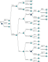
shows the structure of the first stages of the decision model. The [+] indicates sub-branches that have been collapsed for clarity. Nodes are named to show their relationship to one another; nodes with the same letter have identical structures to the branches of the tree beyond, whereas a different number and/or a lower-case letter indicates a different set of probabilities. The prefixes B, L and S denote nodes with probability sets specific to breech presentation or large or small for gestational age infants, respectively.
Model overview. [+], sub-branches of model collapsed for clarity.
At commencement, the scan policy can be set to selective (i.e. status quo), a universal scan for presentation only, or a universal scan for fetal biometry and presentation. The model structure is identical in each case. The difference is in the sensitivity and specificity of the scanning policies and their cost.
A fetus will be in either breech or cephalic presentation (node A1), or be LGA, SGA or AGA (node A2). For ease of modelling, we assume that all four possibilities are mutually exclusive and structured hierarchically, beginning with presentation (breech or cephalic) and followed by size (LGA, SGA or AGA). The implications of this are considered in Discussion. The probability of breech is the prevalence of breech at the time of screening (approximately 4.6%).11 If the scan policy is universal ultrasound (whether for fetal biometry or for presentation only), then, given the ease of interpretation of such a scan, we assume all breeches are detected (i.e. 100% sensitivity and specificity, node B_B). However, under the selective scan policy, approximately 45% of breeches will be undetected11 owing to the mother not having undergone a scan at all (for consistency with the rest of the model, we label these ‘false negatives’). Further outcomes relating to breech presentation are described in Outcomes relating to breech.
If the infant is in cephalic presentation, it may be LGA, SGA or AGA. The probabilities of each is the prevalence of the condition (node A2, by definition 10% for each). If an infant is LGA or SGA, the probability of detection is a function of the sensitivity of the scanning policy (nodes L_B and S_B; LGA: 26.55% under selective and presentation-only scan, 37.85% under universal scan for fetal size;138 SGA: 19.6% under selective and presentation-only scan, 56.53% under universal scan for fetal size8). The sensitivity and specificity of ultrasound for detecting SGA and LGA were derived from the POP study.8,138 The rationale for using the POP study values is that this study was conducted in NHS England, it involved nulliparous women being scanned at 36 weeks’ gestation, it is the only level 1 study of the diagnostic effectiveness of ultrasound to predict SGA and LGA (i.e. where the test result was blinded) and the values of sensitivity and specificity for SGA were similar to those in a 2019 Cochrane review of DTA.23 In addition, the DOR from the POP study for macrosomia was identical to the DOR in the meta-analysis presented in Chapter 8.
If a LGA infant is correctly diagnosed as LGA, the pregnancy is managed in accordance with the defined LGA policy of either IOL or expectant management (node ‘MGT_LGA_TP’), in either case leading to either vaginal delivery or emergency caesarean section (nodes L_C3 and L_C2a; odds ratio of emergency caesarean section compared with otherwise healthy infant, 1.79146). If a LGA infant is misdiagnosed as AGA (i.e. false-negative scan), delivery can be either vaginal or by emergency caesarean section. Further outcomes relating to LGA babies are described in Outcomes relating to large for gestational age infants.
If the infant is SGA and is correctly diagnosed as such, labour is induced, leading to either vaginal delivery or emergency caesarean section (node S_C3). False negatives may lead to vaginal delivery or emergency caesarean section (node S_C2). Further outcomes relating to SGA pregnancies are described in Outcomes relating to small for gestational age infants.
An AGA infant may be misdiagnosed as SGA or LGA (false-positive SGA and LGA, respectively), or correctly diagnosed as AGA (node B). A false-positive SGA infant will be induced unnecessarily, leading to either vaginal delivery or emergency caesarean section (node S_C4). A false-positive LGA infant will be managed in accordance with the defined LGA policy namely either IOL or expectant management (node ‘MGT_LGA_FP’). IOL and expectant management can lead to either spontaneous vaginal or emergency caesarean section delivery (nodes L_C4 and L_C1 respectively). Finally, a correctly diagnosed AGA infant (true negative) can be delivered vaginally or by emergency caesarean section (node C1).
Short- and long-term outcomes
For all parts of the model, different levels of neonatal morbidity and mortality are possible, although these outcomes are structured slightly differently between the model’s subtrees. For the breech, SGA and AGA models, delivery outcomes include no, moderate and severe neonatal morbidity, as well as perinatal death. The risks of each level of adverse outcome differ between specific branches (i.e. are affected by the true status of the infant, the mode of delivery and whether or not labour was induced early). Long-term outcomes are then modelled as a function of the level of neonatal morbidity at delivery. For the LGA model, delivery and long-term outcomes are modelled differently. This is explained in detail in Outcomes relating to large for gestational age infants.
Long-term outcomes include ‘no long-term complications’, ‘SEN’, ‘severe neurological morbidity’ (SNM) and ‘neonatal/infant mortality’. The risk of long-term complications increases with the level of neonatal morbidity (nodes E1, E2 and E3). Unlike delivery outcomes, long-term outcomes are not affected by the actual status of the infant prior to delivery, only by the level of neonatal morbidity at delivery. Importantly, this means that all screening and management options affect long-term outcomes indirectly only as a result of the impact that they have on the outcomes at delivery.
Outcomes relating to breech
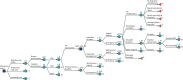
shows the decision tree with outcomes relevant to breech expanded and the remaining branches collapsed. The prevalence of breech refers to the fetal presentation at the time of screening. We assume that sensitivity and specificity for universal ultrasound is perfect at detecting fetal presentation, whether for size or breech presentation only. The sensitivity of selective ultrasound is lower because not all women receive ultrasound screening; however, we assume that all cases of suspected breech presentation would be either confirmed or rejected by ultrasound, so false-positive diagnosis is not an option (i.e. perfect specificity).
Outcomes associated with breech. [+], collapsed sections of the decision tree.
On diagnosis of a breech presentation, an ECV is offered (node B_ECV). If the ECV is successful (node B_ECVs) and the infant remains cephalic (node B_ECVs_rb), no further intervention will be offered (i.e. expectant management). However, the infant may spontaneously revert to breech presentation (node B_ECVs_rb). In either case, there is a probability of emergency caesarean section, which is increased if the infant has reverted to breech presentation (nodes B_C3b and B_C3a respectively). If breech presentation is not diagnosed prior to labour, delivery options include breech vaginal delivery and emergency caesarean section (node B_C2).
Following labour and delivery there is a risk of no, moderate or severe neonatal complications or perinatal death (node D1), subsequently leading to no long-term complications, SEN, SNM or perinatal mortality (node E1). Note that we assume no raised risk of neonatal morbidity associated with cephalic emergency caesarean section compared with cephalic vaginal delivery per se. We do, however, allow for a raised risk of complications with an emergency caesarean section following breech presentation compared with a vaginal breech delivery (nodes B_D2a and B_D2c). If ECV is not accepted, or fails, then elective caesarean section may be offered.
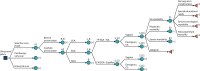
Outcomes relating to large for gestational age infants
shows the decision tree with outcomes relevant to LGA expanded and remaining branches collapsed. When LGA is suspected, the intervention given will be in accordance with the predetermined management strategy (IOL or expectant management) for both true-positive and false-positive LGA diagnoses. The management option will affect the likelihood of the delivery outcome, as well as the mode of delivery, which can be either vaginal or by emergency caesarean section. When LGA is not suspected, delivery can be either vaginal or by emergency caesarean section.
Outcomes associated with LGA. [+], collapsed sections of the decision tree.
Delivery outcomes include ‘no complications’, ‘respiratory morbidity’, ‘shoulder dystocia’, ‘other acidosis’ (i.e. acidosis not caused by shoulder dystocia) and ‘perinatal death’. The risk of each adverse outcome depends on the baseline risk, as well as on the mode of delivery, and whether or not labour was induced early.
Long-term outcomes depend on the outcome at delivery. For ‘no complications’, ‘respiratory morbidity’ and ‘other acidosis’, long-term outcomes included ‘no long-term complications’, ‘SEN’, ‘SNM’ and ‘neonatal/infant mortality’. For ‘no long-term complications’ the risk was equivalent to ‘no neonatal morbidity’ (node E1), and for ‘respiratory morbidity’ and ‘other acidosis’ the risk of long-term complications was equivalent to ‘severe neonatal morbidity’ (node E3). Shoulder dystocia (node L_E1) could result in no complications, brachial plexus injury (BPI) (node L_F1) or acidosis. BPI could be either transient or permanent (node L_G), the latter carrying the same risk of long-term outcomes as no neonatal morbidity (node E1) but with a penalty in terms of quality of life. Permanent BPI, SEN and SNM were long-term events; any other morbidity was expected to be resolved within the first year of life.
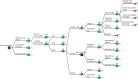
Outcomes relating to small for gestational age infants
shows the decision tree with the outcomes relevant to SGA expanded and the remaining branches collapsed. Labour will be induced early in suspected cases of SGA, whether based on a true or a false SGA diagnosis. Deliveries can be either vaginal or by emergency caesarean section. The probability of each mode of delivery is affected by whether or not labour was induced early. However, to avoid double counting the health effects of early labour induction, the mode of delivery affects only costs and not health outcomes.
Outcomes associated with SGA. [+], collapsed sections of the decision tree.
Delivery outcomes include no, moderate and severe neonatal morbidity, as well as perinatal death. Women with correctly diagnosed SGA pregnancies (true positives) are offered early IOL, which reduces the risk of morbidity and mortality. When SGA is unsuspected (false negatives), pregnancies are managed expectantly, with no risk reduction. Note that early labour induction may also increase the risk of morbidity if initiated needlessly (i.e. in an AGA pregnancy falsely suspected of being SGA). However, in a true SGA pregnancy, early labour induction is expected to reduce the risk of morbidity. The scenario with a false-positive diagnosis is discussed further in Outcomes relating to appropriate for gestational age infants.
Long-term outcomes include ‘no long-term outcomes’, ‘SEN’, ‘SNM’ and ‘neonatal/infant mortality’. Each outcome is possible for all levels of neonatal morbidity. However, the risk of long-term complications increases for moderate and severe neonatal morbidity (nodes E2 and E3).
Outcomes relating to appropriate for gestational age infants
shows the decision tree with the outcomes relevant to AGA expanded and the remaining branches collapsed. An AGA fetus may be either correctly diagnosed or incorrectly diagnosed as either SGA or LGA (node B). If correctly diagnosed, the mode of delivery can be either vaginal or emergency caesarean section (node C1), after which short- and long-term outcomes will follow as described in Short- and long-term outcomes.
Outcomes associated with AGA. [+], collapsed sections of the decision tree.
If an AGA fetus is falsely diagnosed as SGA, early IOL is offered. Unlike in the case of a true SGA, early labour induction of AGA pregnancies increases the risk of morbidity; however, the risk of perinatal death is still reduced.160 Short- and long-term outcomes will then follow as described in Short- and long-term outcomes. If, instead, an AGA fetus is misdiagnosed as LGA, the short- and long-term outcomes depend on the management strategy. Compared with expectant management, early IOL decreases the risk of emergency caesarean section and perinatal death but increases the risk of neonatal morbidity.
Just as for other branches of the model, long-term outcomes include ‘no long-term outcomes’, ‘SEN’, ‘SNM’ and ‘neonatal mortality’. Each outcome is possible for all levels of neonatal morbidity; however, the risk of long-term complications increases for moderate and severe neonatal morbidity (nodes E2 and E3).
Data
We populated the model with data from multiple sources from the literature. Where possible, we prioritised the inclusion of good-quality systematic reviews and meta-analyses, followed by large, good-quality clinical trials or cohort studies, as appropriate. When there was no objective evidence for a parameter, we relied on expert opinion either to judge whether or not a study in a related area provided a sufficient proxy or to provide a central estimate and credible interval representing beliefs about plausible values for the parameter. Data sources were subjectively graded as high, moderate or low, where high represented directly relevant data (i.e. providing the required parameter) from a good-quality source (e.g. RCT for relative effects and high-quality epidemiological study for baseline risks). A low grade represents instances in which evidence on the required parameter was absent from the literature and so is sourced from a related parameter, used as indirect evidence and revised reflecting expert opinion as to the plausible values. Full details of the derivation of model inputs are provided in Appendix 6, Tables 25–30, and all parameters are listed in –.
Model inputs for diagnostic performance
Model inputs for costs and related probabilities
Model inputs for probabilities
Probabilities
Where possible, probabilities were expressed as a baseline (beta or Dirichlet) for an otherwise healthy infant (i.e. neither breech nor LGA or SGA), they were then modified by odds ratios or relative risks, depending on the statistic either reported in, or calculable from, the literature. Odds ratios were selected in preference to risk ratios, as the former are independent of the baseline risk. Where no relative quantities were identified in the literature, probabilities are reported as independent beta distributions. Sampled values for probabilities were inspected to ensure that they were bounded between 0 and 1. Where out-of-range values were sampled, resampling was repeated until within-bounds values were generated.
Where relative effects were expressed as means and 95% CIs, standard error of the log of the mean was estimated by dividing the absolute difference between the log-mean and log-lower or -upper 95% CI by 1.96.
Costs
The price year used in the analysis is 2016/17. The majority of costs were sourced from the English national schedule of reference costs.175 The national schedule of reference costs reports different costs depending on how the service was delivered (e.g. elective inpatient, non-elective inpatient, outpatient procedures). We used costs from total Healthcare Resource Groups (i.e. weighted by each category by the number of yearly activities), except for cases in which only one or a few categories made logical sense. In all categories in the schedule costs were reported as mean and interquartile range. To obtain parameter estimates of costs, we fitted a gamma distribution using these data points. Where multiple cost categories were used, we first calculated a weighted average of the mean and interquartile range by the number of yearly activities in each category before fitting the gamma distribution.
Where no directly applicable cost could be identified from the reference schedule, we first attempted to obtain resource use from literature, and assign costs to this using the reference costs. When insufficient data on resource usage were available, we adopted the costs directly from the literature. Costs reported in currencies other than Great British pounds or in 2016/17 prices were converted to Great British pounds at the exchange rate of the year that the source was published and inflated to 2016/17 prices using the Hospital & Community Health Services (HCHS) index.184 Where no credible estimates could be identified from the literature, we estimated the costs ourselves, assigning a wide credibility interval to represent the uncertainty. Full details on the derivation of all cost parameters are presented in Appendix 6.
All costs presented in Great British pounds and updated to the cost-year of 2016–17 using the Hospital & Community Health Services Index:184 quality of life
We estimated age-specific quality of life for healthy neonates using EuroQol data for a general UK population.185 Age-specific health state utilities were multiplied by age-specific survival,186 the discounted sum over the time horizon of the model yielding the expected QALYs gained for an otherwise healthy neonate. Per definition, the quality of life following mortality is zero, and we made the simplifying assumption that all deaths during a particular year of life occurred on the first day of the year. In the absence of suitable evidence of how SEN affect quality of life, we assumed for our base-case scenario that SEN would affect costs only. In the case of SNM, we adjusted the baseline quality of life with a relative decrease following the methodology of Leigh et al.,187 using cerebral palsy (CP) as a proxy for SNM. Full details on the derivation of quality-of-life parameters are presented in Appendix 6.
Analysis
The model was analysed via Monte Carlo simulation, capturing the overall uncertainty in cost-effectiveness as a function of the uncertainty of the input parameters. Health outcomes were from the fetal perspective only and ultimately presented as QALYs. Cost-effectiveness was explored through incremental cost-effectiveness ratios (ICERs) and net monetary benefits (NMBs), using a WTP threshold of £20,000 per QALY. All costs and QALYs were discounted by 3.5% per annum.188 All costs were from a third-party (payer) perspective (i.e. NHS England plus SEN costs) and the reference case time horizon was 20 years (varied in sensitivity analysis).
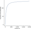
Stability testing was conducted to quantify (and, therefore, minimise) Monte Carlo error as a function of the number of simulations. The model was run 30 times with a given number of simulations. The coefficients of variation of the estimates of the mean and standard error of the mean cost and QALYs for each comparator were calculated. The mean of all of these was used as a summary measure of the Monte Carlo error. We used an arbitrary 2% cut-off point to declare the results stable.
Cost-effectiveness: reference case
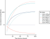
For each of the six discrete strategies, we present mean and 95% credibility intervals for cost and QALYs gained, net benefit at a WTP of £20,000 per QALY, and incremental net monetary benefit (INMB) relative to the assumed status quo (selective scanning with IOL for macrosomia or SGA, offer of ECV for breech). The option with the highest expected NMB was identified as the most cost-effective. Decision uncertainty was expressed as the probability that each decision would be cost-effective at the reference case threshold (i.e. £20,000/QALY). The cost-effectiveness acceptability curve plots decision uncertainty as a function of WTP per QALY (see ).
Cost-effectiveness acceptability curve for the chance that each strategy will be the most cost-effective as a function of WTP for an additional QALY. Mexp, expectant management; MIOL, IOL; Sbre, universal ultrasound for fetal presentation only; Ssel, (more...)
Cost-effectiveness: sensitivity and scenario analyses
In addition to the primary analysis, we report a number of scenario analyses and one-way sensitivity analyses to explore specific uncertainties in more detail. Specifically:
Time horizon.
Cost of scan to assess fetal presentation only.
The cost of a presentation-only scan is dependent on whether it is feasible to incorporate the scan into a routine antenatal visit, with a midwife conducting it using a hand-held unit, or if it can be done only during a dedicated visit by an ultrasonographer in a secondary care setting.
The baseline risks of perinatal death, moderate and severe neonatal morbidity.
The baseline risks of each of these were estimated from different sources, yet they are mutually exclusive events. Ideally, these should be modelled as a Dirichlet distribution, but because the data were from different sources we modelled them as independent betas. We thus explore these further in a one-way sensitivity analysis.
In addition, because of concerns over the validity of input data, we also explore the difference in:
the risk of acidosis and respiratory morbidity associated with vaginal delivery of a LGA infant (vs. AGA)
the odds ratio of perinatal death resulting from delivery by emergency caesarean section of a breech infant (vs. vaginal delivery)
the relative risk of an emergency caesarean section from IOL for a SGA infant (vs. expectant management of an AGA infant)
the relative risk of SEN as a result of inducing labour (vs. expectant management), and the impact that IOL has on health-related quality of life, and the sensitivity of ultrasound scanning at detecting SGA.
Value-of-information analysis
Uncertainty in cost-effectiveness results (i.e. decision uncertainty) was used to conduct a VOI analysis.189 Decision uncertainty arises from parameter uncertainty. The EVPI is the expected value of eliminating all decision uncertainty, which by definition implies eliminating all parameter uncertainty. This therefore provides an upper bound for the value of all research into the decision question. The EVPPI is the expected value of eliminating uncertainty in a single parameter or group of parameters. The EVSI is the expected value of a study of sample size n. The EVSI of a study of size n less the cost of conducting it provides a measure of the expected return on investment in that research project [expected net gain of sampling (ENGS)].190–192 An EVPPI above the plausible cost of a research project is a necessary condition for future research to be economically viable. A positive ENGS is the sufficient condition. The efficient sample size of a study is that which maximises the ENGS.
We estimated that there are approximately 196,297 singleton births at ≥ 37 weeks’ gestation to nulliparous women that are not delivered by elective caesarean section each year. Assuming a time horizon for which the decision question remains valid of 10 years yields a (discounted) beneficial population of 1,689,663. If it is reasonable to assume that our analyses are generalisable to all births in England, the beneficiary population is 5,477,940.
We report the per-patient (i.e. per mother/infant dyad) and population EVPI at a WTP of £20,000 per QALY. We then report the per-patient and population EVPPI for each parameter individually, calculated using the Sheffield Accelerated Value-of-information (SAVI) tool.159 Parameters with a positive EVPPI were grouped into those that could logically be collected in one research study, and the EVPPI for that group of parameters was calculated (also with the SAVI tool159). The EVSI for any parameters or groups of parameters is then calculated using the method of Heath et al.193 Population values are presented as a ‘conservative’ estimate, assuming that the information is of value only to singleton nulliparous pregnancies (i.e. using the 1,689,663 beneficiary population) and a broader estimate that assumes the information is of value to all pregnancies in England (5,477,940 population).