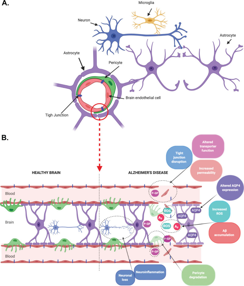
Figure 1. Cellular structure and Alzheimer’s disease (AD) specific changes in the blood–brain barrier (BBB).

Figure 1

Cellular structure and Alzheimer’s disease (AD) specific changes in the blood–brain barrier (BBB). (A) Schematic representation of the BBB structure. The BBB is formed by brain endothelial cells (BECs) with astrocytes and pericytes functioning as key supporting cells. The BBB and other brain cells (neurons and microglia) form the neurovascular unit. (B) AD-specific changes in the BBB include altered tight junction and transporter expression in BECs, pericyte degradation, altered aquaporin-4 (AQP4) expression in astrocytes with these changes leading to increased permeability, reactive oxygen species (ROS) and neuroinflammation, and subsequent amyloid-β (Aβ) accumulation and neuronal loss. Created using BioRender.com.

From: Chapter 7, Modeling the Blood–Brain Barrier to Understand Drug Delivery in Alzheimer’s Disease

Cover of Alzheimer’s Disease: Drug Discovery
Alzheimer’s Disease: Drug Discovery [Internet].
Huang X, editor.
Brisbane (AU): Exon Publications; 2020 Dec 18.
Copyright: The Authors.

Licence: This open access article is licensed under Creative Commons Attribution 4.0 International (CC BY 4.0). https://creativecommons.org/licenses/by-nc/4.0/

NCBI Bookshelf. A service of the National Library of Medicine, National Institutes of Health.