Continuing Education Activity
Miliary tuberculosis is a severe and disseminated form of tuberculosis (TB), a condition arising from Mycobacterium tuberculosis infection. Miliary TB occurs when the bacteria spread through the bloodstream, affecting multiple organs throughout the body. The term "miliary" is derived from the Latin word "miliarius," meaning resembling millet seeds, describing the characteristic appearance of the tiny tubercular lesions in affected organs.
Miliary TB presentations can be complex. Consequently, diagnosis and treatment are challenging. Understanding the tubercular disease process can guide management strategies and improve outcomes.
This activity for healthcare professionals is designed to improve learners' competence in evaluating and managing miliary TB. Valuable insights will be gained that will enable learners to collaborate effectively with an interprofessional team caring for patients with this condition.
Objectives:
Identify the etiology of miliary tuberculosis.
Evaluate a patient with suspected miliary tuberculosis and formulate a clinically appropriate diagnostic plan.
Compare the different miliary tuberculosis treatment regimens and create a personalized managed strategy for a patient diagnosed with the condition.
Collaborate effectively with the interpersonal team in managing miliary tuberculosis by formulating short- and long-term treatment plans, monitoring and addressing antituberculosis treatment's side effects, and preventing infectious spread within the community.
Access free multiple choice questions on this topic.
Introduction
Tuberculosis (TB) is a condition arising from Mycobacterium tuberculosis (MTB) infection. MTB is transmitted from a person with pulmonary TB (PTB) infection. Droplet nuclei containing the tubercle bacilli are aerosolized by speaking, sneezing, or coughing. The droplets dry quickly, remain in the air for several hours, and may be inhaled by other individuals. Other MTB transmission routes are insignificant.
The disease most frequently affects the lungs, though up to a third of TB cases involve other organs. The bacterium is an obligate aerobe and thus typically lodges in the highly oxygenated lung regions, the upper lobe and the lower lobe's superior aspect close to the pleura.
The functional unit of the lungs is the alveolus. The thin alveolar walls are comprised of the capillary endothelium, basement membrane, and alveolar epithelium—components of the blood-air barrier that facilitate gas exchange. Alveolar macrophages are cells derived from monocytes, roaming free inside the alveolar lumen. These phagocytic cells are critical to the lung's immunity but also contribute to TB pathophysiology. Pores of Kohn are alveolar wall perforations where microbes and exudates can spread.
Lung vasculature is comprised of the following:
Pulmonary arteries: arise from the pulmonary trunk and branch into lobar and segmental arteries in the lung parenchyma; these arteries carry deoxygenated blood into the lungs
Pulmonary veins: carry oxygenated blood from the lungs and back into the heart and arterial circulation
Bronchial arteries: arise from the thoracic aorta and posterior intercostal arteries; these vessels supply the root of the lungs and visceral pleura
Bronchial veins: follow the bronchial arteries and drain into the azygos and hemiazygos veins
Pulmonary lymphatic plexuses: the superficial subpleural lymphatic plexus drains into the bronchopulmonary (hilar) lymph nodes; the deep bronchopulmonary lymphatic plexus drains into the intrinsic pulmonary lymph nodes before emptying into the bronchopulmonary lymph nodes; the tracheobronchial lymph nodes drain the bronchopulmonary lymph nodes and ultimately empty into the right lymphatic and thoracic ducts
Parietal pleura lymphatics: parietal pleural lymphatic vessels drain into the thoracic wall and axillary lymph nodes
In immunocompromised patients, failure of the immune system to contain the infection enables MTB to spread from the lungs to the other body organs through the vasculature. Disseminated TB is defined as the simultaneous involvement of at least 2 non-contiguous body organs or infection of the blood, bone marrow, or liver. Miliary TB is a potentially fatal, disseminated form of the disease arising from hematogenous tubercle bacilli spread throughout the lungs and other organs.
The condition results in the formation of millet-seed-sized (1 to 2 mm) tubercular foci. The term "miliary tuberculosis" was first coined by John Jacobus Manget in 1700 while describing a pathological specimen having tiny tubercles resembling millet seeds in appearance. The term originated from the Latin word "miliarius," related to the millet seed. Miliary mottling on a chest radiograph is the classical hallmark that supports the diagnosis of miliary TB. Miliary TB is classified as both pulmonary and extrapulmonary TB.[1][2]
See StatPearls' companion topic, "Tuberculosis," for a comprehensive discussion of the epidemiology, pathophysiology, evaluation, and management of pulmonary tuberculosis.
Etiology
MTB is primarily responsible for TB manifestations in countries where bovine TB has been eradicated. Infected humans serve as a natural MTB reservoir. The organism is a non-spore-forming, non-motile, obligate-aerobic, facultative, catalase-negative, intracellular bacillus. MTB is gram-neutral and may be visualized by Ziehl-Neelsen (ZN) staining. Cell wall mycolic acids impart acid fastness when decolorized with acid-alcohol. Thus, MTB is also known as acid-alcohol-fast-bacillus (AAFB). Other mycobacteria are classified as nontuberculous or atypical mycobacterial organisms.[3]
Epidemiology
According to the World Health Organization (WHO) Global Tuberculosis Report, around 10 million people developed TB, and 1.3 million died from the disease globally in 2017.[4] Most cases are found in the developing world. However, miliary TB's epidemiological patterns continuously change due to the following:
Immunosuppressant use
Immigration from highly endemic countries
Surge of HIV cases
Alcoholism
Other medical conditions manifesting with immunosuppression, such as diabetes mellitus and chronic kidney disease
According to the Centers for Disease Control and Prevention, 8920 new TB cases were reported in the United States in 2019.[5] Miliary TB accounts for about 1% to 2% of all cases of TB and up to 20% of all forms of extrapulmonary TB in immunocompetent individuals.
Miliary TB was thought to be a disease of infants and children before antibiotics were widely used. The disease has a bimodal age distribution, with one peak occurring in young adults and adolescents and the other occurring in older individuals. Miliary TB has a slight male preponderance.[6]
Pathophysiology
MTB infection starts with the lung epithelial macrophages. The organism's replication is initially unchecked, but the T-helper cell response induces macrophages to suppress bacterial proliferation. Failure of cell-mediated immunity in immunocompromised patients results in the organisms reaching the lymphatic ducts through the pulmonary lymphatics. The lymphatic ducts drain into the right side of the heart and the pulmonary arteries. Systemic spread results when infective lung foci seed the pulmonary venous return to the heart, and the bacilli disseminate via the arterial circulation.
The immunopathogenic mechanisms underlying miliary TB development are complex and not fully understood. Containment of the bacteria by effector T-cells is likely compromised. The specific cytokines and other immune-regulatory determinants in the host-pathogen interaction are poorly elucidated.
What is known, however, is that miliary TB is related to lymphohematogenous MTB dissemination from a primary or reactivation focus to various organs. In endemic countries, reinfection can result in miliary TB. Occasionally, miliary TB emerges from simultaneous dormant foci reactivation within previously infected organs.[7]
Histopathology
Grossly, miliary TB is characterized by small, punctate, gray to reddish-brown, rounded lesions with more or less uniform size in the lungs and other organs. In immunocompetent individuals, each microscopic TB focus contains a typical tubercle with central caseation necrosis and tubercle bacilli surrounded by Langhans-type giant cells, epithelioid cells, lymphocytes, and fibrocytes. However, in immunosuppressed individuals, the foci show caseation with tubercle bacilli without granuloma formation. This condition is known as nonreactive miliary TB.[8][9]
History and Physical
The clinical features of miliary TB vary widely, depending on the sites mainly involved, and may remain obscure until late in the disease course. Initial symptoms develop gradually and are usually nonspecific, which include constitutional manifestations such as fever, generalized weakness, anorexia, weight loss, and lassitude. Many patients may initially present with productive coughing, difficulty breathing, chest pain, and hemoptysis. Abdominal symptoms such as nausea, vomiting, and abdominal pain are also common in the early stages.
The most frequent extrapulmonary sites include the lymphatic system, bones, joints, liver, central nervous system (CNS), and adrenal glands. However, virtually any organ system can be infected, and symptoms referrable to organ-specific dysfunction usually appear.
Miliary TB has 2 clinical variants with different demographic profiles and presentations: classical acute and cryptic miliary TB. Both conditions require a high index of suspicion for timely diagnosis and treatment.
Acute Miliary TB
Individuals with acute miliary TB are generally younger than 40 years. Most patients have a history of subacute or chronic constitutional symptoms and organ-specific manifestations, depending on the site affected by the disease. Evening febrile episodes and night sweats of 1 to 2 weeks duration are classically described, although a patient can have early morning temperature spikes. Other common initial symptoms include nonproductive cough and dyspnea. Some patients may observe hemoptysis. Acute respiratory distress syndrome (ARDS) is a rare, fatal presentation of miliary TB.[10][11]
Abdominal TB occurs following hematogenous spread from a pulmonary focus or by local spread from a gastrointestinal or abdominal source. Abdominal TB can occur as hepatic, intestinal, or peritoneal involvement.[12] Hepatic spread presents as right upper quadrant pain, nausea, vomiting, fever, and generalized fatigue. Icterus and hepatosplenomegaly may be seen clinically.[13][14] Intestinal TB usually manifests with fever, micro- and macronutrient deficiencies, altered bowel habits, and subacute to acute intestinal obstruction. Children with intestinal TB may have failure to thrive.
TB peritonitis should be suspected in a patient presenting with complaints of fever, fatigue, abdominal pain, and ascites. Sometimes, the presentation may be confused with an acute abdomen. On surgical exploration, miliary tubercles may be found in the omentum and peritoneal surfaces. Biopsy and histopathology will show tuberculous foci.
Miliary TB can present as overt adrenal insufficiency (Addison disease) at the time of initial presentation or during antitubercular treatment. The manifestations include skin hyperpigmentation, hypotension, hypoglycemia, and electrolyte imbalance.[15]
On fundoscopy, the presence of choroid tubercles is pathognomonic of miliary TB. Choroid tubercles commonly occur in children. These pathological structures are bilateral, pale, and gray-white or yellowish lesions, usually less than a quarter of the optic disc's size, located within 2 cm of the optic nerve.[16]
Musculoskeletal pathology accounts for 10% of extrapulmonary TB cases. The bony site most commonly involved is the spine, manifesting as tubercular spondylitis or Pott spine. On history, patients with Pott spine may have the classic initial TB symptoms such as fever and weight loss. Back pain and tenderness develop insidiously and are often the earliest clues to spine involvement. Paraplegia, paraparesis, kyphosis, or scoliosis develop gradually and may cause complete debility late in the illness.
The joints are the second most frequently involved sites, and septic (tuberculous) arthritis is common. Other musculoskeletal manifestations of miliary TB include osteomyelitis, tenosynovitis, bursitis, and pyomyositis.[17]
Neurological involvement may present as headaches and nuchal rigidity secondary to tubercular meningitis (TBM) with or without tuberculoma formation. TBM has been reported in 10% to 30% of adult patients with miliary TB. Thoracic transverse myelopathy may manifest as sensorimotor abnormalities.[18][19]
Erythematous macules and papules characterize tuberculosis miliaria cutis. These lesions are manifestations of lymphohematogenous MTB spread in the skin.
Clinically significant cardiac or renal involvement is uncommon. However, miliary TB can cause myocarditis, congestive heart failure, endocarditis, mycotic aneurysms, and acute kidney injury either directly or as a syndromic constellation of multiorgan dysfunction syndrome (MODS).[20]
Miliary TB can present similarly in children and adults. However, chills, night sweats, hemoptysis, and productive cough are less frequently reported in children than in adults. In contrast, peripheral lymphadenopathy and hepatosplenomegaly are more common in the pediatric than in the adult population. TBM is more frequently seen in children with miliary TB (20%–40%) than in adults (15% to 30%).[21] Miliary TB develops less frequently in children who have received the Bacillus Calmette-Guérin (BCG) vaccination.
TB's clinical presentation in individuals with HIV depends on their CD4+ count. Patients with HIV having a CD4+ count greater than 200 cells/mm experience disease progression similar to immunocompetent individuals. In contrast, people with HIV with a CD4+ count of less than 200 cells/mm develop atypical manifestations of miliary TB, such as cutaneous lesions, intrathoracic lymphadenopathy, and tuberculin anergy. Profound immunosuppression due to any cause is generally associated with atypical TB presentation.[22]
Cryptic Miliary TB
Most patients with cryptic miliary TB are older than 60 years. The condition may be considered in the differential diagnosis of fever of unknown origin or metastatic carcinoma, as symptoms like fever, progressive weight loss, and general debility can occur without the usual TB signs and symptoms. However, mild hepatosplenomegaly is occasionally observed. Patients often have a normal chest radiograph and negative skin tuberculin test, causing a delay in diagnosis.[23][24]
Atypical manifestations of miliary TB can delay the diagnosis. Such unusual presentations include ARDS, pneumothorax, cytopenia, septic shock, glomerulonephritis, endocarditis, mycotic aortic aneurysm, cholestatic jaundice, and hyponatremia due to syndrome of inappropriate antidiuretic hormone secretion.
Evaluation
Diagnosing miliary TB requires a high suspicion index. A multi-pronged approach, comprised of meticulous history taking, thorough physical examination, and radiological and laboratory investigation, is required for timely diagnosis and adequate treatment.
Laboratory Findings
Hematological changes seen in miliary TB are usually nonspecific. Pancytopenia, anemia, leucopenia, leucocytosis with predominant lymphocytosis, thrombocytopenia, or thrombocytosis may be reported. The most common hematological abnormality encountered in miliary TB is anemia of chronic disease. Elevated acute-phase reactants, particularly erythrocyte sedimentation rate and C-reactive protein, are frequently encountered. Leukaemoid reaction is also described. Thus, miliary TB may also be mistaken for leukemia. Disseminated intravascular coagulation is rare and seen in the setting of MODS and ARDS.[25][26]
Biochemistry panels can be normal or have subtle disturbances. Hyponatremia, secondary to meningeal involvement or abnormal antidiuretic hormone levels, is frequently encountered and is an indicator of neurological damage. Hyperbilirubinemia, hypoalbuminemia (a negative acute-phase reactant), and elevated alkaline phosphatase are also seen. Hypercalcemia, though rare, has also been reported.[27][28]
Imaging Studies
No uniform guidelines exist for diagnosing miliary TB, but the following criteria have been suggested:
Clinical presentation consistent with a diagnosis of TB, such as pyrexia with evening temperature rise, weight loss, anorexia, tachycardia, and night sweats of greater than 6 weeks duration responding to antitubercular treatment
Classical miliary pattern on chest radiograph
Bilateral diffuse reticulonodular lung lesions on a background of miliary shadows demonstrable either on plain chest radiograph or high-resolution computed tomography (HRCT)
Microbiological, cytopathological, histopathological, or molecular evidence of TB
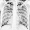
The chest radiograph shows typical miliary mottling, ie, homogenously distributed, discrete, uniform size (1 to 2 mm), and millet-shaped lesions in all lung zones (see Image. Miliary Tuberculosis Radiography). These features are also the radiographic hallmark of miliary TB. However, these x-ray findings are usually absent in early disease or cryptic TB. HRCT may be advisable in such cases to look for parenchymal lesions. Contrast-enhanced CT (CECT) is preferable for evaluating lymphadenopathy, calcifications, and pleural pathology.[29][30]
Extrapulmonary TB lesions may be assessed by ultrasonography, CECT, and magnetic resonance imaging (MRI), which can determine the extent of organ involvement.[31] Positron-emission tomographic CT has been recently used as an investigating tool for evaluating patients with suspected TB.[32][33][34]
Invasive diagnostic procedures are indicated for patients with suspected extrapulmonary TB. The following have a good diagnostic yield in miliary TB:
Image-guided radiological procedures, such as fine-needle aspiration and tissue biopsy, are useful for procuring tissue and body fluids for diagnostic testing. Ultrasound, CT, and MRI are frequently used in these procedures.[35][36]
Immunology-Based Methods
Anergy on tuberculin skin testing (TST) is more frequently reported in miliary TB than PTB, though the test can become positive during antitubercular therapy. A positive interferon-γ release assay indicates infection but does not signify active disease. Thus, this diagnostic modality is of limited use in highly endemic areas.[37]
Serological tests are not advocated for TB detection because of their low sensitivity and specificity. However, adenosine deaminase (ADA) and interferon-γ (IFN-γ) may be used as adjuncts for detecting the presence of MTB in pleural, pericardial, and ascitic fluids.[38][39][40][41] ADA may also be used in diagnosing TBM.[42]
The ADA cutoff value for pleural and pericardial effusion is 40 U/L; for tubercular ascites is 39 U/L; and for CSF is 10 U/L.[43][44][45] ADA levels may be falsely elevated in the presence of empyema, parapneumonic effusions, endometriosis, malignancies like lymphoma, and collagen vascular disorder.[43] High CSF ADA can also be seen in cerebral malaria, brucellosis, neurosarcoidosis, fulminant pyogenic meningitis, AIDS, and CSF lymphoma.[46]
Molecular Studies
Molecular methods such as polymerase chain reaction, Gene Xpert MTB/RIF, and line probe assays may be useful in the early diagnosis of pulmonary and extrapulmonary TB and in detecting drug-resistant tubercle bacilli. Various tissue specimens may be used, and results may be available within hours.[47][48][49][50]
Definitive Diagnosis
Detection of mycobacterial isolates from a clinical specimen provides a definitive diagnosis of disseminated TB. Examples of tissue specimens are sputum, body fluids, tissue, and biopsy samples. The specimens are inoculated on agar-based (eg, Lowenstein-Jensen or LJ) or liquid media with fluorescence detection. Direct visualization of acid-fast bacilli is aided by ZN staining or the more sensitive auramine-rhodamine (fluorochrome dye) staining.[51][52] The specimen should be cultured on standard solid LJ media and then inoculated in liquid media (eg, BACTEC Mycobacterial Growth Indicator Tube or BACTEC™ MGIT™ 960).[53]
Liquid-based media reduces the MTB detection time to 1 to 3 weeks, compared to 6 to 8 weeks on solid media. The method also shortens drug sensitivity testing time.
Blood culture is usually not employed for mycobacterium isolation. This test is usually negative, although positive results may be reported among immunocompromised individuals with hematogenous dissemination.[54] MTB may be differentiated from isolated nontuberculous mycobacteria by hybridization using nucleic acid probes or biochemical methods or by matrix-assisted laser desorption/ionization time-of-flight mass spectrometry (MALDI-TOF).[55]
Histopathological examination of a tissue biopsy specimen should show granulomatous inflammation with central caseation, with or without tubercle bacilli. Demonstration of a tubercle granuloma in a biopsy sample obtained from a patient with typical TB manifestations is suggestive of TB. However, demonstration of bacilli by staining or culture is required to confirm the diagnosis.[56]
Treatment / Management
Standard PTB drug regimen may also be used for treating miliary TB. As per the WHO, a standard 6-month antitubercular drug regimen consists of a 2-month intensive or bactericidal phase with isoniazid, rifampicin, pyrazinamide, and ethambutol and a 4-month continuation or sterilizing phase with isoniazid and rifampicin. Treatment duration may be modified according to the age group affected and primary disease site.
Longer treatment periods are advisable for children, immunocompromised individuals, patients with a slow clinical response, and the presence of TBM, TB lymphadenitis, or skeletal TB. The generally recommended minimum therapeutic duration is 9 months for skeletal TB and 12 months for TBM.[57][58][59]
For abdominal TB, 9 months of therapy used to be the conventional approach. However, a recent multicenter, randomized trial showed that both 6-month and 9-month regimens are equivalent for patients with abdominal TB.[60] Neurological involvement, particularly TBM, must be determined in all miliary TB cases. Neurologic TB requires a longer treatment duration than the standard and concomitant steroid therapy.[61]
For previously treated patients, the WHO guidelines advocate that culture and drug susceptibility testing (DST) specimens be obtained from all previously treated TB patients at or before the start of treatment. DST should be performed for at least isoniazid and rifampicin. In settings where rapid molecular DSTs are available, the DST results should guide the choice of regimen. The duration of treatment may also be individualized according to the clinical setting.
Patients suspected of miliary TB must be screened for diabetes mellitus and HIV before initiating antitubercular treatment and vice-versa. Immune reconstitution inflammatory response (IRIS) is a condition arising from excessive immune reaction to MTB that may develop in patients with HIV during or after completing an antitubercular drug regimen. Antiretroviral treatment (ART) for all patients with HIV and TB must not be initiated within the first 8 weeks of starting TB treatment and within 2 weeks in profoundly immunosuppressed HIV-positive TB patients with CD4+ counts less than 50 to prevent IRIS.[62][63]
Medical and surgical interventions may be warranted for diagnosis and therapy. Examples are mechanical ventilation for ARDS, abdominal surgery for small bowel perforation, and ventriculoperitoneal shunt surgery for TBM, which may be performed to reduce TB complications.
Although substantial evidence is lacking, corticosteroid use in miliary TB has shown clinical efficacy in a few scenarios. Notably, it has been demonstrated to be beneficial in adrenal insufficiency, TB meningitis, large pericardial or pleural effusion, IRIS, ARDS, immune-complex nephritis, and secondary hemophagocytic syndrome.[64][65] Liver function tests (LFTs) should be obtained before and during anti-TB therapy (ATT). Serial LFTs should be performed to check for ATT-induced hepatitis.
The criteria for ATT-induced hepatitis have evolved and include the following:
Transaminases are elevated up to 5 times the upper normal limit without hepatitis symptoms; or
Transaminases are elevated up to 3 times the upper normal limit with hepatitis symptoms; or
Bilirubin rises twice the upper normal limit, and other possible hepatitis causes, like acute viral hepatitis and autoimmune hepatitis, have been ruled out.
All hepatotoxic drugs—isoniazid, rifampicin, and pyrazinamide—should be withdrawn if ATT-induced hepatitis develops. A modified ATT regimen must be initiated to prevent further liver damage. According to guidelines from the British and American Thoracic Societies, ATT rechallenge may be considered once the LFTs have normalized. The decision to restart ATT drugs at incrementally increasing doses or full doses should be guided by the clinical picture.[66][67]
Differential Diagnosis
The protean and nonspecific clinical manifestations of miliary TB often generate a large differential diagnosis. The common constitutional symptoms of fever, chills, night sweats, anorexia, weight loss, and fatigue raise concerns about many potential infectious, autoimmune, and neoplastic etiologies. Headache, seizures, altered sensorium, cough, chest pain, hemoptysis, abdominal pain, lymphadenopathy, hepatosplenomegaly, back pain, and neurologic dysfunction are themselves nonspecific manifestations of a huge number of diseases. The manifestations of miliary TB often develop gradually, and their lack of specificity frequently results in a diagnostic delay. However, the constellation of signs and symptoms and patient demographic and immune status must be taken into account when evaluating patients who may have miliary TB.
Varied clinical etiologies can present with a miliary pattern on chest radiography and CT. Therefore, a thorough workup is required to reach an etiological conclusion. Other causes of miliary shadowing include histoplasmosis, blastomycosis, coccidioidomycosis, nocardiosis, sarcoidosis, lung carcinoma with lymphangitis carcinomatosis, metastatic carcinoma, pyogenic infection spread from a remote site, pulmonary hemosiderosis, and hypersensitivity pneumonitis.[68]
Prognosis
Miliary TB has high morbidity and mortality. Treatment delay appears to be the most significant factor responsible for mortality. The mortality related to miliary TB is approximately 15% to 20 % in children and 25% to 30% in adults.[69][70][71]
In patients with ARDS due to miliary TB, the following Acute Physiology and Chronic Health Evaluation (APACHE II) scores have been identified as mortality predictors:
Greater than 18
Less than or equal to 18 with hyponatremia and a ratio of arterial oxygen tension to the fraction of inspired oxygen (FiO2) less than or equal to 108.5
[72]
Complications
Delayed treatment of miliary TB can produce the following emergent complications:
ARDS
MODS
Tubercular empyema
The air leak syndromes pneumothorax and pneumomediastinum
Tubercular pericardial effusion and pericarditis
Immune reconstitution inflammatory syndrome
Myocarditis, native and prosthetic valve endocarditis, and intracardiac masses
Mycotic aneurysm of the aorta
Tubercular meningitis with focal neurological deficits
Systemic amyloidosis
Immune complex glomerulonephritis
Bone marrow suppression
Disseminated intravascular coagulation
Rapid deterioration may occur in the setting of profound immunosuppression, as in HIV infection with low CD4+ counts, immunosuppressant use, and inborn immune conditions.
Deterrence and Patient Education
Patient education and counseling are the cornerstones of TB management. Proper education materials should be provided to at-risk populations to reduce bacterial transmission and enhance the detection of this condition. Patients diagnosed with TB must be informed about the following:
The disease process in relation to the symptoms
Importance of ATT adherence and follow-up
Monitoring for drug toxicity symptoms
Measures that can help avoid MTB transmission to close contacts
Healthcare providers must monitor patients closely for signs of drug reactions, progress, or lack of response to optimize the antitubercular drug regimen.
Pearls and Other Issues
The most important points to remember when evaluating and managing miliary TB are the following:
Miliary TB is a potentially fatal form of disseminated TB characterized by millet-seed-like granuloma formation in various organs.
Miliary TB often arises from a primary pulmonary infection that spreads hematogenously. However, extrapulmonary primary sites can also give rise to miliary TB.
The clinical presentation can vary widely. The initial manifestations may be nonspecific, including fever, weight loss, fatigue, and respiratory symptoms. Diagnosis can be challenging due to the diversity of presentations.
Miliary TB is more common in individuals with poor immunity, such as people with HIV or AIDS, older individuals, and people on immunosuppressive therapy.
Chest x-rays and CT scans often reveal characteristic miliary patterns, showing widespread, small nodules throughout the lung fields. However, these imaging features may be absent in early and cryptic miliary TB.
Central nervous system involvement, particularly TBM, can occur and presents a serious and life-threatening complication.
Definitive diagnosis often involves a combination of clinical evaluation, imaging studies, and laboratory tests such as sputum culture, polymerase chain reaction, and Gene Xpert MTB/RIF.
The WHO recommends the standard antitubercular regimen for patients diagnosed with miliary TB. However, skeletal and neurological involvement can prolong the treatment course.
Treatment adherence is crucial to prevent the development of drug-resistant strains.
Prognosis depends on factors such as the extent of organ involvement, timeliness of diagnosis, and therapeutic effectiveness. Early detection and intervention improve outcomes.
Preventive measures include vaccination with the BCG vaccine in regions where TB is prevalent. Timely detection and treatment of active TB cases also help prevent the development of disseminated TB.
Miliary TB is a serious condition requiring prompt medical attention and diagnostic and management proficiency. A comprehensive evaluation approach must be pursued to initiate the appropriate treatment.
Enhancing Healthcare Team Outcomes
An interprofessional team approach can ensure that patients with miliary TB receive holistic and integrated care to achieve the best possible outcomes. Judicious evaluation by the physicians, gentle bedside care by the nurses, and utmost diligence by laboratory personnel can help treat the disease promptly and stop its spread in the hospital and community. Effective implementation of the WHO's Directly Observed Treatment, Short-Course strategy by community healthcare personnel can help ensure treatment adherence. Completion of therapy is vital for disease resolution.
Specialists who may be involved in the inpatient care of individuals with miliary TB include emergency physicians, pulmonologists, intensivists, gastroenterologists, endocrinologists, neurologists, infectious disease specialists, surgeons, pain specialists, pathologists, and radiologists. The pharmacist can help educate patients about ATT and manage antitubercular medication dosage to prevent drug-induced complications. Nutritionists can help optimize patient nutrition in aid of recovery. Occupational and physical therapists can help patients improve functional independence during recovery.
Collaboration, shared decision-making, and communication are critical elements for a good outcome. Nothing less than an integrated care pathway and evidence-based diagnostic and management approaches must be rendered to the patient.
Miliary Tuberculosis Radiography. This chest x-ray shows millet-shaped lesions in all lung zones, which are features consistent with miliary tuberculosis. Contributed by Katherine Humphreys
References
- 1.
Sharma SK, Mohan A, Sharma A, Mitra DK. Miliary tuberculosis: new insights into an old disease.
Lancet Infect Dis. 2005 Jul;5(7):415-30. [
PubMed: 15978528]
- 2.
Sahn SA, Neff TA. Miliary tuberculosis.
Am J Med. 1974 Apr;56(4):494-505. [
PubMed: 4206484]
- 3.
Adigun R, Singh R.
StatPearls [Internet]. StatPearls Publishing; Treasure Island (FL): Jul 11, 2023. Tuberculosis. [
PubMed: 28722945]
- 4.
Nguyen HV, Tiemersma EW, Nguyen HB, Cobelens FGJ, Finlay A, Glaziou P, Dao CH, Mirtskhulava V, Nguyen HV, Pham HTT, Khieu NTT, de Haas P, Do NH, Nguyen PD, Cung CV, Nguyen NV. The second national tuberculosis prevalence survey in Vietnam.
PLoS One. 2020;15(4):e0232142. [
PMC free article: PMC7179905] [
PubMed: 32324806]
- 5.
Schwartz NG, Price SF, Pratt RH, Langer AJ. Tuberculosis - United States, 2019.
MMWR Morb Mortal Wkly Rep. 2020 Mar 20;69(11):286-289. [
PMC free article: PMC7739979] [
PubMed: 32191684]
- 6.
Sharma SK, Mohan A, Sharma A. Challenges in the diagnosis & treatment of miliary tuberculosis.
Indian J Med Res. 2012 May;135(5):703-30. [
PMC free article: PMC3401706] [
PubMed: 22771605]
- 7.
Sharma SK, Mohan A. Miliary Tuberculosis.
Microbiol Spectr. 2017 Mar;5(2) [
PubMed: 28281441]
- 8.
Auerbach O. Acute Generalized Miliary Tuberculosis.
Am J Pathol. 1944 Jan;20(1):121-36. [
PMC free article: PMC2033127] [
PubMed: 19970738]
- 9.
de Noronha AL, Báfica A, Nogueira L, Barral A, Barral-Netto M. Lung granulomas from Mycobacterium tuberculosis/HIV-1 co-infected patients display decreased in situ TNF production.
Pathol Res Pract. 2008;204(3):155-61. [
PubMed: 18096327]
- 10.
Kim JY, Park YB, Kim YS, Kang SB, Shin JW, Park IW, Choi BW. Miliary tuberculosis and acute respiratory distress syndrome.
Int J Tuberc Lung Dis. 2003 Apr;7(4):359-64. [
PubMed: 12733492]
- 11.
Mohan A, Sharma SK, Pande JN. Acute respiratory distress syndrome (ARDS) in miliary tuberculosis: a twelve year experience.
Indian J Chest Dis Allied Sci. 1996 Jul-Sep;38(3):157-62. [
PubMed: 8987289]
- 12.
Debi U, Ravisankar V, Prasad KK, Sinha SK, Sharma AK. Abdominal tuberculosis of the gastrointestinal tract: revisited.
World J Gastroenterol. 2014 Oct 28;20(40):14831-40. [
PMC free article: PMC4209546] [
PubMed: 25356043]
- 13.
Rasheed S, Zinicola R, Watson D, Bajwa A, McDonald PJ. Intra-abdominal and gastrointestinal tuberculosis.
Colorectal Dis. 2007 Nov;9(9):773-83. [
PubMed: 17868413]
- 14.
Ramesh J, Banait GS, Ormerod LP. Abdominal tuberculosis in a district general hospital: a retrospective review of 86 cases.
QJM. 2008 Mar;101(3):189-95. [
PubMed: 18234735]
- 15.
Lam KY, Lo CY. A critical examination of adrenal tuberculosis and a 28-year autopsy experience of active tuberculosis.
Clin Endocrinol (Oxf). 2001 May;54(5):633-9. [
PubMed: 11380494]
- 16.
Sharma SK, Mohan A, Pande JN, Prasad KL, Gupta AK, Khilnani GC. Clinical profile, laboratory characteristics and outcome in miliary tuberculosis.
QJM. 1995 Jan;88(1):29-37. [
PubMed: 7894985]
- 17.
Leonard MK, Blumberg HM. Musculoskeletal Tuberculosis.
Microbiol Spectr. 2017 Apr;5(2) [
PubMed: 28409551]
- 18.
Garg RK, Sharma R, Kar AM, Kushwaha RA, Singh MK, Shukla R, Agarwal A, Verma R. Neurological complications of miliary tuberculosis.
Clin Neurol Neurosurg. 2010 Apr;112(3):188-92. [
PubMed: 20031301]
- 19.
Slavin RE, Walsh TJ, Pollack AD. Late generalized tuberculosis: a clinical pathologic analysis and comparison of 100 cases in the preantibiotic and antibiotic eras.
Medicine (Baltimore). 1980 Sep;59(5):352-66. [
PubMed: 7432152]
- 20.
Sharma SK, Mohan A. Extrapulmonary tuberculosis.
Indian J Med Res. 2004 Oct;120(4):316-53. [
PubMed: 15520485]
- 21.
Gurkan F, Bosnak M, Dikici B, Bosnak V, Yaramis A, Tas MA, Haspolat K. Miliary tuberculosis in children: a clinical review.
Scand J Infect Dis. 1998;30(4):359-62. [
PubMed: 9817515]
- 22.
Sharma SK, Mohan A, Kadhiravan T. HIV-TB co-infection: epidemiology, diagnosis & management.
Indian J Med Res. 2005 Apr;121(4):550-67. [
PubMed: 15817963]
- 23.
Proudfoot AT, Akhtar AJ, Douglas AC, Horne NW. Miliary tuberculosis in adults.
Br Med J. 1969 May 03;2(5652):273-6. [
PMC free article: PMC1983145] [
PubMed: 5780453]
- 24.
Yu YL, Chow WH, Humphries MJ, Wong RW, Gabriel M. Cryptic miliary tuberculosis.
Q J Med. 1986 Apr;59(228):421-8. [
PubMed: 3749446]
- 25.
Maartens G, Willcox PA, Benatar SR. Miliary tuberculosis: rapid diagnosis, hematologic abnormalities, and outcome in 109 treated adults.
Am J Med. 1990 Sep;89(3):291-6. [
PubMed: 2393033]
- 26.
Hunt BJ, Andrews V, Pettingale KW. The significance of pancytopenia in miliary tuberculosis.
Postgrad Med J. 1987 Sep;63(743):801-4. [
PMC free article: PMC2428536] [
PubMed: 3444806]
- 27.
Shalhoub RJ, Antoniou LD. The mechanism of hyponatremia in pulmonary tuberculosis.
Ann Intern Med. 1969 May;70(5):943-62. [
PubMed: 5769627]
- 28.
Chan CH, Chan TY, Shek AC, Mak TW, Lui SF, Lai KN. Severe hypercalcaemia associated with miliary tuberculosis.
J Trop Med Hyg. 1994 Jun;97(3):180-2. [
PubMed: 8007059]
- 29.
McGuinness G, Naidich DP, Jagirdar J, Leitman B, McCauley DI. High resolution CT findings in miliary lung disease.
J Comput Assist Tomogr. 1992 May-Jun;16(3):384-90. [
PubMed: 1592920]
- 30.
Lee J, Lim JK, Seo H, Lee SY, Choi KJ, Yoo SS, Lee SY, Cha SI, Park JY, Kim CH. Clinical relevance of ground glass opacity in 105 patients with miliary tuberculosis.
Respir Med. 2014 Jun;108(6):924-30. [
PubMed: 24787005]
- 31.
Yu RS, Zhang SZ, Wu JJ, Li RF. Imaging diagnosis of 12 patients with hepatic tuberculosis.
World J Gastroenterol. 2004 Jun 01;10(11):1639-42. [
PMC free article: PMC4572769] [
PubMed: 15162540]
- 32.
Hara T, Kosaka N, Suzuki T, Kudo K, Niino H. Uptake rates of 18F-fluorodeoxyglucose and 11C-choline in lung cancer and pulmonary tuberculosis: a positron emission tomography study.
Chest. 2003 Sep;124(3):893-901. [
PubMed: 12970014]
- 33.
Heysell SK, Thomas TA, Sifri CD, Rehm PK, Houpt ER. 18-Fluorodeoxyglucose positron emission tomography for tuberculosis diagnosis and management: a case series.
BMC Pulm Med. 2013 Mar 21;13:14. [
PMC free article: PMC3637578] [
PubMed: 23514625]
- 34.
Ichiya Y, Kuwabara Y, Sasaki M, Yoshida T, Akashi Y, Murayama S, Nakamura K, Fukumura T, Masuda K. FDG-PET in infectious lesions: The detection and assessment of lesion activity.
Ann Nucl Med. 1996 May;10(2):185-91. [
PubMed: 8800447]
- 35.
Grieco MH, Chmel H. Acute disseminated tuberculosis as a diagnostic problem. A clinical study based on twenty-eight cases.
Am Rev Respir Dis. 1974 May;109(5):554-60. [
PubMed: 4823410]
- 36.
Munt PW. Miliary tuberculosis in the chemotherapy era: with a clinical review in 69 American adults.
Medicine (Baltimore). 1972 Mar;51(2):139-55. [
PubMed: 5013636]
- 37.
Kim CH, Lim JK, Yoo SS, Lee SY, Cha SI, Park JY, Lee J. Diagnostic performance of the QuantiFERON-TB Gold In-Tube assay and factors associated with nonpositive results in patients with miliary tuberculosis.
Clin Infect Dis. 2014 Apr;58(7):986-9. [
PubMed: 24457341]
- 38.
Riquelme A, Calvo M, Salech F, Valderrama S, Pattillo A, Arellano M, Arrese M, Soza A, Viviani P, Letelier LM. Value of adenosine deaminase (ADA) in ascitic fluid for the diagnosis of tuberculous peritonitis: a meta-analysis.
J Clin Gastroenterol. 2006 Sep;40(8):705-10. [
PubMed: 16940883]
- 39.
Sharma SK, Suresh V, Mohan A, Kaur P, Saha P, Kumar A, Pande JN. A prospective study of sensitivity and specificity of adenosine deaminase estimation in the diagnosis of tuberculosis pleural effusion.
Indian J Chest Dis Allied Sci. 2001 Jul-Sep;43(3):149-55. [
PubMed: 11529433]
- 40.
Sharma SK, Banga A. Pleural fluid interferon-gamma and adenosine deaminase levels in tuberculosis pleural effusion: a cost-effectiveness analysis.
J Clin Lab Anal. 2005;19(2):40-6. [
PMC free article: PMC6808038] [
PubMed: 15756707]
- 41.
Sharma SK, Tahir M, Mohan A, Smith-Rohrberg D, Mishra HK, Pandey RM. Diagnostic accuracy of ascitic fluid IFN-gamma and adenosine deaminase assays in the diagnosis of tuberculous ascites.
J Interferon Cytokine Res. 2006 Jul;26(7):484-8. [
PubMed: 16800787]
- 42.
Rana SV, Chacko F, Lal V, Arora SK, Parbhakar S, Sharma SK, Singh K. To compare CSF adenosine deaminase levels and CSF-PCR for tuberculous meningitis.
Clin Neurol Neurosurg. 2010 Jun;112(5):424-30. [
PubMed: 20347212]
- 43.
Jiménez Castro D, Díaz Nuevo G, Pérez-Rodríguez E, Light RW. Diagnostic value of adenosine deaminase in nontuberculous lymphocytic pleural effusions.
Eur Respir J. 2003 Feb;21(2):220-4. [
PubMed: 12608433]
- 44.
Reuter H, Burgess LJ, Carstens ME, Doubell AF. Adenosine deaminase activity--more than a diagnostic tool in tuberculous pericarditis.
Cardiovasc J S Afr. 2005 May-Jun;16(3):143-7. [
PubMed: 16049586]
- 45.
Gupta BK, Bharat A, Debapriya B, Baruah H. Adenosine Deaminase Levels in CSF of Tuberculous Meningitis Patients.
J Clin Med Res. 2010 Oct 11;2(5):220-4. [
PMC free article: PMC3104661] [
PubMed: 21629544]
- 46.
Egido JA, Gonzales JL, Cubo E. False positive of ADA determination in cerebrospinal fluid.
Acta Neurol (Napoli). 1994 Dec;16(5-6):288-90. [
PubMed: 7709800]
- 47.
Dowdy DW, Steingart KR, Pai M. Serological testing versus other strategies for diagnosis of active tuberculosis in India: a cost-effectiveness analysis.
PLoS Med. 2011 Aug;8(8):e1001074. [
PMC free article: PMC3153451] [
PubMed: 21857810]
- 48.
Boehme CC, Nabeta P, Hillemann D, Nicol MP, Shenai S, Krapp F, Allen J, Tahirli R, Blakemore R, Rustomjee R, Milovic A, Jones M, O'Brien SM, Persing DH, Ruesch-Gerdes S, Gotuzzo E, Rodrigues C, Alland D, Perkins MD. Rapid molecular detection of tuberculosis and rifampin resistance.
N Engl J Med. 2010 Sep 09;363(11):1005-15. [
PMC free article: PMC2947799] [
PubMed: 20825313]
- 49.
Kohli M, Schiller I, Dendukuri N, Dheda K, Denkinger CM, Schumacher SG, Steingart KR. Xpert
® MTB/RIF assay for extrapulmonary tuberculosis and rifampicin resistance.
Cochrane Database Syst Rev. 2018 Aug 27;8(8):CD012768. [
PMC free article: PMC6513199] [
PubMed: 30148542]
- 50.
Ling DI, Zwerling AA, Pai M. Rapid diagnosis of drug-resistant TB using line probe assays: from evidence to policy.
Expert Rev Respir Med. 2008 Oct;2(5):583-8. [
PubMed: 20477293]
- 51.
Strumpf IJ, Tsang AY, Sayre JW. Re-evaluation of sputum staining for the diagnosis of pulmonary tuberculosis.
Am Rev Respir Dis. 1979 Apr;119(4):599-602. [
PubMed: 87141]
- 52.
Stender H, Mollerup TA, Lund K, Petersen KH, Hongmanee P, Godtfredsen SE. Direct detection and identification of Mycobacterium tuberculosis in smear-positive sputum samples by fluorescence in situ hybridization (FISH) using peptide nucleic acid (PNA) probes.
Int J Tuberc Lung Dis. 1999 Sep;3(9):830-7. [
PubMed: 10488893]
- 53.
Tortoli E, Cichero P, Piersimoni C, Simonetti MT, Gesu G, Nista D. Use of BACTEC MGIT 960 for recovery of mycobacteria from clinical specimens: multicenter study.
J Clin Microbiol. 1999 Nov;37(11):3578-82. [
PMC free article: PMC85696] [
PubMed: 10523555]
- 54.
Hanna BA, Walters SB, Bonk SJ, Tick LJ. Recovery of mycobacteria from blood in mycobacteria growth indicator tube and Lowenstein-Jensen slant after lysis-centrifugation.
J Clin Microbiol. 1995 Dec;33(12):3315-6. [
PMC free article: PMC228696] [
PubMed: 8586725]
- 55.
Balada-Llasat JM, Kamboj K, Pancholi P. Identification of mycobacteria from solid and liquid media by matrix-assisted laser desorption ionization-time of flight mass spectrometry in the clinical laboratory.
J Clin Microbiol. 2013 Sep;51(9):2875-9. [
PMC free article: PMC3754642] [
PubMed: 23804379]
- 56.
Lewinsohn DM, Leonard MK, LoBue PA, Cohn DL, Daley CL, Desmond E, Keane J, Lewinsohn DA, Loeffler AM, Mazurek GH, O'Brien RJ, Pai M, Richeldi L, Salfinger M, Shinnick TM, Sterling TR, Warshauer DM, Woods GL. Official American Thoracic Society/Infectious Diseases Society of America/Centers for Disease Control and Prevention Clinical Practice Guidelines: Diagnosis of Tuberculosis in Adults and Children.
Clin Infect Dis. 2017 Jan 15;64(2):111-115. [
PMC free article: PMC5504475] [
PubMed: 28052967]
- 57.
Nahid P, Dorman SE, Alipanah N, Barry PM, Brozek JL, Cattamanchi A, Chaisson LH, Chaisson RE, Daley CL, Grzemska M, Higashi JM, Ho CS, Hopewell PC, Keshavjee SA, Lienhardt C, Menzies R, Merrifield C, Narita M, O'Brien R, Peloquin CA, Raftery A, Saukkonen J, Schaaf HS, Sotgiu G, Starke JR, Migliori GB, Vernon A. Official American Thoracic Society/Centers for Disease Control and Prevention/Infectious Diseases Society of America Clinical Practice Guidelines: Treatment of Drug-Susceptible Tuberculosis.
Clin Infect Dis. 2016 Oct 01;63(7):e147-e195. [
PMC free article: PMC6590850] [
PubMed: 27516382]
- 58.
Thwaites G, Fisher M, Hemingway C, Scott G, Solomon T, Innes J., British Infection Society. British Infection Society guidelines for the diagnosis and treatment of tuberculosis of the central nervous system in adults and children.
J Infect. 2009 Sep;59(3):167-87. [
PubMed: 19643501]
- 59.
Cherian A, Thomas SV. Central nervous system tuberculosis.
Afr Health Sci. 2011 Mar;11(1):116-27. [
PMC free article: PMC3092316] [
PubMed: 21572867]
- 60.
Makharia GK, Ghoshal UC, Ramakrishna BS, Agnihotri A, Ahuja V, Chowdhury SD, Gupta SD, Mechenro J, Mishra A, Mishra A, Pathak MK, Pandey RM, Sharma R, Sharma SK. Intermittent Directly Observed Therapy for Abdominal Tuberculosis: A Multicenter Randomized Controlled Trial Comparing 6 Months Versus 9 Months of Therapy.
Clin Infect Dis. 2015 Sep 01;61(5):750-7. [
PubMed: 25969531]
- 61.
Thwaites GE, Nguyen DB, Nguyen HD, Hoang TQ, Do TT, Nguyen TC, Nguyen QH, Nguyen TT, Nguyen NH, Nguyen TN, Nguyen NL, Nguyen HD, Vu NT, Cao HH, Tran TH, Pham PM, Nguyen TD, Stepniewska K, White NJ, Tran TH, Farrar JJ. Dexamethasone for the treatment of tuberculous meningitis in adolescents and adults.
N Engl J Med. 2004 Oct 21;351(17):1741-51. [
PubMed: 15496623]
- 62.
Churchill D, Waters L, Ahmed N, Angus B, Boffito M, Bower M, Dunn D, Edwards S, Emerson C, Fidler S, Fisher M, Horne R, Khoo S, Leen C, Mackie N, Marshall N, Monteiro F, Nelson M, Orkin C, Palfreeman A, Pett S, Phillips A, Post F, Pozniak A, Reeves I, Sabin C, Trevelion R, Walsh J, Wilkins E, Williams I, Winston A. British HIV Association guidelines for the treatment of HIV-1-positive adults with antiretroviral therapy 2015.
HIV Med. 2016 Aug;17 Suppl 4:s2-s104. [
PubMed: 27568911]
- 63.
Meintjes G, Lawn SD, Scano F, Maartens G, French MA, Worodria W, Elliott JH, Murdoch D, Wilkinson RJ, Seyler C, John L, van der Loeff MS, Reiss P, Lynen L, Janoff EN, Gilks C, Colebunders R., International Network for the Study of HIV-associated IRIS. Tuberculosis-associated immune reconstitution inflammatory syndrome: case definitions for use in resource-limited settings.
Lancet Infect Dis. 2008 Aug;8(8):516-23. [
PMC free article: PMC2804035] [
PubMed: 18652998]
- 64.
Dooley DP, Carpenter JL, Rademacher S. Adjunctive corticosteroid therapy for tuberculosis: a critical reappraisal of the literature.
Clin Infect Dis. 1997 Oct;25(4):872-87. [
PubMed: 9356803]
- 65.
Mayosi BM, Ntsekhe M, Bosch J, Pandie S, Jung H, Gumedze F, Pogue J, Thabane L, Smieja M, Francis V, Joldersma L, Thomas KM, Thomas B, Awotedu AA, Magula NP, Naidoo DP, Damasceno A, Chitsa Banda A, Brown B, Manga P, Kirenga B, Mondo C, Mntla P, Tsitsi JM, Peters F, Essop MR, Russell JB, Hakim J, Matenga J, Barasa AF, Sani MU, Olunuga T, Ogah O, Ansa V, Aje A, Danbauchi S, Ojji D, Yusuf S., IMPI Trial Investigators. Prednisolone and Mycobacterium indicus pranii in tuberculous pericarditis.
N Engl J Med. 2014 Sep 18;371(12):1121-30. [
PMC free article: PMC4912834] [
PubMed: 25178809]
- 66.
Devarbhavi H. Antituberculous drug-induced liver injury: current perspective.
Trop Gastroenterol. 2011 Jul-Sep;32(3):167-74. [
PubMed: 22332331]
- 67.
Abbara A, Chitty S, Roe JK, Ghani R, Collin SM, Ritchie A, Kon OM, Dzvova J, Davidson H, Edwards TE, Hateley C, Routledge M, Buckley J, Davidson RN, John L. Drug-induced liver injury from antituberculous treatment: a retrospective study from a large TB centre in the UK.
BMC Infect Dis. 2017 Mar 24;17(1):231. [
PMC free article: PMC5366108] [
PubMed: 28340562]
- 68.
Dalpiaz G, Piolanti M, Cancellieri A, Barozzi L. Diffuse granulomatous lung disease: combined pathological-HRCT approach.
Radiol Med. 2014 Jan;119(1):54-63. [
PubMed: 24488691]
- 69.
Long R, O'Connor R, Palayew M, Hershfield E, Manfreda J. Disseminated tuberculosis with and without a miliary pattern on chest radiograph: a clinical-pathologic-radiologic correlation.
Int J Tuberc Lung Dis. 1997 Feb;1(1):52-8. [
PubMed: 9441059]
- 70.
Hussain SF, Irfan M, Abbasi M, Anwer SS, Davidson S, Haqqee R, Khan JA, Islam M. Clinical characteristics of 110 miliary tuberculosis patients from a low HIV prevalence country.
Int J Tuberc Lung Dis. 2004 Apr;8(4):493-9. [
PubMed: 15141744]
- 71.
Mert A, Bilir M, Tabak F, Ozaras R, Ozturk R, Senturk H, Aki H, Seyhan N, Karayel T, Aktuglu Y. Miliary tuberculosis: clinical manifestations, diagnosis and outcome in 38 adults.
Respirology. 2001 Sep;6(3):217-24. [
PubMed: 11555380]
- 72.
Sharma SK, Mohan A, Banga A, Saha PK, Guntupalli KK. Predictors of development and outcome in patients with acute respiratory distress syndrome due to tuberculosis.
Int J Tuberc Lung Dis. 2006 Apr;10(4):429-35. [
PubMed: 16602408]
Disclosure: Shekhar Vohra declares no relevant financial relationships with ineligible companies.
Disclosure: Harpal Dhaliwal declares no relevant financial relationships with ineligible companies.