From: Chapter 1, From Odors to Behaviors in Caenorhabditis elegans
Copyright © 2010 by Taylor and Francis Group, LLC.
NCBI Bookshelf. A service of the National Library of Medicine, National Institutes of Health.
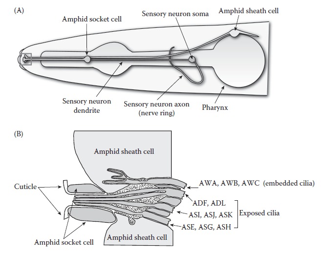
The structure of the amphid sensilla. (A) The soma of amphid sensory neurons are arranged around the pharynx, which is the feeding organ of C. elegans. The axons synapse with interneurons in a structure called the nerve ring. The dendrites extend anteriorly. Only one sensory neuron is shown for clarity. The region indicated by the red box is shown enlarged in panel B. (B) The sensory endings of the so-called wing neurons (AWA, AWB, and AWC) are embedded within the amphid sheath cell. The cilia of the thermosensory AFD neurons (not shown) are also embedded in the sheath cell. Other amphid sensory neurons have exposed cilia. ([A] Redrawn from Hall, D.H. and Z.F. Altun, C. elegans Atlas, Cold Spring Harbor Laboratory Press, Cold Spring Harbor, New York, 2008. [B] Adapted from Perkins, L.A. et al Dev. Biol., 117, 456–87, 1986.)
From: Chapter 1, From Odors to Behaviors in Caenorhabditis elegans
NCBI Bookshelf. A service of the National Library of Medicine, National Institutes of Health.