Continuing Education Activity
Vitiligo is a common acquired skin disorder which results from the loss of melanocytes from the epidermis and clinically manifests as well-demarcated white patches on the body. There are different theories about the pathogenesis of vitiligo but exact etiology is still unknown. It is a form of autoimmune disorder. This activity reviews the role of the interprofessional team in the diagnosis and treatment of this condition.
Objectives:
- Describe the pathophysiology of vitiligo.
- Identify the clinical features of the various clinical types of vitiligo.
- Explain the various treatment options available for vitiligo.
- Outline the importance of collaboration and communication amongst the interprofessional team to ensure the appropriate treatment for vitiligo patients.
Introduction
Vitiligo is an acquired pigmentary skin disorder by the absence of pigmentary cells from the epidermis that results in white macules and patches on the body.[1] The condition is usually associated with few autoimmune disorders, with thyroid abnormalities are the commonest one. The etiology of vitiligo is unknown but there are different theories to explain its pathogenesis. Vitiligo presents clinically with signs and symptoms of white spots on the body distributed symmetrically and more obvious in people with dark skin. The lesions are characterized by well-demarcated pearly white or depigmented macules and patches, oval, round, or linear-shaped, and the borders are convex, range from the size of few millimeters to centimeters and enlarge centrifugally. There are different clinical variants of vitiligo which are Trichrome, Marginal inflammatory, and Quadrichrome vitiligo. Koebner phenomenon(Development of vitiligo at specific trauma prone sites, like cut, burn, or abrasion)is also a common clinical manifestation. Initial lesions occur most frequently on the hands, forearms, feet, and face, favoring a periocular or perioral distribution.[2][3]
On the basis of the distribution, pattern Vitiligo is classified into three types, generalized, segmental, and localized. The severity of the disease is scored by the body surface area affected. The course of the disease is often unpredictable and varies in response to the treatment. Depigmentation often the cause of psychological distress, social stigmatization, and low self-esteem.[3]
Etiology
The exact etiology of vitiligo is unknown. It is frequently associated with multiple autoimmune diseases. There are various theories about its pathogenesis and the etiology is multifactorial. It is characterized by incomplete penetrance, genetic heterogeneity, and multiple susceptibility loci. Family and other twin studies have shown that inheritance is complex and also involves both environmental and genetic factors. Additionally, it is hypothesized that genetic factors can influence the age of onset of vitiligo. The inheritance of vitiligo may also include genes associated with the biosynthesis of melanin, regulation of autoantibodies, and response to oxidative stress.[4]
Recent research studies have not highlighted any associations with any certain HLA type. There is a strong reason to believe that segmental and nonsegmental vitiligo have a unique genetic mechanism, which can account for variable treatment responses.[5]
Epidemiology
Vitiligo is the commonest cause of depigmentation. It can appear at any age from child to adulthood but peak incidence is reported in the second and third decade. The age of onset usually varies between the sexes. Its prevalence is approximately 0.1% to 2% of people including adults and children worldwide and it affects all races equally.[6]
Pathophysiology
Vitiligo is commonly known as multifactorial polygenic disorder and has complex pathogenesis. It is commonly associated with both non-genetic and genetic factors. However various theories have been proposed about its pathogenesis but the exact etiology is still unknown. The generally agreed principles are the absence of melanocytes in vitiligo skin with melanocytes loss, owing to their destruction. The destruction is most results in progressive melanocytes decreases. Theories about the melanocyte destruction include cytotoxic mechanisms, autoimmune mechanisms, intrinsic melanocyte defects, neural mechanisms, and oxidant-antioxidant mechanisms.[5]
In the neural hypothesis, a neurochemical mediator usually destroys the melanocytes and decreases the production of melanin. In oxidant and anti-oxidant mechanism intermediate or metabolic product of melanin synthesis causes the destruction of the melanocyte.[7] In the intrinsic defect of melanocyte, there is an inherent abnormality that impedes their growth and differentiation. Another hypothesis is autoimmune or cytotoxic one where there is an alteration in humoral and cellular immunity that causes the destruction or dysfunction of melanocytes. This theory supports the hypothesis that nonsegmental vitiligo is commonly associated with autoimmune disorders than the segmental type of vitiligo.
Histopathology
The basic histopathological findings in vitiligo is a total loss of functioning melanocytes in association with the complete loss of epidermal pigmentation (determined by Fontana-Masson stain or dohydrophenylalanine. Immunohistochemistry for melanocyte-specific markers like Melan-A and HMB-45 and specific electron microscopy can also be used).[1]
In the margins of lesions, perivascular and perifollicular lymphocytic infiltrate (CD4+ and CD8+ T lymphocytes) are seen, they have specific melanocytic toxicity. Degenerative changes in melanocytes are also visible like cellular enlargement, cytoplasmic vacuolization, and long dendritic processes filled with melanin granules. Other reported changes include epidermal vacuolization, increase number of Langerhans cells, and basement membrane thickening.[8]
History and Physical
In most cases, the diagnosis of vitiligo is very clear based on clinical findings but detailed history and examination are necessary for disease severity.[2]
Important points of history that are helpful in the diagnosis of vitiligo include:
- Age at onset of lesions
- Factors or events that may cause the onset of vitiligo
- Symptoms associated with the lesions
- Progression or spread of lesions
- Changes observed in lesions over time
- Presence of concomitant diseases
- Current medications
- Occupational history/exposure to chemicals or radiation
- Family history of vitiligo and autoimmune diseases
Physical examination of vitiligo reveals these findings:[9]
- Vitiligo manifest as depigmented macules or patches with a convex border without signs of inflammation with normal skin around.
- Macules are milky-white in color, well-demarcated with either round, oval, or linear in shape.
- Ranges in size from a few millimeters to a few centimeters.
- Common sites are face, neck, dorsum of hands, scalp, and trunk. On the face, lesion favors perioral and periocular distribution.
- Lesions can also occur in areas that are subjected to trauma like knees and elbows.
- Koebnerization may occur in 20% to 60% of vitiligo patients.
- Signs of other associated diseases may also be present like Goiter, anemia, generalized wasting, premature grey hairs (leukonychia), alopecia areata and weakness, etc.
Evaluation
The diagnosis of vitiligo is usually made on clinical features, through a wood light examination biopsy is sometimes helpful to differentiate vitiligo from other hypopigmented or depigmented disorders.[10] The histopathological examination of the skin reveals the absence of melanocytes and complete epidermal pigmentation loss. Superficial perifollicular and perivascular lymphocytic infiltrates may be present at the margin of the vitiligo lesions, due to the cell-mediated process that destroys the melanocytes.[11]
Other reported histological findings are:
- Degenerative changes of both melanocytes and keratinocytes located at the border and also of adjacent skin
- Langerhans cells are increased in numbers.
- Vacuolization of epidermis
- Basement membrane thickening
- Pigmentary loss and epidermal melanocytes stained with Fontana-Masson staining and immunohistochemistry testing.
Treatment / Management
Various types of topical and systemic medications, phototherapy, laser therapy, and surgical therapy are used for the treatment of vitiligo. Topical treatment, modalities include corticosteroids, calcineurin inhibitors, and vitamin-D analogs. Phototherapy is an effective treatment option. It induces repigmentation in most of the patients with early and localizes the disease. Narrowband UV-B is widely used, mostly two to three times in a week with 311-312nm wavelength. It has largely replaced the psoralen photochemotherapy because of its toxic side effects. Excimer Laser is used to treating limited, stable patches of vitiligo. In segmental vitiligo which is resistant to most of the treatments, Tacrolimus and systemic corticosteroids can be combined with it.[12] Afamelanotide and JAK inhibitor therapy are emerging treatments. Topical ruxolitinib was also found to be very effective(in 2019 randomized placebo-controlled, double-blind prospective trial)
Surgical treatment options are limited to segmental or localize vitiligo that is limited to a small area. Five basic methods of repigmentation include non cultured epidermal suspensions, thin dermo-epidermal grafts, suction epidermal graft, punch grafting, and cultured epidermis with melanocytes.[13] The following patients are good candidates for surgical treatment.
- Segmental vitiligo
- Localize vitiligo involving small area
- Vitiligo in areas which usually not re-pigment well (hairline, dorsal fingers, forehead, ankles)
- The lesion must be stable.
Differential Diagnosis
- Nevus depigmentosus: It is a circumscribed, segmental depigmented or hypopigmented area present at birth.
- Pityriasis alba: It commonly considered as a spectrum of atopic dermatitis and usually affect the children. It presents as white hypopigmented, scaly macules and patches on the face and other photo exposed areas.
- Idiopathic guttate hypomelanosis: It is characterized by small, asymptomatic pearly white macules on photo exposed areas. Most of these lesions appear in older age groups.
- Tinea (pityriasis) Versicolor: It is a superficial fungal infection that causes loss of pigment. It appears as pale scaly macules present on the back and chest. They give yellow fluorescence under Wood's lamp [
- Halo nevus: It is a type of melanocytic nevus surrounded by an oval halo of depigmentation
- Progressive macular hypomelanosis: It is clinically present as asymptomatic hypopigmented patches on the trunk of young adults
- Drug-induced leukoderma: Potent topical or intralesional corticosteroids can induce hypopigmentation at the site of application. Depigmentation is also noticed in patients treated with the tyrosine kinase and epidermal growth factor receptor inhibitor.
- Hypopigmented mycosis fungoides: It is a variant of early-stage MF commonly seen in children and dark-skinned people. It appears as widespread hypopigmented patches with atrophy and scaling and atrophy.
Toxicity and Adverse Effect Management
Psoralen photochemotherapy; adverse effects include phototoxic effect, nausea, and risk of skin cancer. Short-term adverse effects of NB-UVB include itching, burning, and xerosis.
Prognosis
It is a chronic skin condition with an unpredictable disease course and some patients may notice spontaneous repigmentation over the depigmented areas.[14] The prognosis depends upon the age of onset and the extent of disease. Early disease onset is usually associated with the involvement of greater body surface area and rate of progression. Few types and certain locations may be responsive to treatment. Refractory cases have been noted in patients presenting with segmental vitiligo and younger than 14 years of age. Most of the patients on treatment usually experience intermittent cycles of pigment loss and disease stabilization.[7]
Complications
Complications of vitiligo are social stigmatization and mental stress, eye involvement like iritis, depigmented skin is more prone to sunburn, skin cancer, and hearing loss because of loss of cochlear melanocytes. Other complications are related to medications like skin atrophy after prolonged use of topical steroids.
Consultations
An interprofessional approach is very essential for such patients. Sometimes these patients need special referrals to respective specialties when present with another autoimmune disorder. In addition, for ocular complications consultation with an ophthalmologist and psychological ailments referral to mental health, specialists are important.
Deterrence and Patient Education
As vitiligo is a depigmented and slowly progressive disorder it affects the physical appearance of a patient, and because of this, it has a psychological and social impact. Visible lesions have been associated with more distress and stigmatization than non-visible lesions, lower quality of life is reported in patients with vitiligo. Psychiatric consultation is very important because of psychiatric morbidity. Patient education regarding the slow response of treatment or treatment failure is also necessary because the response to treatment is highly variable in different patients.
Enhancing Healthcare Team Outcomes
A multidisciplinary approach is important because vitiligo is associated with other autoimmune disorders. A proper collaboration and communication are very important. These patients present with multiple psychological disorders due to their appearance and unpredictable course. Psychological interventions are important. Cognitive-behavioral therapy, along with regular conventional treatment is very useful in improving the self-esteem confidence and quality of life of such patients, and it also influences the disease course.

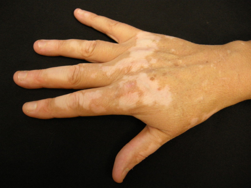
Figure
Vitiligo, Hand DermNet New Zealand
Figure
Woods light for vitiligo patient showed depigmented patch. Contributed by Daifallah Al Aboud, MD
Figure
Vitiligo. Genitals Contributed by Dr. Shyam Verma, MBBS, DVD, FRCP, FAAD, Vadodara, India
Figure
Vitiligo, Facial Contributed by Dr. Shyam Verma, MBBS, DVD, FRCP, FAAD, Vadodara, India
References
- 1.
- Mazzei Weiss ME. Vitiligo: to biopsy or not to biopsy? Cutis. 2020 Apr;105(4):189-190. [PubMed: 32463851]
- 2.
- Eleftheriadou V. Reliability and validity of the Vitiligo Signs of Activity Score. Br J Dermatol. 2020 Nov;183(5):801-802. [PubMed: 32458411]
- 3.
- de Baat C, Phoa KH, Zweers PGMA, Bolling MC, Rozema FR, Vissink A. [Medicaments and oral healthcare. Hyperpigmentation of oral soft tissues due to afamelanotide]. Ned Tijdschr Tandheelkd. 2020 Apr;127(4):237-243. [PubMed: 32459219]
- 4.
- Abdel-Malek ZA, Jordan C, Ho T, Upadhyay PR, Fleischer A, Hamzavi I. The enigma and challenges of vitiligo pathophysiology and treatment. Pigment Cell Melanoma Res. 2020 Nov;33(6):778-787. [PubMed: 32198977]
- 5.
- Henning SW, Jaishankar D, Barse LW, Dellacecca ER, Lancki N, Webb K, Janusek L, Mathews HL, Price RN, Le Poole IC. The relationship between stress and vitiligo: Evaluating perceived stress and electronic medical record data. PLoS One. 2020;15(1):e0227909. [PMC free article: PMC6984686] [PubMed: 31986193]
- 6.
- Bergqvist C, Ezzedine K. Vitiligo: A Review. Dermatology. 2020;236(6):571-592. [PubMed: 32155629]
- 7.
- Cohen BE, Manga P, Lin K, Elbuluk N. Vitiligo and Melanoma-Associated Vitiligo: Understanding Their Similarities and Differences. Am J Clin Dermatol. 2020 Oct;21(5):669-680. [PubMed: 32468356]
- 8.
- Lei TC, Hearing VJ. Deciphering skin re-pigmentation patterns in vitiligo: an update on the cellular and molecular events involved. Chin Med J (Engl). 2020 May 20;133(10):1231-1238. [PMC free article: PMC7249724] [PubMed: 32433056]
- 9.
- Grän F, Emmerich K, Mohme S, Goebeler M, Gesierich A. Vitiligo-like lesions developing upon immune checkpoint inhibition in advanced melanoma. Eur J Dermatol. 2020 Feb 01;30(1):72-73. [PubMed: 32250264]
- 10.
- El-Husseiny R, Abd-Elhaleem A, Salah El-Din W, Abdallah M. Childhood vitiligo in Egypt: Clinico-epidemiologic Profile of 483 patients. J Cosmet Dermatol. 2021 Jan;20(1):237-242. [PubMed: 32320520]
- 11.
- Delgadillo X, Ortega AE, Greco AM. Systemic and Autoimmune Diseases. Clin Colon Rectal Surg. 2019 Sep;32(5):372-376. [PMC free article: PMC6731114] [PubMed: 31507347]
- 12.
- Juntongjin P, Toncharoenphong N. Effectiveness of a combined 308-nm excimer lamp and topical mid-potent steroid treatment for facial vitiligo: a preliminary, randomized double-blinded controlled study. Lasers Med Sci. 2020 Dec;35(9):2023-2029. [PubMed: 32458080]
- 13.
- Kim DS, Ju HJ, Lee HN, Choi IH, Eun SH, Kim J, Bae JM. Skin seeding technique with 0.5-mm micropunch grafting for vitiligo irrespective of the epidermal-dermal orientation: Animal and clinical studies. J Dermatol. 2020 Jul;47(7):749-754. [PubMed: 32452060]
- 14.
- Wen Y, Wu X, Peng H, Li C, Jiang Y, Liang H, Zhong R, Liu J, He J, Liang W. Cancer risks in patients with vitiligo: a Mendelian randomization study. J Cancer Res Clin Oncol. 2020 Aug;146(8):1933-1940. [PubMed: 32462299]
Disclosure: Naila Ahmed jan declares no relevant financial relationships with ineligible companies.
Disclosure: Sadia Masood declares no relevant financial relationships with ineligible companies.
Publication Details
Author Information and Affiliations
Authors
Naila Ahmed jan1; Sadia Masood2.Affiliations
Publication History
Last Update: August 7, 2023.
Copyright
This book is distributed under the terms of the Creative Commons Attribution-NonCommercial-NoDerivatives 4.0 International (CC BY-NC-ND 4.0) ( http://creativecommons.org/licenses/by-nc-nd/4.0/ ), which permits others to distribute the work, provided that the article is not altered or used commercially. You are not required to obtain permission to distribute this article, provided that you credit the author and journal.
Publisher
StatPearls Publishing, Treasure Island (FL)
NLM Citation
Ahmed jan N, Masood S. Vitiligo. [Updated 2023 Aug 7]. In: StatPearls [Internet]. Treasure Island (FL): StatPearls Publishing; 2024 Jan-.