Figure 3

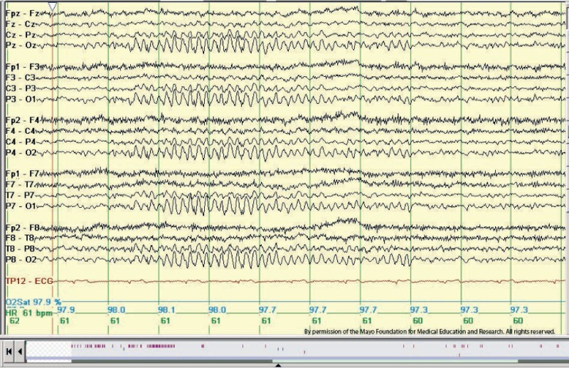
Figure 3. Example of 6-Hz “phantom” spike and wave in an adolescent patient. There are bilaterally symmetrical diffuse tiny spikes with prominent wave components (“mitten-like” morphology) in seconds 3 through 6 below. Longitudinal bipolar montage. This is the posterior variant sometimes referred to as the FOLD (female occipital low-amplitude drowsiness) subtype. Copyright ©2016 by American Epilepsy Society. Copyright of all cited figures: Copyright ©2016 by American Epilepsy Society. No permission is required from the editors, authors, or publisher to reuse or repurpose content, provided the original work is properly cited. Figures, tables, and images included in a publication of “Electroencephalography (EEG): An Introductory Text and Atlas of Normal and Abnormal Findings in Adults, Children, and Infants” are also published under the CC BY-NC-SA license and should be properly cited when reused or repurposed.

From: EEG Benign Variants

Cover of StatPearls
StatPearls [Internet].
Treasure Island (FL): StatPearls Publishing; 2024 Jan-.
Copyright © 2024, StatPearls Publishing LLC.

This book is distributed under the terms of the Creative Commons Attribution-NonCommercial-NoDerivatives 4.0 International (CC BY-NC-ND 4.0) ( http://creativecommons.org/licenses/by-nc-nd/4.0/ ), which permits others to distribute the work, provided that the article is not altered or used commercially. You are not required to obtain permission to distribute this article, provided that you credit the author and journal.

NCBI Bookshelf. A service of the National Library of Medicine, National Institutes of Health.