

NCBI Bookshelf. A service of the National Library of Medicine, National Institutes of Health.
Hall KK, Shoemaker-Hunt S, Hoffman L, et al. Making Healthcare Safer III: A Critical Analysis of Existing and Emerging Patient Safety Practices [Internet]. Rockville (MD): Agency for Healthcare Research and Quality (US); 2020 Mar.

Making Healthcare Safer III: A Critical Analysis of Existing and Emerging Patient Safety Practices [Internet].
Show detailsIntroduction
Background
Diagnostic error, as defined by the National Academy of Medicine in 2015, is “the failure to (a) establish an accurate and timely explanation of the patient’s health problem(s) or (b) communicate that explanation to the patient.”1 This definition focuses on the outcomes of the diagnostic process, recognizing that diagnosis is an iterative process that solidifies as more information becomes available. The diagnosis needs to be timely and accurate so that appropriate treatment is initiated to optimize the patient’s outcome. Any gaps that arise in the diagnostic process can lead to error. In this chapter we discuss four patient safety practices (PSPs) that have the potential to decrease diagnostic errors: the use of clinical decision support (CDS); result notification systems (RNS); education and training; and peer review.
Importance of Harm Area
Diagnostic error is an increasingly recognized threat to public health, with estimates of 5 percent of adults being affected in the outpatient environment.2 In the hospital setting, diagnostic error is responsible for 6 to 17 percent of adverse events.1,3 Diagnostic error has also been shown to be responsible for more closed malpractice claims than other causes.1,4,5 The Institute of Medicine (now the National Academy of Sciences), in their seminal report on diagnostic safety, concluded that “most people will experience at least one diagnostic error in their lifetime.”1
PSP Selection
Using systematic reviews and reports, the Technical Expert Panel, Advisory Group, and Agency for Healthcare Research and Quality developed and reviewed an initial list of 23 PSPs that target diagnostic errors. Studies have uncovered two broad categories of underlying root causes: cognitive-based factors, such as failed heuristics, and systems-based factors, such as lack of provider-to-provider communication and coordination.2,6,7 Therefore, the PSPs selected by consensus for inclusion in this report addressed one or both of these fundamental high-leverage areas.
- CDS offers solutions integrated into the workflow to address diagnostic errors by providing stakeholders with knowledge and person-specific information, intelligently filtered or presented at appropriate times, to improve decision making and communication.8
- Education and training on the diagnostic process enhance clinical reasoning and decrease biases.6
References for Introduction
- 1.
- National Academies of Sciences, Engineering and Medicine. 2015. Improving Diagnosis in Health Care. Washington, DC: National Academies Press. 10.17226/21794. [PubMed: 26803862] [CrossRef]
- 2.
- Singh H, Meyer AN, Thomas EJ. The frequency of diagnostic errors in outpatient care: estimations from three large observational studies involving US adult populations. BMJ Qual Saf. 2014;23(9):727–31. [PMC free article: PMC4145460] [PubMed: 24742777]
- 3.
- Zwaan L, de Bruijne M, Wagner C, et al. Patient record review of the incidence, consequences, and causes of diagnostic adverse events. Arch Intern Med. 2010;170(12):1015–21. [PubMed: 20585065]
- 4.
- Tehrani ASS, Lee H, Mathews SC, et al. 25-Year summary of US malpractice claims for diagnostic errors 1986–2010: an analysis from the National Practitioner Data Bank. BMJ Qual Saf. 2013;22(8):672–80. [PubMed: 23610443]
- 5.
- Schiff GD, Puopolo AL, Huben-Kearney A, et al. Primary care closed claims experience of Massachusetts malpractice insurers. J AM Med Assoc Intern Med. 2013;173(22):2063–8. [PubMed: 24081145]
- 6.
- Graber ML, Kissam S, Payne VL, et al. Cognitive interventions to reduce diagnostic error: a narrative review. BMJ Qual Saf. 2012;21(7):535–57. doi:10.1136/bmjqs-2011-000149. [PubMed: 22543420] [CrossRef]
- 7.
- Kostopoulou O, Delaney BC, Munro CW. Diagnostic difficulty and error in primary care—a systematic review. Fam Pract. 2008;25(6):400–13. [PubMed: 18842618]
- 8.
- Official Website of The Office of the National Coordinator for Health Information Technology (ONC). https://www
.healthit.gov/. - 9.
- Callen JL, Westbrook JI, Georgiou A, et al. Failure to follow-up test results for ambulatory patients: a systematic review. J Gen Intern Med. 2012;27(10):1334–48. [PMC free article: PMC3445672] [PubMed: 22183961]
- 10.
- Davis Giardina T, King BJ, et al. Root cause analysis reports help identify common factors in delayed diagnosis and treatment of outpatients. Health Aff. 2013;32(8):1368–75. [PMC free article: PMC3822525] [PubMed: 23918480]
- 11.
- Butler GJ, Forghani R. The next level of radiology peer review: enterprise-wide education and improvement. J Am Coll Radiol. 2013;10(5):349–53. [PubMed: 23642877]
1.1. Patient Safety Practice: Clinical Decision Support
Authors
Kendall K. Hall, M.D., M.S., Kristen Miller, Dr.P.H., Eleanor Fitall, M.P.H., and Katharine Witgert, M.P.H.1.1.1. Practice Description
Key Findings
- CDS has been shown to improve diagnosis in exploratory and validation studies, but the tools need to be fully implemented and tested in clinical settings.
- CDS is best used as an adjunct to the clinician’s decision-making process and not as a replacement.
- The diagnoses generated by CDS tools are only as good as the information that is put into the system; if the initial assessment of the patient (e.g., physical exam finding) is incorrect, the output is likely to be incorrect.
- Despite their potential, diagnosis generators have had limited use, owing in large part to challenges integrating them into busy clinicians’ workflows.
Diagnostic error is a complex and multifaceted problem that requires systems solutions to achieve the necessary changes. Advancements in health information technology (IT) represent thoughtful and sophisticated ways to reduce delayed, missed, or incorrect diagnoses.1 Contributions of health IT include more meaningful incorporation of evidence-based diagnostic protocols with clinical workflow, and better usability and interfaces in the electronic health record (EHR).
The Office of the National Coordinator for Health Information Technology defines CDS as providing “clinicians, staff, patients or other individuals with knowledge and person-specific information, intelligently filtered or presented at appropriate times, to enhance health and healthcare. CDS encompasses a variety of tools to enhance decision making in the clinical workflow. These tools include computerized alerts and reminders to care providers and patients; clinical guidelines; condition-specific order sets; focused patient data reports and summaries; documentation templates; diagnostic support, and contextually relevant reference information, among other tools.”2
CDS represents a range of different interventions, from documentation templates to interruptive popup alerts. The knowledge bases triggering CDS differ as well. Rules-based or logic-based CDS often takes the form of IF-THEN rules. More advanced CDS leveraging artificial intelligence (AI) and machine learning taps awareness of past experiences and patterns in clinical data. These techniques have generated interest and excitement in their potential to better augment clinician intelligence and support decision making.
Several patient safety researchers have suggested that health IT, including CDS, can be leveraged to improve diagnosis, although the data have been mixed.1,3–7 Therefore, the question of interest for this review is, “Does CDS lead to improved diagnostic performance?” This review’s key findings are located in the box above.
1.1.2. Methods
We searched four databases (CINAHL®, MEDLINE®, PsycINFO®, and Cochrane) for articles published from 2008 through 2018 using the terms “diagnostic errors,” “delayed diagnosis,” “missed diagnosis,” and their synonyms. Terms specific to this PSP include “clinical decision support,” “medical informatics applications,” “artificial intelligence,” “computer-aided decision making,” “computer-assisted diagnosis,” and related terms. The initial search yielded 2,208 results. Once duplicates had been removed and additional relevant referenced articles added, a total of 2,202 articles were screened for inclusion, and 87 full-text articles were retrieved. Of those, 37 studies were selected for inclusion in this review. Articles were excluded if they were not focused on use of CDS specifically for diagnosis (e.g., focus on use of CDS for medication ordering), the outcome was not relevant to this review, the article was out of scope, or the study was of significantly limited rigor.
General methods for this report are described in the Methods section of the full report.
For this patient safety practice, a PRISMA flow diagram and evidence table, along with literature-search strategy and search-term details, are included in the report appendixes A through C.
1.1.3. Evidence Summary
1.1.3.1. CDS To Generate Diagnoses
1.1.3.1.1. Differential Diagnosis Generators
Differential diagnoses (DDX) are a list of diagnostic hypotheses generated by the clinician during the course of the patient interaction, and are based on information such as the history and physical exam. Often several different diagnostic possibilities are initially present, and as the clinician gathers additional information to support or refute the hypotheses, the list can be narrowed until arriving at the correct diagnosis.
DDX generators are “programs which assist healthcare professionals in clinical decision making by generating a DDX based on a minimum of two items of patient data.”8 DDX generators provide a list of potential diagnoses for consideration, sometimes in order of likelihood based on available information, as a means to improve diagnosis.
The first study discussed is a systematic review and meta-analysis conducted by Riches et al. (2016), which included 36 articles investigating the effects of 11 different DDX generators to retrieve accurate diagnoses (i.e., the correct diagnosis appeared in the list of possible diagnoses). Of note, only five of the tools are still in existence. Using different computational approaches, such as pattern matching and Bayesian probabilities, these diagnostic aids generate lists of DDX for consideration based on clinical data that the user inputs. With respect to the effectiveness of the DDX generators at retrieving accurate diagnoses, the authors concluded that the pooled accurate diagnosis retrieval rate was high, although with considerable heterogeneity (pooled rate=0.70, 95% confidence interval [CI], 0.63 to 0.77; I2 = 97%, p<0.0001). In the subgroup analyses examining the accuracy of individual DDX generators, ISABEL, one of the tools under evaluation, outperformed all of the other tools, but again, the heterogeneity was considerable (pooled rate = 0.89, 95% CI, 0.83 to 0.94; I2 = 82%, p<0.0001). When comparing the performance of the DDX tools to that of clinicians, the authors found that the DDX tools were associated with a small, nonsignificant increase in accurate diagnosis retrieval.8
In a study by David et al. (2011), the primary objective was to determine the misdiagnosis rate of cellulitis, an infection of the skin and tissue underneath, but the authors also determined whether or not a visually based, computerized diagnostic decision support system (VCDDSS) could generate an improved DDX based on the presenting signs and symptoms for the misdiagnosed patients. The system requires the user to input relevant patient findings (e.g., clinical information, physical examination findings) to generate a ranked list of potential diagnoses. Using a cellulitis-specific module of the VCDDSS, the authors found that the system included the correct diagnosis in the DDX 64 percent of the time. This was significantly greater than the diagnostic accuracy of the admitting residents, who included the correct diagnosis in their DDX only 14 percent of the time without the use of the VCDDSS (p=0.0003).9 Gegundez-Fernandez et al. (2017) evaluated the diagnostic performance of Uvemaster, a mobile DDX generator that provides a ranked list of syndromes that cause uveitis, a form of eye inflammation, based on clinical findings. The percentage of cases for which a diagnosis included in the DDX by the app matched the original clinician diagnosis was 96.6 percent (95% CI, 84.1 to 96.6). When the diagnoses were ordered by sensitivity, the original diagnosis was listed within the top three diagnoses generated by the app in 90.9% of cases (95% CI, 84.1 to 96.6) and was listed as the first diagnosis in 73.9% of cases (95% CI, 63.6 to 83.0).10
Using real-case vignettes, Segal et al. (2014) and Segal et al. (2016) both evaluated the use of a DDX generator, SimulConsult, on diagnostic performance.11,12 In the first study, pediatric neurologists were asked to read case vignettes and generate a ranked list of DDX and baseline workups (e.g., diagnostic studies). The clinicians then used the tool, and again provided a list of DDX and workups. The authors found the use of the tool significantly reduced the number of missing diagnoses in the DDX (36% to 15%; P<0.0001) across all clinicians and increased the relevance of the diagnoses listed.11 In their second paper, Segal and colleagues evaluated the use of SimulConsult by nonspecialists to diagnose pediatric rheumatologic diseases via case vignettes. Similar to the earlier study, when using the DDX generator, the nonspecialists demonstrated a significant reduction in missed diagnoses in the DDX, which fell from 28 percent unaided to 15 percent using the tool (p<0.0001).12
Three papers provide evidence that DDX generators modestly improve the diagnostic accuracy of clinicians.13–15 Using test patient cases in an exam format, Martinez-Franco et al. (2018) compared the diagnostic accuracy of first-year family medicine residents randomized to the control group with those in the intervention group, which used a DDX generator, DXplain. This tool requires the user to enter patients’ signs, symptoms, and laboratory tests. Using these data, the tool generates a list of possible diagnoses ranked from highest to lowest probability. The mean percent-correct score and standard deviation was 74.1 ± 9.4 for the control group and 82.4 ± 8.5 for the intervention group (p<0.001).13 Kostopoulou et al. (2017) developed a prototype DDX generator integrated with a commercial EHR system for use in general practice and tested it using high-fidelity simulation. As soon as the clinician enters the reason for encounter (RfE), the system generates a list of diagnostic suggestions based on the patient’s RfE, age, and sex, and groups them according to published incidence rates (i.e., common, uncommon, and rare diagnoses). At the time of the study, the prototype supported three RfEs: chest pain, abdominal pain, and shortness of breath. Using standardized patients simulating 12 cases (4 cases per RfE), 34 general practitioners established their baseline performance with half of the cases and then used the DDX tool with the other half. Diagnostic accuracy improved significantly when using the tool, going from 49.5 percent to 58.3 percent accuracy (p<0.003).14 Chou et al. (2017) tested the effect of a VCDDSS on the diagnostic accuracy of medical students and dermatology residents in a dermatology clinic. In this pilot study, the students’ diagnostic accuracy increased significantly, from 62.5 percent without the VCDDSS to 81.25 percent using the VCDDSS (p<0.01).15
1.1.3.1.2. Specific Diagnoses
In addition to the differential diagnosis generators, the search identified papers that describe the development and evaluation of CDS models that determine whether a specific disease is present.
Several papers described rule-based or logic-based CDS for diagnosis where the tool had been integrated into a real clinical setting. Niemi et al. (2009) developed an automated CDS tool to identify patients admitted to the hospital with pneumonia or heart failure (HF) in real time to aid in timely administration of treatment. The system continually monitors data from existing information systems such as the pharmacy information system, laboratory management system, and radiology management system, and applies rules for pneumonia and HF. When the patient accumulates enough points to be diagnosed with either HF or pneumonia, the system looks to see whether appropriate treatment has been provided (e.g., in the case of pneumonia, an antibiotic) within set time limits, and if it has not, the system generates an alert to the clinician and nursing unit. In the emergency department (ED), the sensitivity and specificity of the system to identify pneumonia was 89 percent and 86 percent, respectively, and in the inpatient setting it was 92 percent and 90 percent, respectively. For HF, the sensitivity was 94 percent and the specificity 90 percent. In addition, the system allowed the hospital to increase compliance with national quality indicators for both of these conditions.16
Deleger et al. (2013) developed and tested an automated appendicitis—inflammation of the appendix—risk categorization algorithm for pediatric patients with abdominal pain, based on content from the EHR, and found this system to be comparable to use of physician experts. Using retrospective data, the CDS tool had an average F-measure of 0.867, with a sensitivity (recall) of 0.869 and a positive predictive value (precision) of 0.863.17 Kharbanda et al. (2016) developed and implemented an electronic CDS tool for pediatric patients with abdominal pain that included a standardized abdominal pain order set, a web-based risk stratification tool, and an ordering alert. Compared with in the pre-implementation period, the trend of computed tomography (CT) scan use during the implementation period decreased significantly each month (p=0.007), and showed a 54-percent relative decrease in CT use in the post-implementation period. The authors found that the decrease in CT use was not associated with the potential unintended consequences of decreased use of CT: significant changes to the rates of appendectomies or missed appendicitis cases.18
Chamberlain et al. (2016) developed a mobile smart phone application for screening patients for pulmonary disease and conducted preliminary testing of the algorithms in a clinic setting. The application uses an electronic stethoscope, a method of digitizing peak flow meter readings, and patient questionnaire to identify patients with asthma and chronic obstructive pulmonary disease. The classification algorithms were successful in identifying patients with asthma and chronic obstructive pulmonary disease from the general patient population, with an area under the receiver operating characteristic curve of 0.97. Of note, during the study, patient breath sounds were auscultated using the electronic stethoscope but were evaluated by a pulmonologist. The authors note that they have since been able to develop algorithms to automatically identify abnormal lung sounds, making this technology possible for use by non-pulmonologists and potentially even non-clinicians to assist with diagnosis.19
One study focused on a CDS tool patients could use to aid in the screening of skin lesions. Wolf et al. (2013) investigated the use of four readily available smart phone applications designed to evaluate photographs of skin lesions and provide feedback on the risk of malignancy. Clinical images of previously diagnosed skin lesions were submitted for evaluation through the applications. The application with the highest sensitivity (98.1%) sent images directly to a board-certified dermatologist for analysis—essentially tele-dermatology. The sensitivity of the other three applications ranged from 6.8 percent to 70.0 percent, and they relied on automated algorithms to analyze the images.20
More-advanced CDS tools leveraging AI and machine learning have generated excitement over the potential to better augment clinician intelligence and support decision making. A cohort of the papers in our review describe models based on AI techniques to screen for and diagnose specific disorders and diseases. A systematic review by Wagholikar et al. (2011) includes 220 reports of new decision models or evaluations of existing models. The authors generalized their findings and concluded that these techniques have growing popularity for simple classifications but have yet to achieve an acceptable degree of accuracy, particularly for complex medical problems.21 Other studies of AI identified beyond this systematic review all show promise in identifying disease, although the research continues to be investigational in nature, with a lack of implementation and testing in real clinical settings.17,22–28
1.1.3.2. CDS To Assist With Diagnostic Study Interpretation
Several papers included in this review described investigational studies of CDS tools to assist with diagnostic study interpretation, including imaging studies, electrocardiograms (ECGs), and pathology. Although these CDS tools are proof-of-concept in nature, they demonstrate the potential to augment clinician diagnostic performance but not completely replace it.
1.1.3.2.1. Use in Imaging
Three papers identified through the search focused on techniques to assist with interpretation of imaging studies. All were investigational in nature, describing the development and validation of the models.27,29,30 Herweh et al. (2016) compared the diagnostic performance of an automated machine-learning algorithm to detect acute stroke on CT scans using a standardized scoring method to the performance of stroke experts and novices using the algorithm. Although this study had a small sample size, the automated tool showed similar scoring results to that of experts and better performance than the novices.29 Bien et al. (2018) used deep learning, a subset of machine learning, to model the complex relationships between images and their interpretations. The model was designed to detect general abnormalities and two specific diagnoses (anterior cruciate ligament [ACL] tears and meniscal tears) on knee magnetic resonance imaging (MRI). For general abnormalities, there was no difference between the performance of the model and the general radiologists. For ACL tear detection, the model was highly specific but not significantly different from the specificity achieved by the radiologists. Radiologists achieved a significantly higher sensitivity (p=0.002) in detecting ACL tears. For meniscal tears, the radiologists achieved significantly higher specificity compared with the model (p=0.003). The authors also found that providing the radiologists with the predictions from the model improved their quality of interpretation of the MRI studies.27 Li et al. (2018) developed an AI tool to detect nasopharyngeal malignancies under endoscopic evaluation by oncologists. Results indicate that the tool was significantly better in its performance compared with oncological experts; the overall accuracy was 88.0 percent (95% CI, 86.1 to 89.6) versus 80.5 percent (95% CI, 77 to 84).30
1.1.3.2.2. ECG Interpretation
In the evaluation of cardiac health, 12-lead ECGs are accompanied by computer interpretations to assist the clinician with diagnoses. These interpretations have been shown to often be inaccurate, primarily because of noisy background signals that interfere with automated pattern recognition by the machine algorithms. However, four studies in this review evaluated ECG interpretations by automated systems, and all found that the systems were no better or worse than human performance alone.31–34
Hughes et al. (2017) sought to improve ED workflow and reduce physician interruptions generated by the need to rapidly read triage ECGs for patients with chest pain. The authors examined the accuracy of ECGs identified as normal by the computer with the hypothesis that these normal ECGs would not have clinically significant findings. The negative predictive value of the normal computer interpretations was 99 percent (95% CI, 97 to 99), indicating that there may be a group of ECGs for which rapid physician re-interpretation is not necessary, thereby reducing interruptions.31
Two studies tested the accuracy of the diagnoses generated by the automated systems compared with human interpretation. Given that nonexpert ECG readers are more likely to rely on automated system interpretation for diagnosis, Hakacova et al. (2012) compared the accuracy of two different rhythm analysis software products with the accuracy of nonexpert readers and found no significant difference in performance. The authors also looked at the accuracy of the software for ECGs for which the diagnosis by the nonexpert was incorrect, and found that only 28 percent+/−10 percent (system A) and 25 percent+/−10 percent (system B) of the automated diagnoses were correct.33 Mawri et al. (2016) examined whether the use of automated ECG interpretation would affect time to treatment for patients with ST-elevation myocardial infarction. The authors found that the computer-interpreted ECGs failed to identify 30 percent of patients with ST-elevation myocardial infarction and found significant differences in two quality-of-care measures: immediate emergency physician interpretation led to faster catheterization laboratory activation time (p<0.029) and faster median door-to-balloon time (p<0.001).34 A study by Cairns et al. (2017) tested a semi-automated system that attempts to overcome the accuracy issues of automated systems by leveraging the strengths of human performance (i.e., the ability to recognize patterns through noisy signals). The system integrates a rule-based computer algorithm with interactive questions and prompts for the clinician to generate multiple diagnostic possibilities. The use of this semi-automated system increased the number of correct interpretations, but the increase was not statistically significant.32
1.1.3.2.3. Use in Pathology
Two studies evaluated the use of AI to aid in the diagnostic work of pathologists.35,36 Vandenberghe et al. (2017) developed and evaluated the use of deep learning, an AI method, to identify specific cancer cell types. For 71 breast tumor samples, they found that the use of this computer-aided diagnosis tool had a concordance rate of 83 percent with pathologist review. The pathologist re-reviewed the 12 samples that had discordance between the diagnoses of the pathologist and the computer-aided diagnosis tool, prompting modifications to 8 of the original diagnoses.35 Xiong et al. (2018), also using deep learning, developed and tested an AI-assisted method for the automatic detection of mycobacterium tuberculosis. Results showed high sensitivity (97.9%) and moderate specificity (83.6%), with 2 false negatives and 17 false positive cases due to contaminants.36
1.1.3.3. CDS To Identify Patients at Risk for Diagnostic Errors
Three studies examined the use of CDS tools to identify patients who are at risk of having a diagnostic error.35,37,38 The systems were all effective at identifying at-risk patients and allowed potential diagnostic errors, including missed or delayed diagnoses, to be prevented, while saving the clinicians time by reducing manual workloads and cognitive burden. As previously discussed, the study by Vandenberghe et al. (2017) used discordance between the diagnoses generated by the AI tool and the diagnoses by the pathologist to flag cases where there may be a high risk of diagnostic error.35
Koopman et al. (2015) developed a system to compare final radiology reports with final ED diagnoses to ensure that the ED identified and appropriately treated an abnormality on radiologic examination. A text analysis system first screens radiology reports to identify limb abnormalities, including fractures, dislocations, and foreign bodies. If the system identifies an abnormality, the diagnosis is reconciled with the ED diagnosis, as defined by International Classification of Diseases, 10th Revision (ICD-10) codes. If there is a discrepancy, the chart is flagged as a possible misdiagnosis, allowing immediate review and followup. Across the three settings in which the study took place, 274 of 2,018 patients (13.6%) with radiologic abnormalities were flagged for potentially missed diagnoses, and the chart was reviewed manually. Nine of the cases were identified as truly missed diagnoses, and the other instances were due to the ED ICD-10 discharge diagnoses being ambiguous, and not indicative of a diagnostic error. The value in this method is that clinicians need to review only a small subset of the radiology reports, in this case 11 percent of the total number or radiology studies, to determine whether there were potentially missed diagnoses.37
Murphy et al. (2015) applied electronic triggers to EHR data to identify the presences of “red flags,” exclude records for which further evaluation is not warranted (e.g., patients in hospice), and identify the presence of a delay in diagnostic evaluation for three conditions: colon cancer, lung cancer, and prostate cancer. Examples of red flags include positive fecal occult blood testing for colon cancer, concerning imaging studies for lung cancer, and elevated prostate-specific antigen for prostate cancer. Delayed diagnostic evaluation was defined by the absence of documented followup action. The trigger flagged 1,256 patients out of 10,673 patients with abnormal findings (11.8%) as being high risk for delayed diagnostic evaluation. Of these, 749 were true positives, a positive predictive value of 59.6 percent. Times to diagnostic evaluation were significantly lower in intervention patients compared with control patients flagged by the colorectal trigger and prostate trigger. There was no significant difference for the lung trigger.38
1.1.3.4. Unintended Consequences
In general, the CDS tools have an added benefit of improving access to specialized care by providing the clinician with assistance in diagnosing conditions that would typically fall in the realm of a specialist.12,19,27
Several of the CDS tools identified in this review, in addition to improving diagnostic accuracy, would also allow prioritization of work, creating greater efficiencies and improving workflow once implemented in clinical settings.27,31,38 These systems flagged studies or diagnoses that required followup, allowing the clinicians to prioritize their work.
For the CDS tools that generate DDX, Graber and Mathew (2008) raised the concern that presenting the clinician with a long list of diagnostic possibilities could be distracting or lead to unnecessary testing and procedures.3 Elkin et al. (2010) suggested that these tools actually reduce the cost of care by assisting the clinician with a broader differential diagnosis list, which is more likely to contain the correct diagnosis. In the case of the DXplain tool, providing the list of diagnoses in order of likelihood can lead to the clinicians evaluating the more likely diagnoses earlier.39
1.1.4. Implementation
1.1.4.1. Facilitators
Since many of the studies were conducted to validate algorithms or were exploratory in nature (e.g., testing AI algorithms to determine their ability to predict correct diagnosis), few described experiences with implementation in real clinical settings.
In the meta-review of systematic reviews by Nurek et al. (2015), the authors determined the features and effectiveness of computerized diagnostic decision support systems for medical diagnosis in primary care. The authors identified conditions that need to be met if a fully integrated CDS tool for diagnoses is to be successfully implemented and used: the tool can readily be integrated into EHRs; is based on standard terminologies, such as diagnosis codes (e.g., ICD-10); has the ability to be easily updated; is thoughtfully integrated into the clinicians’ cognitive workflow; and interfaces with the clinicians at appropriate action points.40
1.1.4.2. Barriers
The information generated by CDS for use in diagnosis is only as good as the information that is put into the system. For example, if the clinician interprets the physical exam incorrectly (e.g., saying that a physical sign is absent when it is present) and inputs that incorrect information into the tool, that error may negatively affect any diagnosis that is partially based on the presence of that sign.10,11,25,37 In the study by Koopman et al. (2015), discharge diagnoses, as indicated by ICD-10 codes, are reconciled with the diagnosis from radiology reports. If the ICD-10 code is incorrect, the system may not recognize a potential missed diagnosis.37 Gegundez-Fernandez et al. (2017) commented that accurate diagnosis can be achieved only if the clinician’s assessment of the patients’ signs and symptoms is correct, because the automated system will process only data that humans introduce.10
In the case of ECG interpretation, accurate ECG recording depends on many variables, including lead placement, weight, movement, coexisting electrolyte abnormalities, and symptoms. If the placement is wrong (e.g., leads are placed in wrong location), the interpretation may be wrong.33,34
1.1.5. Resources
Additional information can be found at the HealthIT.gov site, which offers information on how the use of EHRs can improve diagnosis (https://www.healthit.gov/topic/health-it-and-health-information-exchange-basics/improved-diagnostics-patient-outcomes) and through the National Academies report Improving Diagnosis in Health Care (http://www.nationalacademies.org/hmd/Reports/2015/Improving-Diagnosis-in-Healthcare).
1.1.6. Gaps and Future Directions
Although research in the use of CDS for diagnosis has been conducted for many years, there has been a failure to implement these tools widely, and published work continues to be predominantly that of exploratory studies in educational settings, testing of algorithms using retrospective data, or evaluation through simulation.8 Wagholikar et al. (2012), in their systematic review of modeling techniques for diagnostic decision support, provided several suggestions for research and future work in this area, including evaluation of these applications in clinical settings.21
1.1.6.1. Leveraging the “CDS Five Rights” Approach
A useful framework for achieving success in CDS design, development, and implementation is the “CDS Five Rights” approach.41 The CDS Five Rights model states that CDS-supported improvements in desired healthcare outcomes can be achieved if we communicate: (1) the right information: evidence-based, suitable to guide action, pertinent to the circumstance; (2) to the right person: considering all members of the care team, including clinicians, patients, and their caretakers; (3) in the right CDS intervention format, such as an alert, order set, or reference information to answer a clinical question; (4) through the right channel: for example, a clinical information system such as the EHR, a personal health record, or a more general channel such as the Internet or a mobile device; (5) at the right time in workflow, for example, at the time of decision/action/need. CDS has not reached its full potential in driving care transformation, in part because opportunities to optimize each of the five rights have not been fully explored and cultivated.42
Providing the Right Information to the End User: The process of integrating real-time analytics into clinical workflow represents a shift towards more agile and collaborative infrastructure building, expected to be a key feature of future health information technology strategies. As interoperability and big data analytics capabilities become increasingly central to crafting the healthcare information systems of the future, the need to address issues that ease the flow of health information and communication becomes even more important. Without tools that select, aggregate, and visualize relevant information among the vast display of information competing for visual processing, clinicians must rely on cues by “hunting and gathering” in the EHR. Alerts that embody “right information” should provide just enough data to drive end user action, but not so much as to cause overload.43 Overload can create alert fatigue and lead to desensitization to the alerts, resulting in the failure to respond to warnings, both important and less important. Experience from the use of CDS in the medication ordering process has demonstrated this paradoxical increase in risk of harm due to alerts that were intended to improve safety.44,45
Providing Information in the Right Format: Lack of knowledge regarding how to present CDS to providers has impeded alert optimization, specifically the most effective ways to differentiate alerts, highlighting important pieces of information without adding noise, to create a universal standard. The potential solution that CDS represents is limited by problems associated with improper design, implementation, and local customization. In the absence of evidence-based guidelines specific to EHR alerting, effective alert design can be informed by several guidelines for design, implementation, and reengineering that help providers take the correct action at the correct time in response to recognition of the patient’s condition.46
Right Workflow: A well-thought-out user-centered design or equivalent process during the implementation phase includes critical elements of leadership buy-in, dissemination plans, and outcome measurements. Knowledge needs to be gained about how to implement the CDS and how to create an interface between the system and the clinician that takes into consideration the cognitive and clinical workflow.27,47 The optimal approach to CDS should not be focused primarily—or even secondarily—on technology. Implementation is about people, processes, and technology. Systems engineering approaches, including consideration of user experience and improvements in user interface, can greatly improve the ability of CDS tools to reach their potential to improve quality of care and patient outcomes. The application of human factors engineering in determining the right workflow includes but is not limited to ethnographic research including workflow analysis and usability testing.
1.1.6.2. Trust in Automation
CDS is meant to augment clinician performance, not replace it, making it an imperative to carry existing work forward into actual clinical settings.1 CDS has advanced to the point of becoming a “type of automation that supplements the human powers of observation and decision.” Technologies related to big data bring both exciting opportunities and worrying prospects for misinformation, disinformation, and falsified information. Further work is required to demonstrate clinical and economic evidence using data from a population representative of the health system in a way that clinicians find trustworthy.
1.1.6.3. Measurement
Successful CDS deployment requires evaluating not only whether the intended clinicians are using the tool at the point of care, but also whether CDS use translates into improvements in clinical outcomes, workflows, and provider and patient satisfaction. However, success measures are often not clearly enunciated at the outset when developing or implementing CDS tools. As a result, it is often difficult to quantify the extent to which CDS has been effectively deployed, as well as whether it is effective at managing the original diagnostic problem it was designed to address.
References for Section 1.1
- 1.
- El-Kareh R, Hasan O, Schiff GD. Use of health information technology to reduce diagnostic errors. BMJ Qual Saf. 2013;22:(Suppl 2):ii40–ii51. [PMC free article: PMC3786650] [PubMed: 23852973]
- 2.
- Coordinator OotN. Clinical Decision Support. https://www
.healthit .gov/topic/safety/clinical-decision-support. - 3.
- Graber ML, Mathew A. Performance of a web-based clinical diagnosis support system for internists. J Gen Intern Med. 2008;23(1):37–40. [PMC free article: PMC2150633] [PubMed: 18095042]
- 4.
- Schiff GD, Bates DW. Can electronic clinical documentation help prevent diagnostic errors? New Engl J Med. 2010;362(12):1066–9. [PubMed: 20335582]
- 5.
- Bond WF, Schwartz LM, Weaver KR, et al.. Differential diagnosis generators: an evaluation of currently available computer programs. J Gen Intern Med. 2012;27(2):213–9. [PMC free article: PMC3270234] [PubMed: 21789717]
- 6.
- Massalha S, Clarkin O, Thornhill R, et al. Decision support tools, systems, and artificial intelligence in cardiac imaging. Can J Cardiol. 2018;34(7):827–38. [PubMed: 29960612]
- 7.
- Graber ML, Kissam S, Payne VL, et al. Cognitive interventions to reduce diagnostic error: a narrative review. BMJ Qual Saf. 2012;21(7):535–57. [PubMed: 22543420]
- 8.
- Riches N, Panagioti M, Alam R, et al. The effectiveness of electronic differential diagnoses (DDX) generators: a systematic review and meta-analysis. PloS One. 2016;11(3):e0148991. [PMC free article: PMC4782994] [PubMed: 26954234]
- 9.
- David CV, Chira S, Eells SJ, et al. Diagnostic accuracy in patients admitted to hospitals with cellulitis. Dermatol Online J. 2011;17(3). [PubMed: 21426867]
- 10.
- Gegundez-Fernandez JA, Fernandez-Vigo JI, Diaz-Valle D, et al. Uvemaster: a mobile app-based decision support system for the differential diagnosis of uveitis. Invest Ophthamol Vis Sci. 2017;58(10):3931–9. [PubMed: 28772309]
- 11.
- Segal MM, Williams MS, Gropman AL, et al. Evidence-based decision support for neurological diagnosis reduces errors and unnecessary workup. Journal Child Neurol. 2014;29(4):487–92. [PubMed: 23576414]
- 12.
- Segal MM, Athreya B, Son MBF, et al. Evidence-based decision support for pediatric rheumatology reduces diagnostic errors. Pediatr Rheumatol. 2016;14(1):67. [PMC free article: PMC5155385] [PubMed: 27964737]
- 13.
- Martinez-Franco AI, Sanchez-Mendiola M, Mazon-Ramirez JJ, et al. Diagnostic accuracy in Family Medicine residents using a clinical decision support system (DXplain): a randomized-controlled trial. Diagn. 2018;5(2):71–6. [PubMed: 29730649]
- 14.
- Kostopoulou O, Porat T, Corrigan D, et al. Diagnostic accuracy of GPs when using an early-intervention decision support system: a high-fidelity simulation. Br J Gen Pract. 2017;67(656):e201–e8. [PMC free article: PMC5325662] [PubMed: 28137782]
- 15.
- Chou W-Y, Tien P-T, Lin F-Y, et al. Application of visually based, computerised diagnostic decision support system in dermatological medical education: a pilot study. Postgrad Med J. 2017;93(1099):256–9. [PubMed: 27591194]
- 16.
- Niemi K, Geary S, Quinn B, et al. Implementation and evaluation of electronic clinical decision support for compliance with pneumonia and heart failure quality indicators. Am J Health Syst Pharm. 2009;66(4):389–97. [PubMed: 19202049]
- 17.
- Deleger L, Brodzinski H, Zhai H, et al. Developing and evaluating an automated appendicitis risk stratification algorithm for pediatric patients in the emergency department.J Am Med Inform Assoc. 2013;20(e2):e212–e20. [PMC free article: PMC3861926] [PubMed: 24130231]
- 18.
- Kharbanda AB, Madhok M, Krause E, et al. Implementation of electronic clinical decision support for pediatric appendicitis. Pediatr. 2016;137(5):e20151745. [PubMed: 27244781]
- 19.
- Chamberlain DB, Kodgule R, Fletcher RR, et al. A mobile platform for automated screening of asthma and chronic obstructive pulmonary disease. 2016 38th Annual International Conference of the IEEE Engineering in Medicine and Biology Society (EMBC); 2016: IEEE. [PubMed: 28269434]
- 20.
- Wolf JA, Moreau JF, Akilov O, et al. Diagnostic inaccuracy of smartphone applications for melanoma detection. J Am Med Assoc Dermatol. 2013;149(4):422–6. [PMC free article: PMC4019431] [PubMed: 23325302]
- 21.
- Wagholikar KB, Sundararajan V, Deshpande AW. Modeling paradigms for medical diagnostic decision support: a survey and future directions. J Med Syst. 2012;36(5):3029–49. [PubMed: 21964969]
- 22.
- Arthi K, Tamilarasi A. Prediction of autistic disorder using neuro fuzzy system by applying ANN technique. Int J Dev Neurosci. 2008;26(7):699–704. [PubMed: 18706991]
- 23.
- Lee Y-H, Hu PJ-H, Cheng T-H, et al. A preclustering-based ensemble learning technique for acute appendicitis diagnoses. Artificial intelligence in medicine. 2013;58(2):115–24. [PubMed: 23623208]
- 24.
- Lin R-H. An intelligent model for liver disease diagnosis. Artif Intell Med. 2009;47(1):53–62. [PubMed: 19540738]
- 25.
- Farmer N. An update and further testing of a knowledge-based diagnostic clinical decision support system for musculoskeletal disorders of the shoulder for use in a primary care setting. J Eval Clin Pract. 2014;20(5):589–95. [PubMed: 24828447]
- 26.
- Song L, Hsu W, Xu J, et al. Using contextual learning to improve diagnostic accuracy: Application in breast cancer screening. IEEE J Biomed Health. 2016;20(3):902–14. [PubMed: 25807575]
- 27.
- Bien N, Rajpurkar P, Ball RL, et al. Deep-learning-assisted diagnosis for knee magnetic resonance imaging: development and retrospective validation of MRNet. PLoS medicine. 2018;15(11):e1002699. [PMC free article: PMC6258509] [PubMed: 30481176]
- 28.
- Gulshan V, Peng L, Coram M, et al. Development and validation of a deep learning algorithm for detection of diabetic retinopathy in retinal fundus photographs. J Am Med Assoc. 2016;316(22):2402–10. [PubMed: 27898976]
- 29.
- Herweh C, Ringleb PA, Rauch G, et al. Performance of e-ASPECTS software in comparison to that of stroke physicians on assessing CT scans of acute ischemic stroke patients. Int J Stroke. 2016;11(4):438–45. [PubMed: 26880058]
- 30.
- Li C, Jing B, Ke L, et al. Development and validation of an endoscopic images-based deep learning model for detection with nasopharyngeal malignancies. Cancer Commun. 2018;38(1):59. [PMC free article: PMC6156962] [PubMed: 30253801]
- 31.
- Hughes KE, Lewis SM, Katz L, et al. Safety of Computer Interpretation of Normal Triage Electrocardiograms. Acad Emerg Med. 2017;24(1):120–4. [PubMed: 27519772]
- 32.
- Cairns AW, Bond RR, Finlay DD, et al. A decision support system and rule-based algorithm to augment the human interpretation of the 12-lead electrocardiogram. J Electrocardiol. 2017;50(6):781–6. [PubMed: 28903861]
- 33.
- Hakacova N, Trägårdh-Johansson E, Wagner GS, et al. Computer-based rhythm diagnosis and its possible influence on nonexpert electrocardiogram readers. J Electrocardiol. 2012;45(1):18–22. [PubMed: 21816409]
- 34.
- Mawri S, Michaels A, Gibbs J, et al. The comparison of physician to computer interpreted electrocardiograms on ST-elevation myocardial infarction door-to-balloon times. Crit Pathw Cardiol. 2016;15(1):22–5. [PubMed: 26881816]
- 35.
- Vandenberghe ME, Scott ML, Scorer PW, et al. Relevance of deep learning to facilitate the diagnosis of HER2 status in breast cancer. Sci Rep. 2017;7:45938. [PMC free article: PMC5380996] [PubMed: 28378829]
- 36.
- Xiong Y, Ba X, Hou A, et al. Automatic detection of mycobacterium tuberculosis using artificial intelligence. Journal of thoracic disease. 2018;10(3):1936. [PMC free article: PMC5906344] [PubMed: 29707349]
- 37.
- Koopman B, Zuccon G, Wagholikar A, et al., editors. Automated reconciliation of radiology reports and discharge summaries. AMIA Annual Symposium Proceedings; 2015: J Am Med Inform Assoc. [PMC free article: PMC4765582] [PubMed: 26958213]
- 38.
- Murphy DR, Wu L, Thomas EJ, et al. Electronic trigger-based intervention to reduce delays in diagnostic evaluation for cancer: a cluster randomized controlled trial. J Clin Oncol. 2015;33(31):3560. [PMC free article: PMC4622097] [PubMed: 26304875]
- 39.
- Elkin PL, Liebow M, Bauer BA, et al. The introduction of a diagnostic decision support system (DXplain™) into the workflow of a teaching hospital service can decrease the cost of service for diagnostically challenging Diagnostic Related Groups (DRGs). Int J Med Inform. 2010;79(11):772–7. [PMC free article: PMC2977948] [PubMed: 20951080]
- 40.
- Nurek M, Kostopoulou O, Delaney BC, et al. Reducing diagnostic errors in primary care. A systematic meta-review of computerized diagnostic decision support systems by the LINNEAUS collaboration on patient safety in primary care. Eur J Gen Pract. 2015;21(sup1):8–13. [PMC free article: PMC4828626] [PubMed: 26339829]
- 41.
- Campbell RJ. The five rights of clinical decision support: CDS tools helpful for meeting meaningful use. J AHIMA. 2013;84(10):42–7 (web version updated February 2016). [PubMed: 24245088]
- 42.
- Sittig DF, Wright A, Osheroff JA, et al. Grand challenges in clinical decision support. J Biomed Inform. 2008;41(2):387–92. [PMC free article: PMC2660274] [PubMed: 18029232]
- 43.
- Woods DD. The alarm problem and directed attention in dynamic fault management. Ergon. 1995;38(11):2371–93.
- 44.
- Slight SP, Seger DL, Nanji KC, et al. Are we heeding the warning signs? Examining providers’ overrides of computerized drug-drug interaction alerts in primary care. PloS One. 2013;8(12):e85071. [PMC free article: PMC3873469] [PubMed: 24386447]
- 45.
- Wright A, Aaron S, Seger DL, et al. Reduced effectiveness of interruptive drug-drug interaction alerts after conversion to a commercial electronic health record. J Gen Intern Med. 2018;33(11):1868–76. [PMC free article: PMC6206354] [PubMed: 29766382]
- 46.
- Miller K, Mosby D, Capan M, et al. Interface, information, interaction: a narrative review of design and functional requirements for clinical decision support. J Am Med Inform Assoc. 2017;25(5):585–92. [PMC free article: PMC6018977] [PubMed: 29126196]
- 47.
- Noirhomme Q, Brecheisen R, Lesenfants D, et al. “Look at my classifier’s result”: Disentangling unresponsive from (minimally) conscious patients. Neuroimage. 2017;145:288–303. [PubMed: 26690804]
1.2. Patient Safety Practice: Result Notification Systems
Authors
Kendall K. Hall, M.D., M.S., Gordon Schiff, M.D., and Andrea Hassol, M.S.P.H.1.2.1. Practice Description
Failure to communicate test results has been repeatedly noted as a contributing factor to delayed diagnosis and treatment of patients in both ambulatory and inpatient settings.1,2 Due to the negative impact on patients of missed communication of results, The Joint Commission made timely reporting of critical results of tests and diagnostic procedures a National Patient Safety Goal (NPSG.02.03.01) for their Critical Access Hospital and Hospital Programs.3
The laboratory and radiographic testing process has three distinct phases: the pre-analytic phase, during which the test is ordered and that order is implemented; the analytic phase, when the test is performed; and the post-analytic phase, in which results are relayed to the ordering clinician, who acts upon the results, and notifies and follows up with the patient (Figure 1.1).4
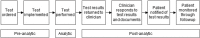
Figure 1.1
Conceptual Framework of the Testing Process.
The post-analytic phase, specifically the step where results, clinically significant test results (CSTR) in particular, are relayed back to the ordering clinician, is a source of diagnostic error.4,5 To reduce errors that occur during this step, experts have advocated for the use of automated alert notification systems to ensure timely communication of CSTR.5–7 RNSs, which are the focus of this review, vary. They can be completely automated, where an abnormal result generates an alert to the ordering clinician; or the RNS may require manual activation by the clinician. There are also a variety of modalities that can be used to alert the practitioner of actionable test results, including short messages relayed via mobile phones; emails; and results (with or without accompanying alerts) in the EHR.8
1.2.2. Methods
The question of interest for this review is, “Do RNSs for radiologic and laboratory tests improve timeliness and reliability of receipt of results and action on the results?” To answer this question, we searched two databases (CINAHL® and MEDLINE®) for articles published from 2008 to 2018 using the terms “diagnostic errors,” “delayed diagnosis,” “missed diagnosis,” and synonyms. Additional terms included “alerts,” “automated systems,” “communication systems,” “critical test results,” “alert notification,” and other similar terms. The initial search yielded 1,965 results. Once duplicates had been removed and additional relevant articles from selected other sources added, a total of 1,981 articles were screened for inclusion, and 46 full-text articles were retrieved. Of those, 17 were selected for inclusion in this review, including 2 systematic reviews. Articles were excluded if the outcomes were not relevant to this review, the article was out of scope (including not quantitative), the study was of limited rigor, or if the study design or results were insufficiently described.
General methods for this report are described in the Methods section of the full report.
For this patient safety practice, a PRISMA flow diagram and evidence table, along with literature-search strategy and search-term details, are included in the report appendixes A through C.
1.2.3. Evidence Summary
Key Findings
- Performance of result notification systems varied by type of test result, setting, synchronous versus asynchronous communication, and manual versus automated alerting mechanisms.
- For both critical and non-critical CSTR of radiologic studies, lab studies and tests pending at discharge, the use of RNS showed some positive but often mixed results in the timeliness and reliability of receipt, action acknowledgment, and action on the test results.
- Policies and procedures that aligned with the system, mindful integration of the RNS into the workflow and the EHR, and appropriate staffing were identified as factors supporting successful RNS.
- Significant barriers to successful implementation include poor system design, the lack of connectivity between hospitals and non-network physicians, challenges associated with changing schedules and providing critical alerts to physicians who may not be available, and variations in clinician response to alerted results.
The papers selected use RNS for CSTR, both life-threatening and nonurgent, for laboratory or radiological studies in inpatient and ambulatory settings. The RNS varied across studies and included both manual and automated mechanisms to generate the alert, and a variety of asynchronous and synchronous modalities to receive the alert. Outcomes included alerts being received (and acknowledged) by a clinician, and alerts being received and/or acted upon by the clinician (Table 1.1).
We reviewed one meta-analysis and one systematic review, both focusing on automated RNSs for laboratory results.8,9 There were also several single studies of high-quality design, with two randomized controlled trials10,11 and three cluster-randomized controlled trials.12–14 Most of the single studies were quasi-experimental, with either pre/post or post-only designs.
1.2.3.1. Use of RNS for Radiologic Studies
Five studies focused on the impact of RNS on the communication of CSTR in radiology. The CSTR ranged from results requiring treatment but not immediately life-threatening to immediately life-threatening results. The impact of the RNS on the communication of results, and action taken on the results, was mixed.
Two studies, both by Lacson and colleagues, evaluated the use of an Alert Notification of Critical Results (ANCR) system to facilitate communication of critical imaging test results to ordering clinicians at a large academic medical center. The ANCR system, integrated into the clinical workflow, allows both synchronous communication (e.g., pagers) for results related to life-threatening conditions, and asynchronous communications (e.g., email). The system relies on radiologists who read and interpret the radiographic images to initiate an alert to the ordering clinician, rather than using a completely automated system. In the first study, the authors evaluated the ANCR system on adherence to a hospital policy for timeliness of notifications that is based on criticality of the imaging result.15 Using a pre/post study design, the authors found a significant improvement in adherence to the timeliness policy, with adherence increasing from 91.3 percent before the ANCR intervention to 95.0 percent after (p<0.0001). In the second study, also using a pre/post study design, the authors evaluated the impact of implementing both the ANCR system and the policy of communication of the critical imaging test result in reducing critical results that lacked documented communication (date, time, and name of ordering clinician contacted). After the implementation of the critical imaging test result policy and the ANCR, critical results lacking documented communication decreased nearly fourfold between 2009 and 2014 (0.19 to 0.05, p<0.0001).16
Table 1.1
Overview of Single Studies.
In a study linked to the work of Lacson and colleagues, O’Connor et al. (2015) integrated an ANCR with an EHR-based results management application and evaluated its adoption and impact on followup of actionable results by primary care providers (PCPs) in the outpatient setting. Prior to integration, PCPs used the EHR application to track and acknowledge results from laboratory studies. The integration of the two systems allowed the PCPs to receive and acknowledge the ANCR-generated non-urgent CSTR alerts in the EHR or through the ANCR system. During the 2 years after implementation, 15.5 percent of the ANCR alerts were acknowledged in the EHR (15.6% year 1, 15.4% year 2). In the post-intervention period, there was a significant difference (p=.03) between the proportion of alerts acted upon that were acknowledged in the EHR application (79%; 95% CI, 52 to 92) compared with the alerts acknowledged in the ANCR system (97%; 95% CI, 90 to 99).17
Singh et al. (2009) evaluated the impact of an EHR-based system to alert clinicians to critical imaging results in a multidisciplinary ambulatory clinic at a large Veterans Administration (VA) medical center and its five satellite clinics. The VA EHR has an embedded notification system for alerting clinicians to CSTR in a “View Alert” window. The system requires that the radiologist reading an image flag abnormal imaging results, and these alerts are then transmitted to the “View Alert” window. During the study period there were 1,196 abnormal imaging alerts generated (0.97% of all imaging studies), and 217 (18.1%) of these alerts remained unacknowledged (i.e., the ordering clinician did not click on and open the alert) after 2 weeks. Using logistic regression, variables associated with a lack of acknowledgement included physician assistants compared with attending physicians (odds ratio [OR]: 0.46; 95% CI, 0.22 to 0.98); resident physicians compared with attending physicians (OR: 5.58; 95% CI, 2.86 to 10.89); and dual communication (i.e., communication with two clinicians) compared with communication with a single clinician (OR: 2.02; 95% CI, 1.22 to 3.36). Notably, 92 alerts, both acknowledged (n=71) and unacknowledged (n=21), lacked followup at 4 weeks.18
Eisenberg et al. (2010) evaluated the use of a Web-based electronic messaging system to communicate non-urgent CSTRs and recommend followup to ordering clinicians. As in the system used in the studies by Lacson and colleagues, the alerts were initiated by radiologists responsible for interpreting images through a web-based application. The request is received by a facilitator, who is then responsible for conveying the results to the ordering clinician. Once the results have been conveyed, the facilitator sends a confirmation back to the radiologist to close the loop. The authors recognized that the study design was weak (post-only with a satisfaction survey). They authors found that 82.2 percent of the alerts were communicated to the ordering clinicians within a 48-hour window, as defined by the time the radiologist submits a communication request to the time the facilitator conveys the communication to the ordering physician. The authors also found that the day of week affected the outcome, with more alerts submitted by the radiologists Monday–Thursday before 3 p.m. communicated within 48 hours (93.7% +/− 2.4), compared with alerts generated on Thursday afternoon through Sunday (73.0% +/− 9.2). The authors incidentally noted that for one-third of communications in which additional imaging or followup had been recommended, the electronic medical record had no documentation that these services were actually performed.19
1.2.3.2. Use of RNS for Laboratory Studies
Nine of the included studies focused on the use of RNS for laboratory studies, including one meta-analysis and one systematic review. As was the case for the RNS for radiologic studies, the evaluated interventions varied across studies and included paging, email, text messages, and EHR alerts. Results of the RNS were mixed.
The meta-analysis and the systematic review examined the effectiveness of automated electronic RNS to alert ordering clinicians to CSTR, and found insufficient/inconclusive evidence for the use of these systems.8,9 The systematic review by Liebow et al. included four studies, two of which were used to calculate a standardized effect size (ES).10,20 Etchells et al. reported results of a randomized controlled trial evaluating an automated RNS that sends critical laboratory values directly from the laboratory information system to a pager carried by the ordering physician. The objective was to evaluate the effect of the system on physician response time, defined as the time from when the critical result is entered into the lab system to the time an order is written in response to the critical value, or the documented time of treatment (whichever is relevant). They found a 23-minute reduction in median response time, 16 minutes (interquartile range [IQR] 2–141) for the automated paging group, and 39.5 minutes (IQR 7–104.5) for the usual care group, but this difference was not statistically significant.10 Park et al. used a pre/post design to test the impact of a short message service and callbacks for action on critical hyperkalemia results. Across all patients in both intensive care units and general wards, the median and interquartile ranges for the clinical response times, defined as the frequency of clinical responses divided by the number of critical value alerts during a given time period, were significantly reduced, going from 213.0 minutes in the intensive care unit and 476.0 minutes in the general wards to 74.5 minutes and 241 minutes, respectively (p<.001).20 Using Cohen’s d, Liebow and colleagues calculated a grand mean reduction of time to communicate critical results for these two studies (d=.42; 95% CI, 0.23 to 0.62), indicating that the time to report a randomly selected CSTR using the automated system will be shorter than with a randomly selected manually reported value 61.8 percent of the time. Liebow et al. gave an overall strength of evidence rating of “suggestive” for automated RNS.8
Liebow et al. also conducted a systematic review of five studies evaluating the use of centralized call centers that communicate critical CSTRs to the ordering clinician.8 Four of the five studies whose primary outcome was percent of calls completed within a specified interval after results were available from the laboratory (either <30 min or <60 min) contained sufficient data to calculate a standardized ES. The results of a random-effects meta-analysis support the implementation of call centers (mean OR=22.2; 95% CI, 17.1 to 28.7). This translates to critical lab values being reported faster with the call system than results reported via usual means (e.g., call to unit by laboratory technologist) approximately 88.6 percent of the time. Liebow et al. consider the overall strength of evidence for call center systems to be “moderate.”
A systematic review by Slovis et al. included 34 articles published through 2016, representing 40 years of research related to asynchronous automated electronic laboratory RNS.9 Although a wide variety of systems were represented and the study designs and outcomes differed, the authors summarized that these systems can be successfully implemented and improve timeliness of result notification and action. On closer examination of the five most recent studies that were included in the review and also identified through our search, the findings neither fully supported nor opposed use of these systems.10,11,20–22
In the first of two randomized controlled trials by Etchells et al. (2010) and included in the systematic review by Slovis et al., an automated paging system to convey critical laboratory results was evaluated in an urban academic medical center.10 As described above, although there was a 23-minute reduction in the median response time, this was not statistically significant. In their second study, Etchells et al. (2011) combined an automated RNS with CDS.11 The alerts, sent via text to a smart phone or to a pager, contained information about the specific patient and the abnormal result, and offered a URL to a webpage with decision support for the specific alert. The primary outcome was the proportion of pre-defined potential actions that were completed in response to the alert. A secondary outcome was the number of adverse events, defined as worsening of the patient’s condition or complications related to the treatment of the condition. The median proportion of potential clinical actions that were completed was 50 percent (IQR 33–75%) with the alerting RNS with CDS and 50 percent (IQR 33–100%) without it, a difference that was not statistically significant, Without the system, there were 111 adverse events (33%) within 48 hours following an alert and with the alerting system on, there were 67 adverse events (42%); a 9 percent increase when using the alerting system that bordered on statistical significance (p=0.06).
In addition to the five studies included in Slovis et al, two additional studies about laboratory RNS were reviewed for this report. In the outpatient department of a large (2,500-bed) tertiary teaching hospital in Taiwan, Lin et al. studied the impact of a phone-based RNS on clinical outcomes of patients taking the anticoagulant warfarin.23 Their RNS automatically generates and delivers text messages about critical lab CSTRs to providers’ mobile phones 24 hours a day, 7 days a week. Using a pre/post study design, the investigators found no significant differences in warfarin-associated adverse events. The rate of major venous thromboembolism events was 1.6 percent for both the manual alert period and the test RNS period. The rate of major hemorrhage requiring an ED visit or hospital admission was 3.1 percent in the manual alert period and 4.2 percent in the RNS alert period (p=0.198). As with the findings in Etchells et al. (2010), the secondary outcome of timeliness of physician followup actions after receipt of an automated critical alert was not significantly improved (11.13 ± 7.65 days for manual alert period vs. 11.32 ± 8.17 days for phone-based RNS period; p=0.814).
Expanding on the work previously described by Lacson and O’Connor, O’Connor et al. (2018) examined the use of the ANCR for communication of non-urgent clinically significant pathology reports indicating new malignancies.24 After a pathologist identifies the CSTR, the ordering physician is contacted via pager about critical or urgent results, and via pager or secure email for non-urgent results, and the CSTR is entered into the ANCR system. For results that the ordering physician does not acknowledge, the system sends reminders to the pathologist and the ordering physician. Acknowledgment of the CTSR within 15 days, the institutional policy for non-urgent CSTR, was documented for 98 of 107 cases (91.6%) before the RNS had been implemented, and for 89 of 103 (86.4%) after the RNS had been implemented, a difference that was not statistically significant. There was also no significant difference in median time to acknowledgment for new malignancies when comparing the pre-RNS period (7 days; IQR 3–11) and post-intervention period (6 days; IQR 2–10). In the post-RNS period, for CTSR using the ANCR, median time to acknowledgment was significantly shorter than when an ANCR alert was not generated (2 vs. 7 days, p=0.0351).
1.2.3.3. Use of RNS for Tests Pending at Discharge
Tests pending at discharge (TPADs) involve transitions, span more than one setting (e.g., the hospital setting to the ambulatory setting), and often involve more than one clinician (e.g., inpatient attending physician and outpatient primary care physician). The risk of missed communication and potential harm to patients is greater during these transitions between settings and clinicians.25,26 Three cluster-randomized controlled studies from a single institution investigated the use of an automated email CSTR notification system for TPAD.12–14 Awareness and confirmed acknowledgement of the test result after discharge were statistically higher in the intervention group, but there was no difference between the intervention and control groups in documented actions taken in response to the test results (i.e., receiving/confirming receipt of a test result did not improve timeliness of acting upon that information).
Two cluster-randomized controlled studies by Dalal et al. (2014 and 2018) included inpatient attending physicians and PCPs whose patients were discharged from inpatient cardiology and medicine units in a large academic medical center, and who had TPAD for both radiology and laboratory studies.12,13 In these studies, a patient’s discharge triggers a series of electronic events that updates the status of any remaining TPADs on a daily basis. As results for these pending tests are finalized, the responsible in-patient attending and outpatient PCP receive an automatic email containing the test result. The primary outcome of the first study was self-reported awareness of the TPAD result by the patient’s inpatient attending physician.12 There was a statistically significant increase in the awareness of TPAD results by attending physicians for patients assigned to the intervention compared with those assigned to usual care (76% vs. 38%, adjusted/clustered OR 6.30, 95% CI, 3.02 to 13.16, p<0.001). The second study was larger, and the primary outcome was the proportion of actionable TPADs with documented action in the EHR.13 For the primary outcome of documentation of action, there was no significant difference between the intervention and usual care groups (60.7% vs. 56.3%). For those that had an action documented, the median days between result notification and documented action was significantly lower in the intervention group (9 days, CI, 6.2 to 11.8) compared with the usual care group (14 days, 95% CI, 10.2 to 17.8) (p=0.04).
In the third study, by El-Kareh et al., the automated RNS described previously was used to alert inpatient and outpatient physicians about positive cultures when the final lab result was returned after patient discharge and the patient was not adequately treated with antibiotics. The alerts included patient identifiers, names and contact information for the physicians involved in their care, the culture results, the discharge medication list, and patient allergy information. Twenty-eight percent of results in the intervention group and 13 percent in the control group met the primary outcome of documented followup (in outpatient chart) within 3 days of receipt of the post-discharge lab result, a statistically significant difference [adjusted OR 3.2, 95% CI, 1.3 to 8.4; p=0.01].27
1.2.3.4. Unintended Consequences
Study authors raised a hypothetical concern about alert fatigue, a potential unintended consequence of implementing alerting RNSs, but only one study measured a related outcome: overuse of the alerting system. Lacson et al. (2016) found that the proportion of reports without critical CSTR and using the ANCR was significantly less than when the ANCR was not used (0.09 vs. 0.20, p<0.002, χ2 test).16 Etchells et al. (2010) noted that critical results, such as those from repeated troponin tests, were viewed as nuisances by receiving clinicians during a pilot of the system.10 They also noted that because physician schedules were not fully automated, it was not possible to consistently route critical results to a responsible and available physician to take action. To compensate for this, physicians handed off “critical value pagers” so that the physician-on-call carried several pagers. Although this could reduce the number of missed alerts, it also created confusion when the on-call physician often could not discern which pager was alerting.
Unexpectedly, Singh et al. (2009) found that dual communication, a duplication intended to ensure that at least one physician received the alert, was associated with delayed followup. This finding was attributed to the lack of clarity about who was responsible for handling the alert.18
1.2.4. Implementation
The studies included in this review demonstrated the critical importance of the local environment and technologies, and circumstances surrounding the success of RNS. Facilitators and barriers to implementation of RNSs are described below.
1.2.4.1. Facilitators
1.2.4.1.1. Integration of RNS Into Workflow
Dalal et al. (2014) attributed the successful implementation of their TPAD email-generating RNS to the existing institutional culture that supports the use of email as a routine part of clinical care. The RNS was integrated into their current practice, which facilitated uptake.12 Two additional studies mention that alignment of the RNS with existing workflows minimized the need to actively seek results, and policies and procedures of the institution supported success.17,27
1.2.4.1.2. Clear Policies and Procedures for RNS Use
Several authors mentioned the need for clear policies and procedures for the RNS. Singh et al. (2009) indicated that institutions need to have clear policies about who is responsible for acknowledging an alert and taking action, so that there is no ambiguity.18 One institution, after much deliberation, established the policy that the responsibility for following up a test rested on the “ordering” clinician, and that this responsibility could be discharged only after a handoff where the “new owner” recipient acknowledged receipt and agreed to take over the followup.6 Other studies mentioned the need for policies establishing which types of alerts warrant use of the RNS and the timeliness of responding to those alerts, based on the criticality of the CSTR.10,15–17,19
1.2.4.1.3. Adequate Staffing To Support the RNS
Two studies mentioned the need for adequate staffing to support the implementation of RNS.19,21 Chen et al. implemented a two-pronged approach to improved communication times, involving increasing the number of staff in the laboratory to improve lab performance and quality, and implementing an RNS with secure messaging.21 Eisenberg et al. noted that their RNS required the hiring of two full-time staff to manage the electronic messaging system.19
1.2.4.2. Barriers
1.2.4.2.1. Unaligned Policies and Procedures
Etchells et al. (2011) found that during weekends and nights there were differences in process between the study sites that involved the receipt of the alerts.11 At one site, the smart phone on which alerts were received was handed from the attending daytime physician to the physician-on-call, so critical alerts could be received after hours. At the other site, the smart phone was not handed off, and the physician-on-call relied on telephone calls from the lab. O’Connor et al. (2018) documented that there were conflicting policies about what could trigger an alert: per local departmental policy, only unexpected malignancies should trigger an alert; but per enterprise policy, any new malignancy should trigger an alert.24
1.2.4.2.2. Lack of Connectivity Between Hospitals and PCPs Outside of Network
The three studies of using RNS to facilitate communication of TPADs during care transitions at hospital discharge all showed challenges in communicating with PCPs outside their hospital system.12,13,27 If RNSs are relied on for TPAD result communication, they must be able to notify non-network and network PCPs.
1.2.4.2.3. Physician Handoffs and Scheduling
Automated physician scheduling is important for optimal performance of automated critical value alerting systems. This barrier to successful implementation was identified by Etchells et al. (2010), who found that when physician schedules are not fully automated, it is impossible to route alerts to the responsible (e.g., on-call) physician who can take action.10,11
1.2.4.2.4. Availability of Resources and Technology Limitations
Lin et al. (2014) indicated that the full implementation of their alert system was challenged by the unavailability of phones for adjunct physician staff, rendering them unable to receive critical alerts sent via the RNS.23 Park et al. (2008) identified that their secure messaging phone reception had inconsistent signal strength in the hospital, but this had a minimal effect, since they had continued to manually call results to the unit in addition to the smartphone alerts.20
1.2.5. Resources
The book “Getting Results: Reliably Communicating and Acting on Critical Test Results,” (Schiff GD, ed., Joint Commission Resources, 2006) is “a collection of articles and case studies on how healthcare organizations are improving communication of critical test results,” as described in the AHRQ Patient Safety Network.28
Pennsylvania passed the Patient Test Result Information Act (2018 Act 112) to ensure that patients with significant abnormalities on imaging exams are notified of the need for medical followup. Information on the law is available through the Pennsylvania General Assembly website: https://www.legis.state.pa.us/cfdocs/legis/li/uconsCheck.cfm?yr=2018&sessInd=0&act=112.]
1.2.6. Gaps and Future Directions
Over half of the studies in this review address the experiences of a small group of researchers from a single large academic institution and its affiliated medical centers. Although these studies are of high quality and some findings are significant, studies in other settings are needed to test and demonstrate generalizability, as well as to engage research in this field more widely. Diagnostic errors due to lapses in communication occur during care transitions, but only three studies (again, all in the same healthcare system) evaluated RNS to improve delivery of results finalized after the transition from the inpatient to the outpatient setting.
As mentioned above, it is challenging when many providers are taking care of a patient, as the RNS needs to discern who is responsible for which patient at any given time. Institutions are establishing policies aimed at addressing this challenge, but how the policies perform needs to be investigated.6
Another area for future study is the development and testing of RNS that are “smart” and use CDS to recognize the difference between critical results that require notification for emergent intervention versus those that do not. Future studies that track the number and types of alerts generated, including synchronous communication for only those CSTR that require urgent action, could include outcomes related to reducing alert fatigue.
References for Section 1.2
- 1.
- Callen JL, Westbrook JI, Georgiou A, et al. Failure to follow-up test results for ambulatory patients: a systematic review. J Gen Intern Med. 2012;27(10):1334–48. [PMC free article: PMC3445672] [PubMed: 22183961]
- 2.
- Davis Giardina T, King BJ, Ignaczak AP, et al. Root cause analysis reports help identify common factors in delayed diagnosis and treatment of outpatients. Health Aff. 2013;32(8):1368–75. [PMC free article: PMC3822525] [PubMed: 23918480]
- 3.
- Commission TJ. 2019 Hospital National Patient Safety Goals. In: Commission TJ, editor. 2019.
- 4.
- Hickner J, Graham D, Elder N, et al. Testing process errors and their harms and consequences reported from family medicine practices: a study of the American Academy of Family Physicians National Research Network. BMJ Qual Saf. 2008;17(3):194–200. [PubMed: 18519626]
- 5.
- Singh H, Wilson L, Reis B, et al. Ten strategies to improve management of abnormal test result alerts in the electronic health record. J Patient Saf. 2010;6(2):121. [PMC free article: PMC2885732] [PubMed: 20563228]
- 6.
- Roy CL, Rothschild JM, Dighe AS, et al. An initiative to improve the management of clinically significant test results in a large health care network. Jt Comm J Qual Patient Saf. 2013;39(11):517–27. [PubMed: 24294680]
- 7.
- Singh H, Vij MS. Eight recommendations for policies for communicating abnormal test results. Jt Comm J Qual Patient Saf. 2010;36(5):226–AP2. [PubMed: 20480756]
- 8.
- Liebow EB, Derzon JH, Fontanesi J, et al. Effectiveness of automated notification and customer service call centers for timely and accurate reporting of critical values: a laboratory medicine best practices systematic review and meta-analysis. Clin Biochem. 2012;45(13–14):979–87. [PMC free article: PMC4518392] [PubMed: 22750773]
- 9.
- Slovis BH, Nahass TA, Salmasian H, et al. Asynchronous automated electronic laboratory result notifications: a systematic review. J Am Med Inform Assoc. 2017;24(6):1173–83. [PMC free article: PMC7787253] [PubMed: 28520977]
- 10.
- Etchells E, Adhikari NK, Cheung C, et al. Real-time clinical alerting: effect of an automated paging system on response time to critical laboratory values—a randomised controlled trial. BMJ Qual Saf. 2010;19(2):99–102. [PubMed: 20351157]
- 11.
- Etchells E, Adhikari NK, Wu R, et al. Real-time automated paging and decision support for critical laboratory abnormalities. BMJ Qual Saf. 2011;20(11):924–30. [PubMed: 21725046]
- 12.
- Dalal AK, Roy CL, Poon EG, et al. Impact of an automated email notification system for results of tests pending at discharge: a cluster-randomized controlled trial. J Am Med Assoc. 2014;21(3):473–80. [PMC free article: PMC3994865] [PubMed: 24154834]
- 13.
- Dalal AK, Schaffer A, Gershanik EF, et al. The impact of automated notification on follow-up of actionable tests pending at discharge: a cluster-randomized controlled trial. J Gen Intern Med. 2018;33(7):1043–51. [PMC free article: PMC6025668] [PubMed: 29532297]
- 14.
- El-Kareh R, Hasan O, Schiff GD. Use of health information technology to reduce diagnostic errors. BMJ Qual Saf. 2013;22:(Suppl 2):ii40–ii51. [PMC free article: PMC3786650] [PubMed: 23852973]
- 15.
- Lacson R, Prevedello LM, Andriole KP, et al. Four-year impact of an alert notification system on closed-loop communication of critical test results. Am J Roentgenol. 2014;203(5):933–8. [PMC free article: PMC4426858] [PubMed: 25341129]
- 16.
- Lacson R, O’Connor SD, Sahni VA, et al. Impact of an electronic alert notification system embedded in radiologists’ workflow on closed-loop communication of critical results: a time series analysis. BMJ Qual Saf. 2016;25(7):518–24. [PubMed: 26374896]
- 17.
- O’Connor SD, Dalal AK, Sahni VA, et al. Does integrating nonurgent, clinically significant radiology alerts within the electronic health record impact closed-loop communication and follow-up? J Am Med Inform Assoc. 2016;23(2):333–8. [PMC free article: PMC5009922] [PubMed: 26335982]
- 18.
- Singh H, Thomas EJ, Mani S, et al. Timely follow-up of abnormal diagnostic imaging test results in an outpatient setting: are electronic medical records achieving their potential? Arch Intern Med. 2009;169(17):1578–86. [PMC free article: PMC2919821] [PubMed: 19786677]
- 19.
- Eisenberg RL, Yamada K, Yam CS, et al. Electronic messaging system for communicating important, but nonemergent, abnormal imaging results. Radiol. 2010;257(3):724–31. [PubMed: 20884911]
- 20.
- Park H-i, Min W-K, Lee W, et al. Evaluating the short message service alerting system for critical value notification via PDA telephones. Ann Clin Lab Sci. 2008;38(2):149–56. [PubMed: 18469361]
- 21.
- Chen T-C, Lin W-R, Lu P-L, et al. Computer laboratory notification system via short message service to reduce health care delays in management of tuberculosis in Taiwan. Am J Infect Control. 2011;39(5):426–30. [PubMed: 21496958]
- 22.
- Singh H, Thomas EJ, Sittig DF, et al. Notification of abnormal lab test results in an electronic medical record: do any safety concerns remain? Am J Med. 2010;123(3):238–44. [PMC free article: PMC2878665] [PubMed: 20193832]
- 23.
- Lin S-W, Kang W-Y, Lin D-T, et al. Comparison of warfarin therapy clinical outcomes following implementation of an automated mobile phone-based critical laboratory value text alert system. BMC Med Genomics. 2014;7(1):S13. [PMC free article: PMC4101312] [PubMed: 25079609]
- 24.
- O’Connor SD, Khorasani R, Pochebit SM, et al. Semiautomated System for Nonurgent, Clinically Significant Pathology Results. Appl Clin Inform. 2018;9(02):411–21. [PMC free article: PMC5996775] [PubMed: 29874687]
- 25.
- Roy CL, Poon EG, Karson AS, et al. Patient safety concerns arising from test results that return after hospital discharge. Annals of internal medicine. 2005;143(2):121–8. [PubMed: 16027454]
- 26.
- Gandhi TK. Fumbled handoffs: one dropped ball after another. Ann Intern Med. 2005;142(5):352–8. [PubMed: 15738454]
- 27.
- El-Kareh R, Roy C, Williams DH, et al. Impact of automated alerts on follow-up of post-discharge microbiology results: a cluster randomized controlled trial. J Gen Intern Med. 2012;27(10):1243–50. [PMC free article: PMC3445692] [PubMed: 22278302]
- 28.
- AHRQ Patient Safety Network. Getting Results: Reliably Communicating and Acting On Critical Test Results. Book/Report. Agency for Healthcare Research and Quality. Accessed Feb. 18, 2020. https://psnet
.ahrq.gov /issue/getting-results-reliably-communicating-and-acting-critical-test-results.
1.3. Patient Safety Practice: Education and Training
Authors
Kendall K. Hall, M.D., M.S., Gordon Schiff, M.D., and Katharine Witgert, M.P.H.1.3.1. Practice Description
In the 2015 National Academies of Sciences, Engineering, and Medicine (NASEM) report Improving Diagnosis in Health Care, one of the recommended strategies for improving diagnosis is to enhance healthcare professional education and training in the diagnostic process.1 The content of this education can be guided by an understanding of the root causes of diagnostic errors. Studies have uncovered two broad categories of underlying root causes: cognitive-based factors, such as failed heuristics; and systems-based factors, such as lack of provider-to-provider communication and coordination.2–4 In the realm of cognitive-based errors, there are also two main streams of thought about causes: heuristics failures and shortcomings in disease-specific knowledge and experience. These sets of broad conceptual factors are by no means mutually exclusive, and ideally system redesign and educational efforts can leverage overlaps and synergies. How to best provide education and training to change these underlying factors and thereby improve diagnostic accuracy and reduce diagnostic errors leads to a more fundamental question that this review attempts to address, “Do education and training lead to improved diagnostic performance?”
1.3.2. Methods
Key Findings
- Although there are a limited number of studies, the literature suggests that training on metacognitive skills may improve diagnostic accuracy, particularly as clinical experience increases.
- Online training, either didactic or via simulation, can be successfully used as a mode of delivery for educational interventions targeting clinical reasoning and diagnostic safety.
- There are several promising training interventions to improve visual perception for radiology practice.
- Limitations include a dearth of studies that examine the transfer of learning from the educational setting into the clinical setting and actual patient care.
We searched four databases (CINAHL®, MEDLINE Cochrane, and PsycINFO®) for articles published from 2008 to 2018 using the terms “diagnostic errors,” “delayed diagnosis,” “missed diagnosis,” and synonyms. Terms specific to this PSP include “education, professional,” “training,” “simulation training,” “structured practice,” and related terms. The initial search yielded 211 results. Once duplicates had been removed and additional relevant referenced articles added, 187 articles were screened for inclusion and 29 full-text articles were retrieved. Of those, 22 studies were selected for inclusion in this review. Articles were included if the intervention being tested was training and an outcome was diagnostic accuracy. Articles were excluded if the article was out of scope, or the study provided limited detail or was of limited rigor.
General methods for this report are described in the Methods section of the full report.
For this patient safety practice, a PRISMA flow diagram and evidence table, along with literature-search strategy and search-term details, are included in the report appendixes A through C.
1.3.3. Evidence Summary
A majority of the selected studies focused on training directed at the cognitive aspects of diagnostic errors, such as clinical reasoning and biases. Other studies focused on training in visual perception skills for radiologists and specific diagnostic skills. Few studies involved experienced clinicians, with medical students and residents being the predominant types of learners.
Overall, the quality of evidence was moderate, with some strong study designs, such as randomized controlled trials, but with low numbers of subjects, making generalization of findings challenging. The educational interventions varied in both their content and the mode by which the content was delivered, and in several cases the distinction between the testing of the content versus the testing of the mode of delivery was difficult to ascertain.
1.3.3.1. General Training in Clinical Reasoning
Clinical reasoning is the process by which clinicians collect data, process the information, and develop a problem representation, leading to the generation and testing of a hypothesis to eventually arrive at a diagnosis.5,6
Cook et al. (2010) conducted a meta-analysis and systematic review of the effects on training outcomes of using virtual patients, including the effects on clinical reasoning. The learners interact with a computer program that simulates real-life clinical scenarios to obtain a history, conduct a physical exam, and make diagnostic and treatment decisions. In comparing virtual patients to no intervention, the pooled ES for the five studies with an outcome of clinical reasoning was 0.80 (95% CI, 0.52 to 1.08). Pooled ESs for the outcomes of knowledge (N=11 studies) and other skills (N=9 studies) were also large. When comparing the use of virtual patients to noncomputer instruction (e.g., didactic instruction, standardize patients, routine clinical activities), the pooled ES for the outcome of clinical reasoning was −0.004 (95% CI, −0.30 to 0.29, N=10 studies), and it was also low for satisfaction, knowledge, and other skills. The main takeaway from this meta-analysis and review was that the use of virtual patients is associated with large positive effects on clinical reasoning and other learning outcomes when compared with no intervention and is associated with small effects in comparison with noncomputer instruction.7
Graber et al. (2012) in their systematic review identified papers that reported testing interventions aimed at reducing cognitive errors.8 Three broad categories of interventions emerged: interventions to improve knowledge and experience, interventions to improve clinical reasoning, and interventions that provide cognitive support. Several papers examined the use of training in metacognitive skills to improve clinical reasoning, which is below. Wolpaw et al. (2009) studied the use of a learner-centered technique by third-year medical students to present clinical cases in a structured manner (SNAPPS: Summarize, Narrow, Analyze, Probe, Plan, Select). Although the authors did not assess whether the DDX were accurate, they found that students using the SNAPPS technique performed better on all outcomes, including analyzing possibilities of the DDX, expressing uncertainties, and obtaining clarification.9
1.3.3.2. Training in Metacognitive Skills To Reduce Biases
Cognitive biases can affect clinical reasoning and influence the diagnostic process, contributing to a large proportion of misdiagnoses.6,8,10,11 Metacognition, the understanding, control, and monitoring of one’s cognitive processes, can be used to gain better insight and counteract these biases.12,13 Nine studies focused on techniques to enhance metacognitive skills, specifically training on the use of cognitive forcing strategies (CFS) and the use of reflection during the diagnostic process. The results of the studies are mixed, but overall suggest the use of training metacognitive strategies to improve diagnostic performance.
The use of CFS, a metacognitive strategy, is a technique to bring about self-monitoring of decision making and to force the consideration of alterative diagnoses.13 Three studies (Sherbino et al., 2011, Sherbino et al., 2014, Smith and Slack, 2015) provided medical students and residents with training on the use of CFS and measured its impact on diagnosis.14–16 The results did not show any appreciable improvement in diagnostic accuracy. In a preliminary and followup study, Sherbino et al. employed a 90-minute, standardized, interactive, case-based teaching seminar on CFS for medical students during their emergency medicine rotation.14,15 Neither study showed any improvement in diagnostic errors. In the first study, they found that fewer than half of the students could use the CFS to debias themselves, and that 2 weeks post-training the students’ knowledge of debiasing was no longer present. In the second study, there was no difference in the diagnostic accuracy between the control and intervention groups. In the study by Smith and Slack (2015), family medicine residents participated in a debiasing workshop that included training on CFS. They found that the residents’ ability to formulate an acceptable plan to mitigate the effect of cognitive biases significantly improved after the training (p=0.02), although the residents were not able to translate the plan into practice, as evidenced by no change in the outcomes of preceptor concurrence with the residents’ diagnoses, residents’ ability to recognize their risk of bias, and the preceptors’ perception of an unrecognized bias in the residents’ presentations. This study was limited in that CFS targets biases related to nonanalytic reasoning (so-called pattern recognition). Novice diagnosticians, such as medical students, may lack sufficient experience to employ nonanalytic reasoning, rendering these methods increasingly more useful as experience increases.16
In a frequently cited study, Mamede et al. (2010) investigated whether recent diagnostic experiences elicit availability bias (i.e., judging a diagnosis that comes to mind more readily as being correct), and then tested a simple instructional procedure to reduce that bias. The training consisted of a five-step procedure to induce structured reflection and improve diagnostic accuracy in first- and second-year internal medicine residents. The use of reflection did reduce availability bias and improved diagnostic accuracy.17 Two additional studies by this group of investigators (Mamede 2012, Mamede 2014), furthered this work on the use of structured reflection as a tool to facilitate diagnosis. The first of these studies found that the use of structured reflection after providing an immediate diagnosis when practicing with clinical cases fostered the learning of clinical knowledge more effectively than providing an immediate diagnosis only, or generating an immediate diagnosis followed by a differential diagnosis.18 In the second study, the use of this technique enhanced learning of the diagnosis practiced as well as its alternative diagnoses.19
Coderre et al. (2010) tested the effectiveness of questioning a medical student’s initial hypothesis as a means to induce cognitive reflection. The authors found that the questioning of an initial correct diagnosis did not change the final diagnosis; the students tended to retain the initial diagnosis. If the student’s initial diagnosis was incorrect, the questioning provided an opportunity for the students to recognize and react to their error, and correct their diagnosis.20
In a randomized controlled study, Nendaz et al. (2011) studied the impact of weekly in-person case-based clinical reasoning seminars incorporating diagnostic reflection, during which the students were prompted to reflect on their reasoning process and were provided feedback on each step of that process. They found no difference in the accuracy of the medical students’ final diagnoses between intervention and control groups (74% vs. 63%), although the students in the intervention group were more likely to have mentioned the correct diagnosis somewhere on their working list of DDX under consideration (75% vs. 97%, p=0.02).21
Reilly et al. (2013) incorporated the promotion of reflection on past experiences where a cognitive bias led to a diagnostic error, as part of a longitudinal curriculum on cognitive bias and diagnostic errors for residents. Residents who completed the curriculum significantly improved their ability to recognize cognitive biases when compared with their baseline performance (p=0.002) and when compared with the control group (p<0.0001). The study was limited in that it did not evaluate the impact of the intervention on diagnostic accuracy.22
1.3.3.3. Training on the Use of Heuristics
Heuristics are decision strategies, or mental shortcuts, that allow fast processing of information to arrive at a decision or judgment. One type of heuristic is representativeness; the use of the degree to which an event is representative of other, similar events to assess the probability of an event occurring.23,24 Although the literature around the use of heuristics in medicine tends to focus on the biases they introduce, there is a recognized potential for training with heuristics to achieve better diagnostic accuracy.25,26
Mohan et al. (2018) conducted a randomized controlled trial comparing two training interventions designed to improve the use of the representativeness heuristic to improve trauma triage by emergency physicians. The authors developed two serious video games to train in the use of the heuristic. The first was an adventure game, based on the theory of narrative engagement, and the second was a puzzle-based game, based on the theory of analogical reasoning, using comparisons to help train the learners on applying decision principles. Both games incorporated feedback on diagnostic errors and how they could be corrected. Results showed that both games had positive effects on trauma triage, whereas traditional medical education had none.26
1.3.3.4. Training To Improve Visual Perception Skills
In radiology, diagnostic errors fall into two broad categories: perceptual errors, in which an abnormality on an image is not seen or identified, and interpretive errors, in which an abnormality is seen but the meaning or the importance of the finding is not correctly understood.27,28 Perceptual errors account for a majority of misdiagnoses in radiology,27,29,30 and can be rooted in faulty visual processing or, to a lesser extent, cognitive biases.31
Improving visual perception skills, which predominate the diagnostic process in radiology, requires methods of training different from those to improve clinical reasoning.28 Four studies were identified through our search that evaluated the impact of educational interventions on perceptive skills, with three showing improvement in perceptive performance.32–34 The studies involved subjects early in their medical training, and each tested a different intervention to improve perceptive performance, making aggregation of findings challenging.
A novel study by Goodman and Kelleher (2017) took 15 first-year radiology residents to an art gallery, where experts with experience in teaching fine art perception trained the residents on how to thoroughly analyze a painting. The trainees were instructed to write down everything they could see in the painting, after which the art instructor showed the trainees how to identify additional items in the painting that they had not perceived. To test this intervention, the residents were given 15 radiographs pre-intervention and another 15 post-intervention and asked to identify the abnormalities. At baseline, the residents scored an average of 2.3 out of a maximum score of 15 (standard deviation [SD] 1.4, range 0–4). After the art training, the residents’ scores significantly improved, with an average score of 6.3 (SD of 1.8, range 3–9, p<.0001), indicating that perception training may improve radiology residents’ abilities to identify abnormalities in radiographs.32
In a small randomized crossover study by van der Gijp et al. (2017), 19 first- and second-year radiology residents received training on two different visual search strategies to determine their effect on accuracy of detecting lung nodules on CT scans. The first search strategy was “scanning,” in which the resident views all the visible lung tissue on a single image and slowly scrolls down, image by image, through the entire study. The second search strategy, “drilling,” has the resident mentally divide each lung into three regions and scroll through each region individually while keeping the eyes fixed on that region. Perceptual performance for both scanning and drilling strategies and a pre-test using a free search strategy was determined by the mean numbers of true positives and false positives. There was a significant effect of year of residency on the true positive score (p<0.01) but not for false positives. Drilling (p<0.001) and free search (p<0.001) both resulted in significantly higher true positive scores (i.e., lung nodules identified appropriately) than did scanning. There was no difference between the drilling or free search strategies. The free search strategy resulted in higher false positive scores than drilling (p<0.001) and scanning (p<0.001). The authors concluded that drilling outperforms scanning for detecting lung-nodules and should be the preferred strategy when teaching perceptive skills.33
In a randomized controlled trial, Soh et al. (2013) used an online e-learning tutorial to train 14 first-year medical radiation sciences students (i.e., radiology technologists) in Australia to improve their ability to detect breast lesions on mammographic images. The 1-hour tutorial focused on anatomy, image positioning, mammogram viewing and analysis, and the appearance of normal breast tissue and asymmetric densities and masses. The students were randomized to the intervention (tutorial) or control group, with their performance evaluated by their viewing normal and abnormal mammograms. The study used eye-tracking technology to determine when and how often the student fixated on a lesion. The intervention group demonstrated improvement in the mean number of fixations per case (p=.047), and decreased time to first fixation on a lesion by 49 percent (p=.016). The intervention increased students’ ability to identify lesions (i.e., sensitivity) by 30 percent (p=.022).34
The fourth study evaluated different proportions of normal and abnormal radiographs in image training sets to determine the best case-mix for achieving higher perceptive performance.35 For the intervention, Pusic et al. (2012) used three different 50-case training sets, which varied in their proportions of abnormal cases (30%, 50%, 70%). One hundred emergency medicine residents, pediatric residents, and pediatric emergency medicine fellows were randomized to use one of the training sets. After the intervention, all participants completed the same post-test. All three groups showed improvement after the intervention, but with varying sensitivity-specificity trade-offs. The group that received the lowest proportion (30%) of abnormal radiographs had a higher specificity and was more accurate with negative radiographs. The group that trained on the set with the highest proportion of abnormal radiographs (70%) detected more abnormalities when abnormalities were present, achieving higher sensitivity. These findings have significant implications for medical education, as it may be that case mix should be adjusted based on the desired sensitivity or specificity for a given examination type (e.g., screening exams vs. diagnostic test).35
1.3.3.5. Other Education and Training Interventions
In the systematic review of patient safety strategies targeted at diagnostic errors, McDonald et al. (2013) identified 11 studies, ranging in dates from 1981 to 2011, that involved a variety of interventions. Two randomized trials targeted patients and families, and found that the interventions improved performance: the first found that parent education improved parents’ ability to identify serious symptoms requiring a physician office visit, and the second showed that patient education, in addition to reminders, improved breast cancer screening rates.36
Medical teaching typically involves error avoidance training (EAT), in which the focus is on how to perform a task correctly rather than on how to manage errors. However, evidence suggests that in the early stages of learning a skill, errors are necessary in order to avoid them in the future.37 In their randomized trial with 56 medical students, Dyre et al. (2017) compared the use of error management training (EMT), in which students were given “permission” to make errors while conducting simulation-based ultrasound examinations, with EAT. Two outcomes were measured: the objective structured assessment of ultrasound scale, a measure of ultrasound performance; and diagnostic accuracy. The scale scores showed a significant improvement by the EMT group compared with the EAT group (p<0.001). Although diagnostic accuracy showed some improvement, this was not statistically significant. The study is limited in that the authors cannot determine whether the outcomes were a result of EMT, the positive framing of errors, or the combination of these two components.38
In a quasi-randomized controlled trial, Schwartz et al. (2010) tested the use of in-person didactic sessions to teach fourth-year medical students skills in contextualizing patient care. The authors based this work on the premise that there are both biomedical and contextual data that must be ascertained during the diagnostic process and incorporated into the treatment plan. Students who participated in the didactic sessions were significantly more likely to probe for contextual issues and significantly more likely to develop appropriate treatment plans for the standardized patients with contextual issues.39
Two studies investigated the use of online training to improve specific diagnostic skills, both resulting in significant improvements in diagnostic accuracy.40,41 Smith et al. (2009) conducted a 4-month online didactic continuing education program to improve the ability of radiographers in rural areas to interpret plain musculoskeletal radiographs. The results showed a significant improvement in image interpretation accuracy for more complex cases (p<0.05), although there was no change in accuracy for less complex cases.40 McFadden et al. (2016), using convenience sampling, compared a traditional in-person training with an on-line simulation-based training, both designed to improve the diagnosis by primary care practitioners of the etiology of joint pain. The online training included interactive practice opportunities and feedback delivered by an AI-driven tutor. The intervention group’s diagnostic performance was significantly improved from baseline (p<0.02) compared with the group that received the traditional training.41
1.3.4. Implementation
Many of the studies were conducted in training and simulated environments. As such, there were limited discussions regarding facilitators and barriers to implementation in the clinical practice environment.
1.3.4.1. Facilitators
Several of the studies used interventions that brought the education to the learner, such as those using information technology–based platforms.34,35,40,41 Although there are costs in both time and money associated with the set-up of these online learning systems, once implemented, the training is more easily administered to more learners and is more flexible to being customized.42
1.3.4.2. Barriers
The use of cognitive training interventions, such as reflective practice, may yield the greatest improvements for only the most complex diagnostic cases.17,21 This makes application of appropriate strategies in actual clinical settings difficult, as whether a case is complex is often not determined until after the diagnostic process has begun.
In addition, some of these teaching techniques, such as those using standardized patients or requiring development of simulations, are labor intensive and may not be generalizable.
1.3.5. Resources
The Society to Improve Diagnosis in Medicine offers a clinical reasoning toolkit that contains resources to help clinicians and educators. The toolkit can be accessed at the Society’s website: https://www.improvediagnosis.org/clinicalreasoning/.
1.3.6. Gaps and Future Directions
Graber et al. (2012) noted that the research directed at improving cognition as a means to reduce diagnostic errors was immature, and recommended more research to study different approaches and interventions, including training strategies.8 Since the publication of that paper, as evidenced in this review, there has been a modest increase in research focused on education and training to improve diagnostic accuracy, but gaps persist and many questions remain unanswered and untested. There is a particularly strong need to be able to take the educational work out of the realm of the classroom and into the real and complex world that busy diagnosticians work in to reliably make accurate and timely diagnoses.
It should be noted that interventions, such as education and training, that address human error directly are considered to be weaker at driving effective and sustained improvement compared with stronger actions that remove dependence on the human.43 There is an opportunity to identify and investigate the use of system supports (e.g., process changes) to complement and solidify these educational efforts. The supports should prevent the need for clinicians to rely solely on their human, hence fallible, memory to recall what they learned both about diseases and about heuristics pitfalls.
Many of the studies engaged medical students or first-year residents, who were relatively new in their careers and lacked clinical experience. Improving clinical reasoning through the use of metacognitive strategies, particularly CFS, is targeted at reducing biases associated with the use of nonanalytic reasoning. Expanding training on these strategies to more experienced clinicians, as opposed to trainees, may yield stronger results.14,15 In contrast, visual perceptive skills develop earlier and faster than interpretive skills.28,44 Therefore, educational interventions directed at improving perception would more likely benefit medical students and residents early in their training. Understanding the best timing for different educational strategies to maximize their effectiveness in the continuum of medical education, from student through experienced clinician, would be beneficial.
A variety of methodological aspects of the studies could have been improved to strengthen the evidence base. Several of the studies occurred outside of clinical settings (e.g., online training and testing), and did not involve transferring the skills to diagnostic accuracy outcomes in actual clinical practice. Some of the studies were limited due to the inability to untangle the effect of the mode of the training (e.g., online didactic training) from the content that was being delivered.
Finally, although research indicates that the root causes of diagnostic errors include both cognitive factors and systems-based factors,2–4 nearly all the identified studies targeted cognitive-based training strategies. The 2015 NASEM report on improving diagnosis in healthcare included a call for training in systems-based factors. This is an opportunity to conduct studies on the impact of team-training and communication on diagnostic errors, which is lacking, and training to support patient integration into the diagnostic process.
References for Section 1.3
- 1.
- National Academies of Sciences, Engineering and Medicine. 2015. Improving Diagnosis in Health Care. Washington, DC: National Academies Press. 10.17226/21794. [PubMed: 26803862] [CrossRef]
- 2.
- Graber ML, Franklin N, Gordon R. Diagnostic error in internal medicine. Arch Intern Med. 2005;165(13):1493–9. [PubMed: 16009864]
- 3.
- Kostopoulou O, Delaney BC, Munro CW. Diagnostic difficulty and error in primary care—a systematic review. Fam Pract. 2008;25(6):400–13. [PubMed: 18842618]
- 4.
- Singh H, Giardina TD, Meyer AN, et al. Types and origins of diagnostic errors in primary care settings. J Am Med Assoc Intern Med. 2013;173(6):418–25. [PMC free article: PMC3690001] [PubMed: 23440149]
- 5.
- Bowen JL. Educational strategies to promote clinical diagnostic reasoning. New Engl J Med. 2006;355(21):2217–25. [PubMed: 17124019]
- 6.
- Norman GR, Monteiro SD, Sherbino J, et al. The causes of errors in clinical reasoning: cognitive biases, knowledge deficits, and dual process thinking. Acad Med. 2017;92(1):23–30. [PubMed: 27782919]
- 7.
- Cook DA, Erwin PJ, Triola MM. Computerized virtual patients in health professions education: a systematic review and meta-analysis. Acad Med. 2010;85(10):1589–602. [PubMed: 20703150]
- 8.
- Graber ML, Kissam S, Payne VL, et al. Cognitive interventions to reduce diagnostic error: a narrative review. BMJ Qual Saf. 2012;21(7):535–57. [PubMed: 22543420]
- 9.
- Wolpaw T, Papp KK, Bordage G. Using SNAPPS to facilitate the expression of clinical reasoning and uncertainties: a randomized comparison group trial. Acad Med. 2009;84(4):517–24. [PubMed: 19318792]
- 10.
- Norman GR, Eva KW. Diagnostic error and clinical reasoning. Med Educ. 2010;44(1):94–100. [PubMed: 20078760]
- 11.
- van den Berge K, Mamede S. Cognitive diagnostic error in internal medicine. Euro J Intern Med. 2013;24(6):525–9. [PubMed: 23566942]
- 12.
- Colbert CY, Graham L, West C, et al. Teaching metacognitive skills: helping your physician trainees in the quest to’know what they don’t know’. Am J Med. 2015;128(3):318. [PubMed: 25446293]
- 13.
- Croskerry P. The importance of cognitive errors in diagnosis and strategies to minimize them. Acad Med. 2003;78(8):775–80. [PubMed: 12915363]
- 14.
- Sherbino J, Dore KL, Siu E, et al. The effectiveness of cognitive forcing strategies to decrease diagnostic error: an exploratory study. Teach Learn Med. 2011;23(1):78–84. [PubMed: 21240788]
- 15.
- Sherbino J, Kulasegaram K, Howey E, et al. Ineffectiveness of cognitive forcing strategies to reduce biases in diagnostic reasoning: a controlled trial. Can J Emerg Med. 2014;16(1):34–40. [PubMed: 24423999]
- 16.
- Smith BW, Slack MB. The effect of cognitive debiasing training among family medicine residents. Diagn. 2015;2(2):117–21. [PubMed: 29540027]
- 17.
- Mamede S, van Gog T, van den Berge K, et al. Effect of availability bias and reflective reasoning on diagnostic accuracy among internal medicine residents. J Am Med Assoc. 2010;304(11):1198–203. [PubMed: 20841533]
- 18.
- Mamede S, van Gog T, Moura AS, et al. Reflection as a strategy to foster medical students’ acquisition of diagnostic competence. Med Educ. 2012;46(5):464–72. [PubMed: 22515754]
- 19.
- Mamede S, Van Gog T, Sampaio AM, et al. How can students’ diagnostic competence benefit most from practice with clinical cases? The effects of structured reflection on future diagnosis of the same and novel diseases. Acad Med. 2014;89(1):121–7. [PubMed: 24280855]
- 20.
- Coderre S, Wright B, McLaughlin K. To think is good: querying an initial hypothesis reduces diagnostic error in medical students. Acad Med. 2010;85(7):1125–9. [PubMed: 20592507]
- 21.
- Nendaz M, Gut A-M, Louis-Simonet M, et al. Bringing explicit insight into cognitive psychology features during clinical reasoning seminars: a prospective, controlled study. Educ Health. 2011;24(1):496. [PubMed: 21710417]
- 22.
- Reilly JB, Ogdie AR, Von Feldt JM, et al. Teaching about how doctors think: a longitudinal curriculum in cognitive bias and diagnostic error for residents. BMJ Qual Saf. 2013;22(12):1044–50. [PubMed: 23955466]
- 23.
- Kahneman D, Tversky A. Subjective probability: A judgment of representativeness. Cogn Psychol. 1972;3(3):430–54.
- 24.
- Kahneman D. Thinking, fast and slow: Macmillan; 2011.
- 25.
- Marewski JN, Gigerenzer G. Heuristic decision making in medicine. Dialogues Clin Neurosci. 2012;14(1):77. [PMC free article: PMC3341653] [PubMed: 22577307]
- 26.
- Mohan D, Fischhoff B, Angus DC, et al. Serious games may improve physician heuristics in trauma triage. Proc Natl Acad Sci. 2018;115(37):9204–9 [PMC free article: PMC6140476] [PubMed: 30150397]
- 27.
- Bruno MA, Walker EA, Abujudeh HH. Understanding and confronting our mistakes: the epidemiology of error in radiology and strategies for error reduction. Radiographics. 2015;35(6):1668–76. [PubMed: 26466178]
- 28.
- Norman GR, Coblentz CL, Brooks L, et al. Expertise in visual diagnosis: a review of the literature. Acad Med. 1992;67(10):S78–83. [PubMed: 1388563]
- 29.
- Berlin L. Radiologic errors, past, present and future. Diagn. 2014;1(1):79–84. [PubMed: 29539959]
- 30.
- Donald JJ, Barnard SA. Common patterns in 558 diagnostic radiology errors. J Med Imag Radiat On. 2012;56(2):173–8. [PubMed: 22498190]
- 31.
- van der Gijp A, Ravesloot C, Jarodzka H, et al. How visual search relates to visual diagnostic performance: a narrative systematic review of eye-tracking research in radiology. Adv Health Sci Educ. 2017;22(3):765–87. [PMC free article: PMC5498587] [PubMed: 27436353]
- 32.
- Goodman TR, Kelleher M. Improving novice radiology trainees’ perception using fine art. J Am Coll Radiol. 2017;14(10):1337–40. [PubMed: 28838750]
- 33.
- van der Gijp A, Vincken KL, Boscardin C, et al. The effect of teaching search strategies on perceptual performance. Academic Radiol. 2017;24(6):762–7. [PubMed: 28242103]
- 34.
- Soh BP, Reed WM, Poulos A, et al. E-tutorial improves students’ ability to detect lesions. Radiologic technology. 2013;85(1):17–26. [PubMed: 24029881]
- 35.
- Pusic MV, Andrews JS, Kessler DO, et al. Prevalence of abnormal cases in an image bank affects the learning of radiograph interpretation. Med Educ. 2012;46(3):289–98. [PubMed: 22324528]
- 36.
- McDonald KM, Matesic B, Contopoulos-Ioannidis DG, et al. Patient safety strategies targeted at diagnostic errors: a systematic review. Ann Intern Med. 2013;158(5_Part_2):381–9. [PubMed: 23460094]
- 37.
- Rogers DA, Regehr G, MacDonald J. A role for error training in surgical technical skill instruction and evaluation. Am J Surg. 2002;183(3):242–5. [PubMed: 11943119]
- 38.
- Dyre L, Tabor A, Ringsted C, et al. Imperfect practice makes perfect: error management training improves transfer of learning. Med Educ. 2017;51(2):196–206. [PubMed: 27943372]
- 39.
- Schwartz A, Weiner SJ, Harris IB, et al. An educational intervention for contextualizing patient care and medical students’ abilities to probe for contextual issues in simulated patients. J Am Med Assoc. 2010;304(11):1191–7. [PubMed: 20841532]
- 40.
- Smith T, Traise P, Cook A. The influence of a continuing education program on the image interpretation accuracy of rural radiographers. Rural Remote Health. 2009;9(2). [PubMed: 19392573]
- 41.
- McFadden P, Crim A. Comparison of the effectiveness of interactive didactic lecture versus online simulation-based CME programs directed at improving the diagnostic capabilities of primary care practitioners. J Contin Educ Health Prof. 2016;36(1):32–7. [PubMed: 26954243]
- 42.
- O’Doherty D, Dromey M, Lougheed J, et al. Barriers and solutions to online learning in medical education–an integrative review. BMC Med Educ. 2018;18(1):130. [PMC free article: PMC5992716] [PubMed: 29880045]
- 43.
- Foundation NPS. RCA2: improving root cause analyses and actions to prevent harm. National Patient Safety Foundation Boston, MA; 2015.
- 44.
- Brazeau-Lamontagne L, Charlin B, Gagnon R, et al. Measurement of perception and interpretation skills during radiology training: utility of the script concordance approach. Med Teach. 2004;26(4):326–32. [PubMed: 15203845]
1.4. Patient Safety Practice: Peer Review
Authors
Kendall K. Hall, M.D., M.S., Gordon Schiff, M.D., and Katharine Witgert, M.P.H.1.4.1. Practice Description
Key Findings
- Second reviews of radiology or pathology interpretations by peers consistently uncover small but significant numbers of misread tests.
- The existence of any positive outcomes from increasing awareness of this general vulnerability and its effects on personal accountability—knowing that readings are being scrutinized—cannot be determined from the published studies.
- There is a lack of evidence to show that traditional random peer review and feedback mechanisms, which are used to maintain compliance with accreditation requirements, improve diagnostic quality over time or prevent diagnostic errors from reaching the patient.
- When nonrandom peer review is conducted prospectively, there is an opportunity to identify and remediate the diagnostic error before it reaches the patient.
- Limiting peer review to specific case types where it was most impactful was identified as a factor supporting implementation.
- Significant barriers to successful implementation include the increased staffing needs, workload, associated costs, concern over fairness, and maintenance of confidentiality of clinician performance.
Peer review is the systematic and critical evaluation of performance by colleagues with similar competencies using structured procedures.1 Peer review in clinical settings has two recognized objectives: data collection and analysis to identify errors; and feedback with the intention of improving clinical performance and practice quality.2,3 It also serves to fulfill accreditation requirements, such as The Joint Commission requirement that all physicians who have been granted privileges at an organization undergo evaluation of and collect data relating to their performance, or the American College of Radiology physician peer review requirements for accreditation.4–6 When done systematically and fairly, peer review contributes to and derives from a culture of safety and learning.7
For this PSP, we are focusing the use of peer review as a tool to help identify, analyze, and discuss failures in establishing timely and accurate diagnoses, as well as a method to reduce diagnostic errors in the future.
Peer review, when designed appropriately, has the potential to achieve patient safety goals by having an impact on care either directly at the time of testing (e.g., identifying and resolving the error before it affects the patient) or indirectly by improving physician practice through continual learning and feedback. Thus, the question of interest for this review is, “Do peer review and feedback lead to improved diagnostic performance, i.e., fewer diagnostic errors?”
1.4.2. Methods
We searched three databases (CINAHL®, MEDLINE, and PsycINFO®) for articles published from 2008 to 2018 using the terms “diagnostic errors,” “delayed diagnosis,” “missed diagnosis,” and synonyms. Terms specific to this PSP include “peer review,” “performance review,” “performance feedback,” “feedback,” “quality assurance,” and related terms. The initial search yielded 426 results. Once duplicates had been removed and additional relevant referenced articles added, 334 articles were screened for inclusion and 42 full-text articles were retrieved. Of those, 16 studies were selected for inclusion in this review. Articles were excluded if the focus was on the use of peer review in medical student or resident education, the outcome was not relevant to this review, the article was out of scope, or the study was of significantly limited rigor.
General methods for this report are described in the Methods section of the full report.
For this patient safety practice, a PRISMA flow diagram and evidence table, along with literature-search strategy and search-term details, are included in the report appendixes A through C.
1.4.3. Evidence Summary
A summary of key findings related to the use of peer review and feedback to reduce diagnostic error is located in the text box above. After the inclusion and exclusion criteria are applied, the preponderance of published literature about peer review related to diagnosis is from the specialties of radiology and pathology, likely due to findings that are “fixed” in images or specimens, leaving an artifact of the error that the review process can identify. It is also likely a testament to the leadership in these specialties, who have engaged their practitioners in responsibly reviewing their peers and caring for their patients.
Overall, the quality of evidence was moderate, with many descriptive study designs characterizing the rate and types of missed diagnoses using peer review. Studies also noted that scoring of radiological discrepancies is subjective and has significant variations in interrater reliability.8
The selected studies were categorized into two types of peer review, random and nonrandom, based on the methods of case selection.5 Random peer review is characterized by the random selection of cases for review. There are several types of nonrandom review, including double reading of selected cases, review of diagnostically complex cases, and review of cases where potential diagnostic errors have been identified.
1.4.3.1. Traditional Peer Review: Random Versus Nonrandom Selection
Evaluation of professional practice, which can be accomplished through peer review, is a requirement for accreditation by organizations such as the American College of Radiology (ACR) and The Joint Commission, and recommended by professional associations such as the College of American Pathologists. The best-known example is that used in radiology, the ACR’s RADPEER™ program, which is a standardized process with a set number of cases targeted for review (typically 5%) and a uniform scoring system.9 The cases, which are originally interpreted images being used for comparison during a subsequent imaging exam by the reviewing “peer” radiologist, are randomly selected and scored.5,6 Scores are assigned based on the clinical significance of the discrepancy between the initial radiologist’s interpretation and the review radiologist’s interpretation: (1) concur with interpretation; (2) discrepancy in interpretation, correct interpretation is not ordinarily expected to be made (i.e., an understandable miss); and (3) discrepancy in interpretation and the correct interpretation should be made most of the time. Scores of 2 and 3 can be modified with an additional designation of (a) unlikely to be clinically significant or (b) likely to be clinically significant. Scores of 2b, 3a, or 3b are reviewed by a third party, typically a department chair, medical director, or quality assurance committee.9 Discrepancy rates can then be calculated for individual radiologists and used for comparison against peer groups or national benchmarks, and for improving practice.
Six studies involved the use of random peer review strategies similar to that of RADPEER. Each of these studies calculated discrepancy rates of case interpretation between the initial physician’s diagnosis and the peer reviewer’s diagnosis, with some studies using a third party to adjudicate the presence and severity of a diagnostic error.10–13 Four of the studies compared random versus nonrandom approaches.11–14 Table 1.2 lists the studies by case type, case selection, and discrepancy (i.e., diagnostic error) rates. The definition of discrepancy varied slightly across studies, but typically ranged from minor disagreements between reviewers that would necessitate a change to a report but are incidental to treatment, to major disagreements that may directly affect a patient’s outcome.
Table 1.2
Discrepancy Rates for Peer Review in Pathology and Radiology.
Cases selected for review by a random process consistently had lower discrepancy rates between the initial interpretation and the peer review interpretation (0.8%–3.8%) than the cases selected nonrandomly (1.6%–13.2%). The more focused the case selection criteria, the higher the yield of identified diagnostic errors.
In their study of the effectiveness of random and focused reviews in anatomic pathology, Raab et al. found that the 5 percent targeted random selection method identified significantly fewer cases with diagnostic errors than the focused review of case types known to be diagnostically challenging or where there is a lack of standardization (2.6% vs. 13.2%, p<.001).12 In practical terms, the focused review detected approximately 4 times the number of errors compared with the random reviews, which involved 20 times the number of specimens. Layfield and Frazier compared four different methods of anatomic pathology case selection and found that randomly targeted cases had the lowest rate of identified diagnostic errors (0.8%) compared with the three non-random methods. The focused review, in which all cases of a specific type are reviewed (all dermatopathology cases), identified the greatest percentage of diagnostic errors (8.5%).14 In a study by Itri et al., at an institution where radiologists are required to review 20 randomly selected cases per month, the discrepancy rate was found to be 2.6 percent, with all identified errors being considered minor discrepancies.11 The authors also found that, among 190 additional cases selected for review because of concern about potential errors, 130 (68.4%) had significant discrepancies: 94 were significant discrepancies that may affect treatment but not outcomes, and 36 were major discrepancies that may affect outcomes. In a study conducted at a large, urban, multidisciplinary children’s hospital, Swanson et al. (2012) describe discrepancy rates of 3.6 percent using their mandatory random peer review process, where each radiologist reviews four cases per shift. Radiologists could also conduct nonrandom reviews on cases of interest or concern, or if they were referenced in a clinical consultation or part of a review conference. There was a 12-percent discrepancy rate using this method.13
One study combined the use of random case selection with a prospective review approach, where the peer review occurred prior to report finalization.15 The rate of discrepancies using this method was 2.6 percent, aligning with the findings of the other studies using random case review. There was one discrepancy considered to be of major significance identified, which was resolved prior to patient care decisions being made.
1.4.3.2. Double Reading
A common form of nonrandom peer review, particularly in radiology practice, is the use of double reading, in which a second clinician reviews a recently completed case.5 With this method the review is integrated into the diagnostic process rather than conduced retrospectively, allowing errors to be identified and resolved prior to a report being transmitted to the ordering provider or the patient.
Geijer and Geijer (2018) reviewed 46 studies to identify the value of double reading in radiology. The studies fell into two categories: those that used two radiologists of similar degree of subspecialization (e.g., both neuroradiologists) and those that used a subspecialized radiologist only for the second review (e.g., general radiologist followed by hepatobiliary radiologist). Across both types of studies included in the review, double reading increased sensitivity at the expense of reduced specificity. In other words, double reading tended to identify more disease, while also identifying disease in cases that were actually negative (i.e., false positives). With discrepancy rates in studies between 26 and 37 percent, the authors suggest that double reading might be most impactful for trauma CT scans, for which there are a large number of images generated that need to be read quickly under stressful circumstances. The authors also suggest that it may be more efficient to use a single subspecialized radiologist rather than implement double reading, as using a subspecialist as a second reviewer introduced discrepancy rates up to 50 percent. This was thought to be a result of the subspecialist changing the initial reports and the bias introduced by having the subspecialist being the reference standard for the study.16
Pow et al. (2016) reviewed 41 studies to assess the use of double reading on diagnostic efficacy for screening and diagnostic imaging studies. As with the previously described systematic review, the use of double reading was found to increase sensitivity and reduce specificity, making it more desirable for tests, such as cancer screening, where high sensitivity is desired.17 Also consistent with Geijer and Geijer (2018), the authors recommended the use of double reading in trauma due to the large number of images generated and emergent need for results. They also found that the level of expertise of the reviewers influences the error rate, with those review processes using a subspecialist for the second review having higher rates of error detection than those using two radiologists with similar training.
Four studies evaluated the use of double reading, in which the second reading occurred either concurrently with or in immediate proximity to the first reading.18–21 In each of these studies, significant numbers of discrepancies were determined to be clinically significant by RADPEER scoring criteria. In Agrawal et al, dual reporting identified 145 errors (3.8%; 95% CI, 3.2 to 4.4) that led to report modification, with 69 determined to be clinically significant.18 Lauritzen et al. identified 146/1,071 (14%, 95% CI, 11.6% to 15.8%) of changes to abdominal CT exam reports that were clinically significant.19 In a similar study of dual reading for thoracic CT, 91/1,023 (9%) of the report changes were clinically significant, including 3 that were critical and required immediate action and 15 that were major and required a change in treatment.20 In both studies, the authors found that double reading uncovered errors with less delay and during the time when patient treatment was still able to be affected. Murphy et al. (2010), unlike the other prospective double-reading studies, evaluated blinded double reporting for patients undergoing colon CT scans. They found that, of the 24 significant findings, 7 were identified by only one of the two observers. Although this is counter-intuitive, the probability that a patient with a finding on CT examination had colon cancer was 69 percent for single reporting (11 true positives, 5 false positives) and 54.5 percent for double reporting (12 true positives, 10 false positives). For double reporting, one extra true-positive colon cancer was detected at the expense of five unnecessary colonoscopies (false positives), reducing the positive predictive value.21
Lian et al. (2011) compared the diagnostic error rates in a study in which CT angiograms of the head and neck were initially read by a staff neuroradiologist alone (n=144), double-read by staff and a diagnostic radiology resident (n=209), or double-read by staff and a neuroradiology fellow (n=150). Retrospectively, the CT angiograms were then blindly reviewed by two neuroradiologists to detect errors; 503 cases were included, with 26 significant discrepancies discovered in 20/503 studies (4.0%), and all errors were missed diagnoses. Ten of the 26 discrepancies were originally missed by staff alone (6.9% of studies read), 9 by staff and a resident (4.3%), and 7 by staff and a fellow (4.7%). The authors concluded that double reading with a resident or fellow reduces error.22
1.4.3.2.1. Economic Outcomes
In their systematic review, Pow et al. (2016) identified six studies from different countries that evaluated cost-effectiveness of double reading for screening mammography. The authors concluded that double reading is a cost-effective strategy. The increased early cancer detection rates outweigh the costs incurred by the double reading, such as infrastructure and additional clinician resources necessary to carry out double reading.17
Natarajan et al. (2017) quantified the hospital charges associated with the dual reading by an orthopedist and a radiologist for radiographs at a hospital-based orthopedic clinic. The authors calculated that the total charges for the radiology interpretations for the 2,264 radiographs was $87,362, or $39/study. There were 23 cases where the radiology report provided additional clinically relevant diagnoses not noted by the orthopedist, at the average cost of $3,798 in hospital charges per occurrence.23
1.4.3.3. Reinterpretation of Studies Conducted at Outside Institutions
Two studies examined the effect of reinterpretations of radiology studies done at outside institutions. Onwubiko and Mooney (2016) found that out of 98 reinterpreted CT scans of the abdomen and pelvis done in the context of pediatric blunt trauma, 12 significant new injuries were identified, 3 patients had their solid organ injuries upgraded, and 4 patients were downgraded to no injury. The benefit of reinterpreting scans extends beyond identifying potential diagnostic errors to limiting radiation exposure and unnecessary testing in the pediatric population.24 Lindgren et al. (2014) determined the clinical impact and value of having outside abdominal imaging exams reinterpreted by subspecialized radiologists. Twenty of the 398 report comparison discrepancies (5.0%) had high clinical significance and 30 (7.5%) medium clinical significance. Over half of these discrepancies were due to overcalls, where the outside institution placed more importance on the significance of a finding than was warranted by the second review.25
1.4.3.4. Unintended Consequences
1.4.3.4.1. Negative
In the case of dual reading, Natarajan et al. (2017) found that the addition of the radiologist interpretation to the orthopedic interpretation of musculoskeletal films in pediatric orthopedic practice added clinically relevant information in 1.0 percent of the cases, yet misinterpreted 1.7 percent of the cases, potentially adding diagnostic errors into the process.23 Murphy et al. (2010) found that double reading of colon CT scans increased the number of individuals falsely diagnosed with colon pathology. The protocol found one extra-colonic cancer, but at the expense of five unnecessary endoscopic procedures.21
1.4.3.4.2. Positive
Harvey et al. (2016) identified that their group-oriented consensus review method had a secondary effect of fostering a culture of safety in their department, where radiologists feel comfortable identifying and openly discussing diagnostic errors.10 This finding was supported by Itri et al. (2018), who recognized that peer learning conferences, during which diagnostic errors were reviewed, supported a culture of safety where clinicians learned from their mistakes.11
1.4.4. Implementation
1.4.4.1. Facilitators
1.4.4.1.1. Limiting Peer Review to Specific Case Types
Several studies found that certain more complex radiology cases, such as trauma scans or MRIs, benefited more from double reading when compared with examinations such as plain musculoskeletal radiographs.16,17 Recommendations include the use of subspecialty reinterpretation of high-risk cases, such as in patients with history of cancer or trauma, or using data from peer review to identify areas where there are more likely to be missed diagnoses and focusing peer review on those areas. Raab et al. (2008) recommended a similar approach in pathology, using focused peer review for specific subspecialty cases.12
1.4.4.2. Barriers
1.4.4.2.1. Concern Over Maintenance of Confidentiality and Medical Malpractice
Concerns over maintenance of confidentiality by the physicians and fears about the impact of peer review findings on medical malpractice litigation have been identified as a barrier to participation in peer review.1,26 Several of the studies identified these concerns as barriers to implementing their peer review systems.10,19 As a way to overcome this challenge, Harvey et al. (2016) described deliberately designing their program to ensure that all information disclosed through the process of peer review is protected under their State’s statutory peer review privilege, preventing the information from being used against a clinician in malpractice claims.10,27 At this time, all 50 States and the District of Columbia have privilege statutes that protect peer review records of medical staff members, although how the privilege is applied may vary by State.
1.4.4.2.2. Increased Staffing Needs, Workload, and Associated Costs
Several studies mentioned the need for increased staffing for peer review activities, requiring additional funds and departmental leadership support and engagement.10,14–16,21,23,24 One study posited that error rates will depend on the workload of the clinician, with greater workloads leading to greater error rates.22
1.4.5. Resources
There are limited resources related to conducting peer review to prevent diagnostic error. As mentioned previously, the ACR offers information regarding RADPEER, their peer review system, on their association’s website.28
1.4.6. Gaps and Future Directions
Based on the literature identified in this review, traditional random peer review mechanisms employed to maintain compliance with accreditation requirements have not consistently been demonstrated to improve diagnostic accuracy. The studies focus on the rates of discrepancies as detected by the peer review process, but lack follow-through to examine the direct effects on patient harm and clinician performance over time. In addition to uncovering discrepancy rates, there is also a need to identify the root causes of the discrepancies so that they can be understood and prevented. Discrepancies that are generated because of poor image or specimen quality will be addressed very differently from those that are a result of a lack of time or knowledge by the clinician.
The studies did not address the impact of peer feedback, a critical component of peer review.2,29 There is a missed opportunity to learn from errors, both at the individual and practice levels.2 It would be beneficial to understand how to best deliver performance feedback and how the feedback is then used to change clinical practice.
There is also a need to design and test different types of peer review systems to maximize their value for improving care while maximizing limited resources. From the literature reviewed, it appears that there is benefit, at least in the field of radiology, to using both random and nonrandom case selection, subspecialist involvement, and prospective and retrospective reviews. Finding the right balance between the different modes of review in terms of clinical effectiveness and cost-effectiveness would be of use.
The available literature on peer review and its impact on diagnosis is focused on the fields of pathology and radiology, areas where peer review has been used the longest as part of quality assurance programs. It would be valuable to expand the breadth of studies to include other forms of peer review, including group consensus or conferences that could potentially be used to improve diagnostic accuracy in other fields, such as primary care, as these methods might be suitable for diagnostic dilemmas encountered in a variety of settings.
Lastly, even with the use of peer review in any of its forms, patients continue to experience errors in diagnostics, some significant and with a real potential for harm. In the Improving Diagnosis in Health Care report, the Committee on Diagnostic Error in Health Care, after reviewing the evidence, concluded that “most people will experience at least one diagnostic error in their lifetime, sometimes with devastating consequences.”3 This is disconcerting and speaks to the need for considering “upstream” measures as well—not just relying on the inspection mode at the point of care but also looking at re-engineering the entire process for more- accurate diagnosis.30 In order to start this process, efforts should be directed to elucidate the root causes of diagnostic errors. This knowledge can then be used to guide the development of strategies aimed at improving the underlying system of care.
References for Section 1.4
- 1.
- Kaewlai R, Abujudeh H. Peer review in clinical radiology practice. Am J Roentgenol. 2012;199(2):W158–W62. [PubMed: 22826416]
- 2.
- Butler GJ, Forghani R. The next level of radiology peer review: enterprise-wide education and improvement. J Am Coll Radiol. 2013;10(5):349–53. [PubMed: 23642877]
- 3.
- National Academies of Sciences, Engineering and Medicine. 2015. Improving Diagnosis in Health Care. Washington, DC: National Academies Press. 10.17226/21794. [PubMed: 26803862] [CrossRef]
- 4.
- Commission TJ. Ongoing Professional Practice Evaluation (OPPE) - Low Volume Practitioners - Data Use From Another Organization. https://www
.jointcommission .org/en/standards /standard-faqs/critical-access-hospital /medical-staff-ms/ka02s000000loqc/. - 5.
- Moriarity AK, Hawkins CM, Geis JR, et al. Meaningful peer review in radiology: a review of current practices and potential future directions. J Am Coll Radiol. 2016;13(12):1519–24. [PubMed: 28233533]
- 6.
- Larson DB, Donnelly LF, Podberesky DJ, et al. Peer feedback, learning, and improvement: answering the call of the Institute of Medicine report on diagnostic error. Radiol. 2016;283(1):231–41. [PubMed: 27673509]
- 7.
- Schiff GD, Ruan EL. The Elusive and lllusive quest for diagnostic safety Metrics. J Gen Intern Med 2018;33(7):983–5.10.1007/s11606-018-4454-2. 10.1007/s11606-018-4454-2. [PMC free article: PMC6025651] [PubMed: 29713882] [CrossRef]
- 8.
- Mucci B, Murray H, Downie A, et al. Interrater variation in scoring radiological discrepancies. The Brit J Radiol. 2013;86(1028):20130245. [PMC free article: PMC3745061] [PubMed: 23833035]
- 9.
- Goldberg-Stein S, Frigini LA, Long S, et al. ACR RADPEER committee white paper with 2016 updates: revised scoring system, new classifications, self-review, and subspecialized reports. J Am Coll Radiol. 2017;14(8):1080–6. [PubMed: 28551339]
- 10.
- Harvey HB, Alkasab TK, Prabhakar AM, et al. Radiologist peer review by group consensus. J Am Coll Radiol. 2016;13(6):656–62. [PubMed: 26908200]
- 11.
- Itri JN, Donithan A, Patel SH. Random versus nonrandom peer review: a case for more meaningful peer review. J Am Coll Radiol. 2018;15(7):1045–52. [PubMed: 29807816]
- 12.
- Raab SS, Grzybicki DM, Mahood LK, et al. Effectiveness of random and focused review in detecting surgical pathology error. Am J Clin Pathol. 2008;130(6):905–12. [PubMed: 19019767]
- 13.
- Swanson JO, Thapa MM, Iyer RS, et al. Optimizing peer review: a year of experience after instituting a real-time comment-enhanced program at a children’s hospital. Am J Roentgenol. 2012;198(5):1121–5. [PubMed: 22528902]
- 14.
- Layfield LJ, Frazier SR. Quality assurance of anatomic pathology diagnoses: Comparison of alternate approaches. Pathol Res Pract. 2017;213(2):126–9. [PubMed: 28040330]
- 15.
- Kamat S, Parwani AV, Khalbuss WE, et al. Use of a laboratory information system driven tool for pre-signout quality assurance of random cytopathology reports. J Pathol Inform. 2011;2. [PMC free article: PMC3169920] [PubMed: 21969923]
- 16.
- Geijer H, Geijer M. Added value of double reading in diagnostic radiology, a systematic review. Insights Imaging. 2018;9(3):287–301. [PMC free article: PMC5990995] [PubMed: 29594850]
- 17.
- Pow RE, Mello-Thoms C, Brennan P. Evaluation of the effect of double reporting on test accuracy in screening and diagnostic imaging studies: a review of the evidence. J Med Imag Radiat On. 2016;60(3):306–14. [PubMed: 26996634]
- 18.
- Agrawal A, Koundinya D, Raju JS, et al. Utility of contemporaneous dual read in the setting of emergency teleradiology reporting. Emerg Radiol. 2017;24(2):157–64. [PubMed: 27858233]
- 19.
- Lauritzen PM, Andersen JG, Stokke MV, et al. Radiologist-initiated double reading of abdominal CT: retrospective analysis of the clinical importance of changes to radiology reports. BMJ Qual Saf. 2016;25(8):595–603. [PMC free article: PMC4975845] [PubMed: 27013638]
- 20.
- Lauritzen PM, Stavem K, Andersen JG, et al. Double reading of current chest CT examinations: Clinical importance of changes to radiology reports. Euro J Radiol. 2016;85(1):199–204. [PubMed: 26724666]
- 21.
- Murphy R, Slater A, Uberoi R, et al. Reduction of perception error by double reporting of minimal preparation CT colon. Br Journal Radiol. 2010;83(988):331–5. [PMC free article: PMC3473446] [PubMed: 19651707]
- 22.
- Lian K, Bharatha A, Aviv R, et al. Interpretation errors in CT angiography of the head and neck and the benefit of double reading. Am J Neuroradiol. 2011;32(11):2132–5. [PMC free article: PMC7964376] [PubMed: 21903915]
- 23.
- Natarajan V, Bosch P, Dede O, et al. Is there value in having radiology provide a second reading in pediatric orthopaedic clinic? J Pediatr Orthop. 2017;37(4):e292–e5. [PubMed: 28009800]
- 24.
- Onwubiko C, Mooney DP. The value of official reinterpretation of trauma computed tomography scans from referring hospitals. J Pediatric Surg. 2016;51(3):486–9. [PubMed: 26342629]
- 25.
- Lindgren EA, Patel MD, Wu Q, et al. The clinical impact of subspecialized radiologist reinterpretation of abdominal imaging studies, with analysis of the types and relative frequency of interpretation discrepancies. Abdominal Imaging. 2014;39(5):1119–26. [PubMed: 24748211]
- 26.
- Mahgerefteh S, Kruskal JB, Yam CS, et al. Peer review in diagnostic radiology: current state and a vision for the future. Radiographics. 2009;29(5):1221–31. [PubMed: 19564252]
- 27.
- Commonwealth of Massachusetts General Laws. https:
//malegislature .gov/laws/generallaws - 28.
- Radiology ACo. RADPEER. https://www
.acr.org/Clinical-Resources /RADPEER. - 29.
- Sheu YR, Feder E, Balsim I, et al. Optimizing radiology peer review: a mathematical model for selecting future cases based on prior errors. J Am Coll Radiol. 2010;7(6):431–8. [PubMed: 20522396]
- 30.
- Berwick DM. Continuous Improvement as an Ideal in Health Care. New Engl J Med. 1989;320(1):53–6.10.1056/nejm198901053200110. 10.1056/NEJM198901053200110. [PubMed: 2909878] [CrossRef]
Conclusion and Comment
The PSPs reviewed in this chapter aim to reduce diagnostic errors by targeting cognitive-based factors and systems-based factors. The evidence in support of these practices varied in depth and consistency.
CDS offers solutions to address diagnostic errors through incorporation of evidence-based diagnostic protocols, and improve communication and integration with clinical workflow. This review found that CDS may improve diagnosis, although the studies tend to be exploratory in nature, validating the decision algorithms. The use of AI and machine learning has generated excitement over its potential, but they are also exploratory and lack testing during the care of actual patients. These systems need to be reassessed once fully implemented and iteratively improved in real clinical settings on patients actively undergoing diagnosis. Studies included in the review also support the notion that CDS tools are best used as adjuncts to the clinician’s decision making process and not as replacements. This was particularly true for CDS tools that assist with diagnostic study interpretation, such as ECG interpretation. The literature also identified that the diagnoses generated by CDS tools are only as good as the information that is put into the system; if the initial assessment of the patient (e.g., physical exam finding) is incorrect, it is likely that the output will be incorrect.
RNSs aim to address lapses in communication, a contributing factor to delayed diagnosis and treatment of patients in both ambulatory and inpatient settings. There was considerable variability in the findings of the included studies, with the results being dependent on many factors, including the type of the test, the type of communication (i.e., synchronous or asynchronous), and whether the alert was manual or automated. Studies were conducted in a surprisingly limited number of institutions. For both critical and non-critical CSTR of radiologic studies, lab studies, and tests pending at discharge, the use of RNS showed mixed results in the timeliness of receipt and in action on the test results. Policies and procedures that aligned with the system, mindful integration of the RNS into the existing workflow, and appropriate staffing were identified as factors supporting successful implementation of the systems. Barriers to successful implementation, particularly when results are conveyed across transitions from inpatient to outpatient settings, include the lack of connectivity between hospitals and non-network physicians. Additionally, there were operational challenges associated with providing critical alerts to physicians who may not be available at the time the result is available (e.g., not on call). Ultimately, they have a central role to play in closed-loop systems to ensure reliability and tracking of critical test results.
Evidence to support education and training on the diagnostic process to enhance clinical reasoning and decrease biases showed generally positive results, with study designs being strong (e.g., randomized controlled trials), although there was some lack of generalizability, as many of the studies had low numbers of subjects. Training on metacognitive skills as a way to reduce biases may improve diagnostic accuracy, particularly as clinical experience increases. Online training, either didactic or simulation based, was shown to be successful at improving clinical reasoning skills. Of note, there was a dearth of studies that examined the transfer of learning from classroom or simulated settings into the clinical setting and actual patient care, where there is a critical need for future research.
For the PSP of peer review, studies show significant numbers of missed or misread test interpretations. However, there is a lack of evidence to show that traditional random peer review and feedback mechanisms used in radiology or pathology to maintain compliance with accreditation requirements improve diagnostic quality over time or prevent diagnostic errors from reaching the patient. For both radiology and pathology, nonrandom peer review appears to be more effective at identifying diagnostic errors than random peer review; and when peer review is conducted prospectively, there is an opportunity to identify diagnostic errors before they reach or harm the patient.
Since the previous Making Health Care Safer Report was published, studies examining the use of these four PSPs are increasing in both number and quality. Overall, there is still a relative dearth of studies focused on diagnostic error prevention and methods to improve diagnostic accuracy compared with other patient safety topics. General considerations for future research in diagnostic safety include the use of consistent measures and definitions of diagnostic error to allow comparisons of studies and aggregation of data across smaller studies (i.e., meta-analyses), moving from exploratory studies to studies conducted in real clinical settings in real time, and understanding how to best integrate technology with the current workflow to support diagnosis-related activities. There is also a need to design and test innovative and more refined versions of the past interventions using more advanced educational, quality improvement, and health information technology tools in the future.
- Diagnostic Errors - Making Healthcare Safer III: A Critical Analysis of Existing...Diagnostic Errors - Making Healthcare Safer III: A Critical Analysis of Existing and Emerging Patient Safety Practices
Your browsing activity is empty.
Activity recording is turned off.
See more...