Acoustic waves with frequencies ξ between 16 Hz and 20 kHz can be sensed by the human hearing and are thus called audible waves or audible sound.
11.1. Introduction
Acoustic waves with frequencies ξ between 16 Hz and 20 kHz can be sensed by the human hearing and are thus called audible waves or audible sound. If ξ > 20 kHz, one speaks of ultrasound (). Some animal species such as bats can perceive ultrasound and use it for echolocation: by measuring the time between sending and receiving (after partial reflection on a surface) ultrasonic waves, the distance of an object (e. g., a wall or prey) to the sender (bat) can be computed accurately, assuming that the sound velocity is known. In the previous century, modern technology started to make use of this technique with applications ranging from marine distance measurement (1920: SONAR) to medicine (1958: first ultrasound device in clinical use). A typical system is shown in .
Clinical Ultrasound System in action. Image courtesy of Siemens Healthineers AG.
Today, medical ultrasound often is the first-resort clinical imaging modality due to its cost-effectiveness and lack of ionizing radiation. Typical medical ultrasound frequencies are between 2 MHz < ξ < 40 Mhz. Traditionally, medical ultrasound is mainly put to use in diagnostic applications, however, more therapeutic applications are emerging.
11.2. Physics of Sound Waves
This section introduces the basic underlying physics of ultrasound imaging.
11.2.1. Sound Waves
Acoustic signals emerge from organized movement of molecules or atoms, which cause local periodic compression of matter (gas, liquids, solid objects). Such spatially propagating, periodically repeating processes are commonly known as waves. Based on the direction of propagation, a distinction between transverse and longitudinal waves is made, where the nature of sound waves is the one of the latter class.
Sound waves are mainly characterized by frequency, velocity, wavelength, and intensity. Frequency ξ is measured in Hertz (Hz) and denotes the oscillation count per second. Sound velocity ν within a medium, measured in meters per second (m s−1), is independent of ξ, but varies with material properties such as elasticity and density. For some prominent examples see . The wavelength λ is the distance between two oscillation maxima and measured in meters (m). Recall the fundamental wave equation relates wavelength λ with sound velocity c and frequency ξ:
Finally, the intensity
J of a sound wave is measured in Watts per area (W m
−2) and denotes the acoustic power density. Typical values for
J in ultrasound diagnostics are between 1 and 10 mW cm
−2.
Sound velocity v and impedance Z of various media occurring in the human body.
11.2.2. Sound Wave Characteristics at Boundaries
The human body contains various kinds of boundaries between different materials, for instance, at borders between organs and liquids or other tissues. At such boundaries between two media, sound waves are partially reflected and partially transmitted.
11.2.2.1. Reflection
The well known law of reflection states that the angle of incidence equals the angle of reflection. This also holds for reflection of sound waves (cf. ). For perpendicular incidence (cf. ), the reflection R and transmission T coefficients write:
where
Jr,
Jt, and
J0denote the wave intensity of the reflected, transmitted, and incident sound, respectively.
Z1and
Z2denote the acoustic impedance of two different media. Acoustic impedance
Z, which is measured in g cm
−2 s
−1, can be computed from the tensile modulus
E (elasticity) and the density
D of the given medium:
For some prominent examples see .
(a) Reflected Jrand transmitted Jtwave intensity at border between two different materials with impedance Z1and Z2, respectively. (b) Reflection of sound waves at smooth surfaces (α1 = α2).
From Eq. (11.2), it is interesting to see that for two media with equal impedance Z1= Z2, no reflection happens. With similar impedance Z1 ≈ Z2, as often occurring inside the human body at boundaries between similar types of tissue, the reflection coefficient R is rather small, while for |Z1 – Z2| ≫ 0, e. g., at boundaries between air (low impedance) and soft tissue (high impedance), almost the entire wave is reflected (total reflection). The latter immediately leads to the conclusion that organs containing air, such as the lungs, cannot be examined via medical ultrasound. For more details see .
Reflectivity at boundaries between two materials.
11.2.2.2. Scattering
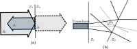
Scattering means diffuse reflection of small portions of a wave in various directions. It should be noted that the law of reflection (see above) holds for each of those portions. Small inhomogeneities in the material cause scattering of sound waves, cf. . The same holds for boundaries with rough surfaces as shown in , where the width of the reflection cone increases with decreasing wavelength λ and increasing roughness of the surface. Scattering at rough surfaces is highly relevant in medical ultrasound, because in the case of perfectly smooth boundaries, waves are only reflected towards the sender if the direction of the wave is perpendicular to the surface (no diffusion), whereas for rough boundaries, the reflections in various directions enable imaging of tilted boundaries.
Scattering of sound waves at (a) a rough boundary (diffuse reflection) between two different media with impedance Z1and Z2; and scattering at (b) inhomogeneities (depicted as blue dots) in a medium.
11.2.2.3. Diffraction
When sound waves pass barriers, obstacles, or openings on their path, they get diffracted. Diffracion involves a change in direction of the sound wave. Increasing wavelength λ yields an increased amount of diffraction (sharpness of bending), and vice versa. If λ is smaller than the size of the barrier, obstacle, or opening, the occurring diffraction becomes negligible.
11.2.2.4. Refraction
Snell’s law of refraction known from optics states
where α
1and α
2denote the angle of refraction in two different media, also applies to sound waves. However, since sound velocities (
υ1,
υ2) in human soft tissue differ only marginally (see ), the little effects of refraction in medical ultrasound are negligible and therefore not considered further in this chapter.
11.2.3. Attenuation
Attenuation is the reduction in sound wave intensity J that occurs when a wave penetrates a medium. It follows the well-known exponential law of attenuation:
where
J0denotes the initial intensity. The attenuation coefficient
μ denotes the attenuation that occurs with each cm the sound wave travels inside a medium. It depends on material (tissue type) and ultrasound frequency
ξ and is measured in decibel (dB). The attenuation coefficient mainly consists of two additive components
μ =
μa+
μs, namely absorption
μaand scattering
μs(see above). Absorption
μacauses tissue to heat.
From Eq. (11.6), it can be easily seen that the acoustic intensity J decreases with increasing penetration depth x. For a high maximum penetration depth, low frequencies are necessary as shown in . However, the resolution of the acquired images decreases with decreasing frequency (cf. Sec. 11.3.3). Thus, the deeper the tissue penetration, the lower the spatial resolution.
Maximum penetration depth dmax for various frequencies f.
11.3. Image Acquisition for Diagnostics
11.3.1. Transducers
An ultrasound transducer functions as both: a generator and a detector of ultrasonic waves. It converts mechanical energy into electrical energy and vice versa. When the transducer is pressed against the skin, it directs high-frequency sound waves into the body. Since sound waves produced by the transducer can barely penetrate air (cf. ), gel is applied to the skin to help to minimize the amount of air between the transducer and the skin. As the waves penetrate the body, sound echoes are generated from the body’s fluids and tissues due to (diffuse) reflection and scattering. The strength and character of these sound echoes are recorded by the transducer and, depending on the type of transducer, can be transformed into 1-D, 2-D or 3-D images, which can be rendered and viewed to the user.
11.3.2. Piezoelectric Effect
In order to generate and detect ultrasonic waves, transducers rely on the so-called piezoelectric effect. It describes the conversion of electrical energy into mechanical energy and vice versa in piezoelectric materials. On the one hand, mechanical pressure (pressure translates to “piezo” (gr.)) is converted to electric polarization, which generates electric voltage. The electric voltage can be measured using two electrodes, as shown in . On the other hand, electrical fields cause contraction or stretching of the piezoelectric material. This contraction and stretching can be used to generate ultrasound waves by applying a high frequency alternating voltage.
Typical piezoelectric materials used in medical ultrasound transducers are barium titanate (BaTiO3) and lead zirconium titanate (PZT).
11.3.3. Spatial Resolution
In ultrasound imaging, a distinction is made between two different kinds of spatial resolutions, in particular axial and lateral resolution (cf. ).
Axial and lateral resolution of ultrasound devices. Minimal distance between two structures (blue dots) in axial/lateral direction that allows for distinguishing between them in the ultrasound image.
11.3.3.1. Axial Resolution
Axial resolution concerns structures lying behind each other w. r. t. the direction of the ultrasound waves. The better the axial resolution, the smaller the distance between two structures can be such that they can be distinguished by the transducer. Axial resolution is highly dependent on the ultrasound wave frequency f. The illustration in explains that dependency based on a simple example, where an ultrasonic pulse generated by the transducer consists in a single wave only (shortest possible pulse). The distance d between the structures needs to be d ≥ λ/2 in order to be able distinguish between them.
Axial resolution: illustration of dependence on wave frequency f. Left and right shows two different timesteps, where d denotes the distance between two structures (blue dots). The high frequency (top) wave allows for distinguishing between the two structures, (more...)
11.3.3.2. Lateral Resolution
Lateral resolution concerns the distinguishability of structures located next to each other in the same lateral distance to the transducer (same penetration depth). Lateral resolution is always inferior to axial resolution.
11.3.3.3. Frequency Trade-off
As described above, axial and lateral transducer resolution depend on the ultrasound frequency ξ, and thus on the wavelength λ. As a rule of thumb:
where
∆z and
∆x denote the minimum distance between to structures in axial/lateral direction such that the ultrasound echo is distinguishable. Hence, high
ξ yields high resolution, whereas low
ξ yields low resolution. However, the frequency is also directly related to attenuation (cf.
Sec. 11.2.3), where high
ξ yields high attenuation and vice versa. Thus, with high frequency
f, the penetration depth is low but the images will have high resolution. At low
ξ, deeper penetration is possible, but the resolution will be lower. Depending on the application, a trade-off between the desired properties (deep penetration versus high resolution) needs to be found, and the transducer frequency be adjusted accordingly.
11.3.4. Imaging Modes
Ultrasound offers a large variety of different imaging modes. The most common ones include A-mode, B-mode, and M-mode. A- and M-mode generate one-dimensional (1-D) images (signals), whereas B-mode can be used to acquire 2-D or even 3-D images (cf. Geek Box 1.1). Doppler (cf. Geek Box 1.2) can be acquired in 1-D and 2-D, and with the most recent generations of transducers also in 3-D.
11.3.4.1. A-Mode (Amplitude Mode)
A-mode is the simplest scanning mode. The height of the amplitude of the reflected ultrasound is displayed over the sonic runtime in the sonic ray direction. Extractable measurements are: frequency, modulated frequency, height of the impulse/amplitude, runtime, wave phase, phase shift, and attenuation. However, the major disadvantage is that only very localized information (one single line through the body) is acquired.
The backward scattered ultrasound intensity along a single ray is called A-mode. From a continuously running high-frequency generator, a wave packet is cut out with a “gate” and is passed to the transducer. The returning echo is given through a duplexer to a time-dependent amplifier (Time Gain Compensation). Later arriving echoes, which are weaker because of the absorption, are more amplified than the signals from the surface. Signals of high depth (15 cm) are raised up to 120 dB. The signal height of an interface reflected signal is independent of the penetration depth from which the echo comes. The signal-to-noise ratio becomes worse with increasing depth. The next sonic impulse will be emitted if all echoes of the preliminary sonic impulse are decayed. The repeat rate depends on the penetration depth and therewith on the used frequency.
11.3.4.2. B-Mode (Brightness Mode)
B-mode is the most common ultrasound mode. B-mode images are generated by systematically combining a multitude of A-mode (1-D) scans into a single 2-D image, where the intensity of a pixel is defined by the amplitude of the corresponding ultrasonic ray. In brief, in order to acquire 2-D images of the inner body, the ultrasound device has to sample not only on a 1-D ray (as in A-mode), but on a 2-D plane in 3-D space. Hence, various rays are sent in different directions. To achieve this, two techniques are commonly used: the mechanical and the electronic method.
Mechanical scanners The transducer librates in front of the patient, without any external movement of the gaging head. Thus, a slice of the human body is represented in the form of a circle segment. The intensity of the echo is transformed into gray scales and is inserted into an image matrix (B-Mode). An image consists of a fan of typically 100 lines.
Electronic scanners (linear/curved arrays) Here, many (60 to 100) and very small (0.5 mm to 1 mm) transducers are used, which are arranged in a row (“array”). A group of transducers is activated simultaneously. For scanning, the whole group of elements is shifted. With a curved arrangement of transducers, an image detail can be represented as a circle segment.
Electronic scanners (phased arrays) Every transducer element of an array can be accessed for both sending and receiving with an individual adjustable delay.
Geek Box 11.13-D Ultrasound
In 3-D ultrasound imaging, several 2-D images (B-mode) at different angles (w. r. t. axial direction) are combined into one 3-D volume. Real-time processing and visualization (rendering) of 3-D ultrasound images (volumes) requires high computational power, where graphics processing units can be used. Arbitrary section planes and “virtual travels through the body” are possible. The first 3-D ultrasound system was reported by Kazunori Baba in 1984. Slowly but steadily, 3-D ultrasound is becoming the standard of care in various medical fields (e. g., echocardiography), where 2-D imaging was traditionally used. One common application is to show their children to parents even before birth. An example is found right below this text.
11.3.4.3. M-Mode (Motion Mode)
In motion mode, ultrasonic pulses are emitted from the transducer in quick succession without movement of the transducer. Either an A-mode or a B-mode image is acquired each time. This allows for time-dependent measurement of organ movement relative to the probe. Thus, the velocity of specific organ structures can be obtained. This can be useful, for instance, when the movement of the cardiac wall (myocardium) is to be analyzed (echocardiography).
11.4. Safety Aspects
Ultrasound imaging o ers many benefits over other imaging techniques, including:
Non-invasiveness (no injections or needles in most cases) and mostly painless.
Geek Box 11.23-D Ultrasound
Medical Doppler ultrasonography enables the measuring and visualization of blood flow (blood velocities). Two modes are frequently used: continuous wave (CW) Doppler and pulsed wave (PW) Doppler. In CW Doppler, half of the transducer array emits, and the other half detects pulses. It has the advantage that it allows for continuous imaging due to simultaneous emission and detection. However, no distance information can be measured. In PW Doppler, which is pulse-based, distance information can be obtained using time-gating. However, no continuous imaging is possible.
Doppler ultrasonography exploits the well-known Doppler effect. The Doppler effect is named after its discoverer Christian Johann Doppler (1803–1853) and can be observed in various situations, for instance, the noise of the siren of the ambulance when an ambulance passes at high speed. Other examples can be found in astronomy: the astronomical red-shift. Most relevant to medical Doppler ultrasonography, however, is blood flow, i. e., Doppler ultrasonography can visualize blood velocities. The Doppler effect describes the change in wave frequency by a relative movement between source and observer. A characteristic frequency shift appears, which is proportional to the relative velocity. Doppler ultrasonography aims at measuring the shift in frequency to estimate velocities (e. g., of blood in vessels).
In Doppler blood flow imaging, the source are the moving blood cells, at which the waves scatter. The observer is the ultrasound transducer. The smaller the angle between the direction of blood flow in the vessel and the ultrasound wave direction, the better the Doppler effect can be exploited.
Image acquisition is fast and relatively easy to learn.
No ionizing radiation (contrary to X-ray/CT).
Large number of potential applications: ultrasound can visualize structure, movement, and function of the body’s organs and blood vessels.
However, ultrasound waves can harm the body:
through heating, proportional to absorbed acoustic intensity, or
through cavitation, which means gas bubbles that emerge in the low pressure phases of sound waves and collapse at high pressure phases.
Since acoustic intensities for medical diagnostics are rather low, the potentially harmful effects described above have proven to be harmless. Medical ultrasound is considered one of the least harmful imaging techniques available today and is even used during pregnancy.
Therapeutical use of ultrasound can be found in gallstone and kidney stone therapies, where high intensity localized ultrasound is used to break up the stones. The heating effect of ultrasound waves can further be used to destroy diseased or cancerous tissue.
Further Reading
- [1]
Olaf
Dössel. Bildgebende Verfahren in der Medizin: Von der Technik zur medizinischen Anwendung. Springer, 1999. isbn: 978-3-540-66014-9.
- [2]
G
Goretzki. Medizinische Strahlenkunde: physikalisch-technische Grundlagen. Elsevier, Urban und Fischer, 2004. isbn: 978-3-437-47200-8.
- [3]
Paul
Suetens. Fundamentals of Medical Imaging. Cambridge University Press, 2009. isbn: 978-0-521-51915-1.


 1 and
1 and