This book is distributed under the terms of the Creative Commons Attribution-NonCommercial-NoDerivatives 4.0 International (CC BY-NC-ND 4.0) ( http://creativecommons.org/licenses/by-nc-nd/4.0/ ), which permits others to distribute the work, provided that the article is not altered or used commercially. You are not required to obtain permission to distribute this article, provided that you credit the author and journal.
NCBI Bookshelf. A service of the National Library of Medicine, National Institutes of Health.
StatPearls [Internet]. Treasure Island (FL): StatPearls Publishing; 2024 Jan-.
This publication is provided for historical reference only and the information may be out of date.

StatPearls [Internet].
Show detailsIntroduction
Meningomyelocele or myelomeningocele, commonly known as open spina bifida is a devastating congenital malformation of the central nervous system and is associated with significant morbidity and mortality. Neural tube defects are of two types:
- Open neural tube defect means that the defect is either not covered at all or just covered by a membrane. It comprises about 80% of all neural tube defects.
- Closed neural tube defect: No apparent defect externally, usually result from a defect either in the fat, bone, or membranes covering the spinal cord.
Myelomeningocele is the most common open neural tube defect. It is characterized by failure of the neural tube to close in the lumbosacral region during embryonic development (fourth-week post-fertilization), leading to the herniation of the meninges and spinal cord through a vertebral defect.[1] The neural tube fusion starts at the level of the hindbrain (medulla and pons) and progresses rostrally and caudally. Incomplete fusion caudally leads to the formation of meningomyelocele around day 26 of gestation.[2]
Etiology
Most open neural tube defects occur sporadically. however, several risk factors have been linked to neural tube defect development, including [1][3][4][5]
- Chromosomal and genetic conditions:
- Parent or sibling with neural tube defect
- Trisomies 18 and 13
- Meckel-Gruber syndrome ( renal cysts, neural tube defects, and polydactyly).
- Roberts, Jarcho-Levin ( high incidence in Puerto Rican, present with multiple defects in the spine and fan-like ribs anomalies)
- HARD syndrome (Hydrocephalus, agyria and retinal dysplasia),
- VACTERAL and VATER associations
- X-linked neural tube defects, among others.
- Amniotic bands that disrupt normal neural tube formation.
- Maternal environmental factors and exposure: alcohol use, caffeine intake, smoking, air pollution, disinfectant byproducts in drinking water, exposure to organic solvents, pesticides, nitrate-related compounds, polycyclic aromatic hydrocarbons, maternal fever or hyperthermia ( especially in the first trimester) from febrile illness or external sources like sauna, hot tub.
- Maternal medical conditions: elevated glycemic index, and gestational diabetes mellitus infections, obesity, and stress
- Maternal nutritional deficiencies, folate, methionine, zinc, vitamin C, vitamin B12, and choline.
- Maternal medications: various folic acid antagonists like valproic acid, carbamazepine, and methotrexate.
Epidemiology
Myelomeningocele is the most common disorder of neurulation that results in viable infants. Its incidence in the United States is about 0.2 to 0.4 per 1000 live births. The rates are higher in the Latino population. Females have a 3-7x higher risk compared to males. There are marked racial differences; for example, the incidence is several times higher in some regions in China, parts of Africa, the middle east, Thailand, and India. Folate supplementation guidelines in these countries may play a role in these racial differences.[1][6]Neural tube defects incidence increases with lower socioeconomic status and older maternal age. The recurrence rate in subsequent pregnancies is about 2% to 3%.[7]
Pathophysiology
Failure of the closure of the neural tube leads to exposure of the neural tube to amniotic fluid. Although -early on- the neuroepithelium, neuronal differentiation, and function develop normally, these neurons die over time because of toxicity from exposure to amniotic fluid. The failed neural tube closure and neurodegeneration in utero is described as a "Two-hit" process.[8]
History and Physical
The diagnosis in a newborn is usually apparent because of the grossly visible lesion in the back. Protruding membrane-covered sac-containing meninges, cerebrospinal fluid (CSF), and nerve tissue are seen through a vertebral column defect. The clinical features of myelomeningocele depend on the:
- Level of involvement
- The presence of hydrocephalus
- Associated brain abnormalities
Newborns may remain asymptomatic up to 6 weeks of age. In the presence of hydrocephalus; Clinical signs of increased intracranial pressure (increase in head circumference, irritability, lethargy, and limited upward gaze) may be present. [9] Impairment in sensory, motor and sphincter function depends on the lesion level. Bowel and bladder function is impaired in almost 97% of the population with spina bifida. Due to loss of function in antigravity muscles like iliopsoas and quadriceps, ambulation problems are common and usually progress with age. Most individuals with myelomeningocele have complete paralysis and loss of sensation in their lower extremities and trunk, below the lesion level.[10]
Spina bifida can also be associated with Chiari-II malformation, characterized by downward displacement of the cerebellar tonsils and medulla. This malformation leads to obstruction of the CSF flow through the posterior fossa leading to hydrocephalus.[11] These associated neurological abnormalities are responsible for most of the major neurological morbidity and mortality. If brainstem dysfunction is present, these patients can have swallowing difficulties, vocal cord paresis leading to apnea and stridor.[12][13]
Evaluation
Most neonates are diagnosed prenatally by maternal screening by ultrasound and/or serum levels of alpha-fetoprotein. The screening test of choice is a high-quality second-trimester ultrasound as it is more accurate in detecting neural tube defects than the serum level of alpha-fetoprotein. A positive screening will require further evaluation, including a complete anatomy scan, genetic testing like fetal karyotype, and fetal magnetic resonance imaging if ultrasound imaging is indeterminate. Once the diagnosis is confirmed, extensive prenatal counseling must be undertaken to discuss the natural history of spina bifida, offer additional prenatal testing, and provide management choices, including termination of pregnancy, postnatal surgery, or fetal surgery if available. Serial ultrasounds are also done to monitor head growth, ventricular size, and help with delivery planning.[14]
Treatment / Management
Fetal surgery has increasingly become common in the treatment of myelomeningocele. Animal studies have shown that the myelomeningocele lesion's intrauterine closure will prevent the neural tissue's progressive intrauterine damage due to exposure to amniotic fluid. [15] A randomized control trial comparing prenatal surgery's efficacy vs. postnatal repair was done and was stopped early due to the evident better outcomes with prenatal surgery. The trial showed that prenatal surgery was associated with a decreased need for shunt placement (40% in the prenatal group and 82% in the postnatal surgery group). It also showed that children who underwent prenatal surgery had improved mental development and motor function at 30 months. Complications associated with prenatal surgery were increased risk of prenatal delivery and uterine dehiscence at the time of delivery.[16]
When the infant is born, careful assessment of the lesion should be done while laying the infant in the lateral or prone position. Non-latex gloves should be used to prevent latex sensitization[17]. The lesion should be covered by a moist dressing or plastic wrap to prevent heat loss. In postnatal surgery, closure of myelomeningocele should be done as early as possible to reduce infection risk (ideally within the first 48 hours after birth).[18] In patients with associated Chiari II malformation, presenting with brainstem dysfunction, decompressive upper cervical laminectomy may be needed to reduce brainstem and cerebellar tonsillar decompression.[19] Most of these patients benefit from a multidisciplinary team approach for coordinating their management. In patients with urinary dysfunction, daily catheterization decreases urine stasis and therefore reduce urinary tract infections. An orthopedic follow-up should be arranged to evaluate and manage associated complications, like contractures and scoliosis.[20] Delivery should preferably be done at a center with a high-care-level neonatal intensive care unit where pediatric neurosurgery services are available. Delivery should be performed at term to prevent complications related to prematurity. When in utero surgery is performed, a cesarean section is recommended at 37 weeks of gestation or earlier ( if maternal indication) to decrease the uterine rupture's associated risk.[21]
Differential Diagnosis
The differential diagnosis of neural tube defects include:[22]
- Caudal regression syndrome (sacral agenesis)
- Sacrococcygeal teratoma
- Multiple vertebral segmentation disorder
- VACTERL (vertebral anomalies, anal atresia, cardiac abnormalities, tracheoesophageal atresia/ esophageal atresia, renal anomalies, and limb defect).
- Spine segmental dysgenesis
Prognosis
Patients with spina bifida have a mortality rate of about 1% per year from 5 to 30 years. The higher the lesion, the greater the morbidity and mortality. The consequences of neural tube defects massively impact the quality of life among survivors. Psychosocial issues like increased incidence of depression, anxiety, and risk-taking behaviors are common in this group. Studies have shown that the areas most likely to be affected are employment, romantic relationships, and financial independence.[14]
Complications
Complications associated with myelomeningocele include: [23]
- Learning disabilities and cognitive impairments
- Seizures
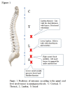
- Paralysis and loss of sensation below the site of the lesion, figure 1
- Decreased mobility due to associated muscle weakness
- Neurogenic bladder and frequent urinary tract infections
- Bowel dysfunction
- Pressure ulcers due to sensory loss
- Orthopedic problems associated with paralysis, for example, scoliosis, contractures, hip dislocation, among others
Deterrence and Patient Education
Spina bifida is a congenital disability in which the backbone or spine does not typically form in a baby while developing in the mother’s womb. It is also known as myelomeningocele or open neural tube defect. It will appear an opening in their skin on their back through which part of the spine and nerve tissue can protrude out.
Neural tube defects can cause various long-term problems in your child, and they depend on how severe the defect is. Many babies with spina bifida can have the following:
- A weakness of their leg and may not be able to walk
- Loss of feeling below their waist
- Problems controlling their urine and stools
- Issues of learning or memory
- Some children may also develop too much fluid in their brain and may need surgery to relieve the pressure on their brain
After receiving a prenatal diagnosis of spina bifida, some parents chose to terminate the pregnancy, while others prefer to continue their pregnancy. If you decide to continue the pregnancy, some centers can offer fetal surgery to correct the defect, or scheduled surgery to correct the congenital disability immediately after birth. After the initial treatment, your baby will need medical management throughout their life.
They may need:
- Wheelchair or assist devices to help them walk
- Other surgeries to fix their bones
- They may have learning problems and require additional testing and support at school
- If their bladder does not work, they may need a tube to empty their bladder from time to time.
A child’s diagnosis will affect their quality of life. The best way to support the child will be to take them to a multidisciplinary team of medical professionals who specialize in taking care of patients with this diagnosis.
Enhancing Healthcare Team Outcomes
Problems with urinary continence are common in patients with myelomeningocele. They are at increased risk of urinary tract infections and, subsequently, renal dysfunction. Intermittent catheterization reduces the risk of renal disease. These patients also have an increased risk of bowel incontinence and issues with bowel motility. A comprehensive bowel management routine, including routine use of laxatives, enemas, and suppositories, should be adopted to avoid complications.
Routine surveillance in an orthopedic clinic also helps promote ambulation and manage deformities like scoliosis and contractures. Worsening scoliosis could be a sign of complications like tethered cord, shunt malfunction, hydromyelia, among others, and must trigger immediate evaluation.
Almost 75% of patients with spina bifida survive to early adulthood. an interprofessional, patient-centered, team-based approach to provide medical, educational, social, and developmental services can enhance these patients' quality of life by improving their overall health and functioning.[24]
Review Questions

Figure
Figure 1. Predictors of outcomes according to the spinal cord level involvement in meningomyelocele. C: Cervical, T: Thoracic, L: Lumbar, S: Sacral. Contributed by Mahdi Alsaleem MD
References
- 1.
- Copp AJ, Adzick NS, Chitty LS, Fletcher JM, Holmbeck GN, Shaw GM. Spina bifida. Nat Rev Dis Primers. 2015 Apr 30;1:15007. [PMC free article: PMC4898641] [PubMed: 27189655]
- 2.
- Brody BA, Kinney HC, Kloman AS, Gilles FH. Sequence of central nervous system myelination in human infancy. I. An autopsy study of myelination. J Neuropathol Exp Neurol. 1987 May;46(3):283-301. [PubMed: 3559630]
- 3.
- Sepulveda W, Corral E, Ayala C, Be C, Gutierrez J, Vasquez P. Chromosomal abnormalities in fetuses with open neural tube defects: prenatal identification with ultrasound. Ultrasound Obstet Gynecol. 2004 Apr;23(4):352-6. [PubMed: 15065184]
- 4.
- Au KS, Ashley-Koch A, Northrup H. Epidemiologic and genetic aspects of spina bifida and other neural tube defects. Dev Disabil Res Rev. 2010;16(1):6-15. [PMC free article: PMC3053142] [PubMed: 20419766]
- 5.
- Shimoji K, Kimura T, Kondo A, Tange Y, Miyajima M, Arai H. Genetic studies of myelomeningocele. Childs Nerv Syst. 2013 Sep;29(9):1417-25. [PubMed: 24013315]
- 6.
- Zaganjor I, Sekkarie A, Tsang BL, Williams J, Razzaghi H, Mulinare J, Sniezek JE, Cannon MJ, Rosenthal J. Describing the Prevalence of Neural Tube Defects Worldwide: A Systematic Literature Review. PLoS One. 2016;11(4):e0151586. [PMC free article: PMC4827875] [PubMed: 27064786]
- 7.
- Papp C, Adám Z, Tóth-Pál E, Török O, Váradi V, Papp Z. Risk of recurrence of craniospinal anomalies. J Matern Fetal Med. 1997 Jan-Feb;6(1):53-7. [PubMed: 9029387]
- 8.
- Greene ND, Massa V, Copp AJ. Understanding the causes and prevention of neural tube defects: Insights from the splotch mouse model. Birth Defects Res A Clin Mol Teratol. 2009 Apr;85(4):322-30. [PubMed: 19180568]
- 9.
- Avagliano L, Massa V, George TM, Qureshy S, Bulfamante GP, Finnell RH. Overview on neural tube defects: From development to physical characteristics. Birth Defects Res. 2019 Nov 15;111(19):1455-1467. [PMC free article: PMC6511489] [PubMed: 30421543]
- 10.
- Williams EN, Broughton NS, Menelaus MB. Age-related walking in children with spina bifida. Dev Med Child Neurol. 1999 Jul;41(7):446-9. [PubMed: 10454227]
- 11.
- Naidich TP, McLone DG, Fulling KH. The Chiari II malformation: Part IV. The hindbrain deformity. Neuroradiology. 1983;25(4):179-97. [PubMed: 6605491]
- 12.
- Nagler J, Levy JA, Bachur RG. Stridor in an infant with myelomeningocele. Pediatr Emerg Care. 2007 Jul;23(7):478-81. [PubMed: 17666932]
- 13.
- Holinger PC, Holinger LD, Reichert TJ, Holinger PH. Respiratory obstruction and apnea in infants with bilateral abductor vocal cord paralysis, meningomyelocele, hydrocephalus, and Arnold-Chiari malformation. J Pediatr. 1978 Mar;92(3):368-73. [PubMed: 632975]
- 14.
- Shaer CM, Chescheir N, Schulkin J. Myelomeningocele: a review of the epidemiology, genetics, risk factors for conception, prenatal diagnosis, and prognosis for affected individuals. Obstet Gynecol Surv. 2007 Jul;62(7):471-9. [PubMed: 17572919]
- 15.
- Danzer E, Flake AW. In utero Repair of Myelomeningocele: Rationale, Initial Clinical Experience and a Randomized Controlled Prospective Clinical Trial. Neuroembryology Aging. 2008 Mar;4(4):165-174. [PMC free article: PMC2810532] [PubMed: 22479081]
- 16.
- Adzick NS, Thom EA, Spong CY, Brock JW, Burrows PK, Johnson MP, Howell LJ, Farrell JA, Dabrowiak ME, Sutton LN, Gupta N, Tulipan NB, D'Alton ME, Farmer DL., MOMS Investigators. A randomized trial of prenatal versus postnatal repair of myelomeningocele. N Engl J Med. 2011 Mar 17;364(11):993-1004. [PMC free article: PMC3770179] [PubMed: 21306277]
- 17.
- Rendeli C, Nucera E, Ausili E, Tabacco F, Roncallo C, Pollastrini E, Scorzoni M, Schiavino D, Caldarelli M, Pietrini D, Patriarca G. Latex sensitisation and allergy in children with myelomeningocele. Childs Nerv Syst. 2006 Jan;22(1):28-32. [PubMed: 15703967]
- 18.
- McLone DG. Care of the neonate with a myelomeningocele. Neurosurg Clin N Am. 1998 Jan;9(1):111-20. [PubMed: 9405769]
- 19.
- Messing-Jünger M, Röhrig A. Primary and secondary management of the Chiari II malformation in children with myelomeningocele. Childs Nerv Syst. 2013 Sep;29(9):1553-62. [PubMed: 24013325]
- 20.
- Hopson B, Rocque BG, Joseph DB, Powell D, McLain ABJ, Davis RD, Wilson TS, Conklin MJ, Blount JP. The development of a lifetime care model in comprehensive spina bifida care. J Pediatr Rehabil Med. 2018;11(4):323-334. [PMC free article: PMC6924509] [PubMed: 30507593]
- 21.
- Wilson RD, Lemerand K, Johnson MP, Flake AW, Bebbington M, Hedrick HL, Adzick NS. Reproductive outcomes in subsequent pregnancies after a pregnancy complicated by open maternal-fetal surgery (1996-2007). Am J Obstet Gynecol. 2010 Sep;203(3):209.e1-6. [PubMed: 20537307]
- 22.
- Chen CP. Prenatal diagnosis, fetal surgery, recurrence risk and differential diagnosis of neural tube defects. Taiwan J Obstet Gynecol. 2008 Sep;47(3):283-90. [PubMed: 18935990]
- 23.
- Mummareddy N, Dewan MC, Mercier MR, Naftel RP, Wellons JC, Bonfield CM. Scoliosis in myelomeningocele: epidemiology, management, and functional outcome. J Neurosurg Pediatr. 2017 Jul;20(1):99-108. [PubMed: 28452655]
- 24.
- Beuriat PA, Poirot I, Hameury F, Szathmari A, Rousselle C, Sabatier I, di Rocco F, Mottolese C. Postnatal Management of Myelomeningocele: Outcome with a Multidisciplinary Team Experience. World Neurosurg. 2018 Feb;110:e24-e31. [PubMed: 28987842]
Disclosure: Mitali Sahni declares no relevant financial relationships with ineligible companies.
Disclosure: Mahdi Alsaleem declares no relevant financial relationships with ineligible companies.
Disclosure: Abhinav Ohri declares no relevant financial relationships with ineligible companies.
- Neural Tube Disorders.[StatPearls. 2024]Neural Tube Disorders.Bhandari J, Thada PK. StatPearls. 2024 Jan
- Use of 'stacked' dermal template: Biodegradable temporising matrix to close a large myelomeningocele defect in a newborn.[Scars Burn Heal. 2024]Use of 'stacked' dermal template: Biodegradable temporising matrix to close a large myelomeningocele defect in a newborn.Hasham S, O'Boyle C, Alexander S. Scars Burn Heal. 2024 Jan-Dec; 10:20595131241270220. Epub 2024 Sep 2.
- Review Spina bifida and other neural tube defects.[Curr Probl Pediatr. 2000]Review Spina bifida and other neural tube defects.Northrup H, Volcik KA. Curr Probl Pediatr. 2000 Nov-Dec; 30(10):313-32.
- Review Neural tube defects--disorders of neurulation and related embryonic processes.[Wiley Interdiscip Rev Dev Biol...]Review Neural tube defects--disorders of neurulation and related embryonic processes.Copp AJ, Greene ND. Wiley Interdiscip Rev Dev Biol. 2013 Mar-Apr; 2(2):213-27. Epub 2012 May 29.
- Review [Spina bifida].[Radiologe. 2018]Review [Spina bifida].Mühl-Benninghaus R. Radiologe. 2018 Jul; 58(7):659-663.
- Meningomyelocele - StatPearlsMeningomyelocele - StatPearls
Your browsing activity is empty.
Activity recording is turned off.
See more...