Continuing Education Activity
Bisphosphonates are synthetic pyrophosphate analogs used to treat hypercalcemia secondary to bone resorption conditions such as malignancy, osteoporosis, multiple myeloma, Paget disease, osteosclerosis, and fibrous dysplasia. Infrequent side effects associated with bisphosphonate use include pyrexia, renal function impairment, hypocalcemia, and, more recently recognized, avascular osteonecrosis of the jaw. According to the American Society of Bone and Mineral Research, bisphosphonate-related osteonecrosis of the jaw (BRONJ) is described as exposed bone in the maxillofacial region that does not heal within 8 weeks of being identified by a healthcare provider in a patient that is currently or has been on bisphosphonates who does not have a history of radiation therapy in the craniofacial region. This activity describes the evaluation and management of patients with bisphosphonate-related jaw osteonecrosis and highlights the role of the interprofessional team in improving care for affected patients.
Objectives:
Identify the pathological mechanism of action involved in bisphosphonate-related osteonecrosis of the jaw.
Describe the examination and evaluation procedures necessary to diagnose bisphosphonate-related jaw necrosis.
Summarize the various treatment strategies available for treating bisphosphonate-related osteonecrosis of the jaw.
Describe risk factors associated with bisphosphonate-related osteonecrosis of the jaw.
Access free multiple choice questions on this topic.
Introduction
The widespread use of bisphosphonate (BP) to treat various medical conditions led to increased recognition of their possible association with osteonecrosis (ON) of the jaw.[1] Bisphosphonates are highly efficient antiresorptive drugs used to treat diseases with increased osteoclast activity such as cancer-related conditions, osteoporosis, multiple myeloma, Paget disease, osteosclerosis, and fibrous dysplasia.[2] Although rare, avascular osteonecrosis of the jaw has been recently recognized as a complication of bisphosphonate use. Bisphosphonate-related osteonecrosis of the jaw (BRONJ) is defined as a current or previous treatment with BPs that leads to an exposed bone or bone that can be probe through a fistula in the maxillofacial region that does not heal within eight weeks. The patient has no history of radiation therapy in the craniofacial region.[3][4] Eight weeks is considered because most surgical and infectious sites heal in this time frame, even if complications such as postsurgical infection, chemotherapy, or systemic diseases are present.
Etiology
Bisphosphonates inhibit bone resorption by causing osteoclast cell apoptosis, impairing the osteoclast’s resorptive capacity, and preventing osteoclast formation.[5] They have a high affinity for bone minerals and accumulate mainly in the sites of osteoclast activity.[5] Without resorption and new bone formation, old bone survives beyond its lifespan, and its capillary network is not maintained, leading to avascular necrosis of the jaw. Also, high potency biphosphonates can lead to necrosis by the toxicity of soft tissue and bone cells, further complicated by infection.[6] Due to altered wound healing, delayed epithelial closure of a mucosal opening in the mouth leads to chronic infection and the necrosis of bone.[7] Osteonecrosis develops in the jaw because this bone has a higher remodeling rate than other bones, making it more prone to the effect of bisphosphonates.
Epidemiology
Intravenous (IV) Versus Oral Biphosphonates
Osteonecrosis of the jaw is mainly reported with the use of more potent nitrogen-containing BPs like pamidronate and zoledronic acid. However, the incidence is higher with the latter - zoledronic acid causes a higher antiresorptive activity, leading to decreased bone turnover.[8]
Oral biphosphonates rarely cause osteonecrosis of the jaw. They are less aggressive than intravenous BP, and the osteonecrosis caused by oral BP responds better to treatment. Oral BPs are less liposoluble, limiting their intestinal absorption, resulting in a lower accumulation in the bone.[9]
Dose and Duration
The incidence of osteonecrotic events increases with a higher dose of potent BPs administered for a longer duration.[10][11][12][13][14] The risk ranges from greater than 1% at 12 months to 11% after four years of treatment - taking zoledronic acid alone increases the risk of osteonecrosis to 21% after the third year. Due to the slow accumulation of oral BPs, no clinically exposed bone appears until after 3-year treatment, and incidence and severity increase with each additional year of use.
Osteoporosis and Oncology Patients
The frequency of biphosphonate-induced osteonecrosis in osteoporosis is very low, ranging from 0.15% to less than 0.001% person-years of exposure, and is only slightly higher than in non-osteoporotic patients. In oncology patients with bone metastasis, the risk is much higher as they are exposed to more intensive osteoclast inhibition and high-dose intravenous BPs. Also, the incidence varies according to the underlying condition. Wang et al. conducted a 5-year retrospective study of 292 patients treated with IV BPs that develop osteonecrosis. They found that 3% to 8% of patients had multiple myeloma, 2% to 5% breast cancer, and 2.9% prostate cancer.[15] Abu-Id et al.'s retrospective study found that osteonecrosis developed in 2 to 11% of multiple myeloma patients, 1% to 7% of breast cancer patients, and 6% to 15% of prostate cancer patients.[16]
Location
Osteonecrosis occurs most frequently in the mandible than the maxilla. It almost always begins in the alveolar bone due to its greater bone turnover rate, which relies on osteoclast-related remodeling due to occlusion and denture wearing pressure and tension forces.[17] The most common affected sites are nonhealing dentoalveolar sites, traumatized palatal and mandibular tori, and exposed portions of the mylohyoid ridge.
Incidence of BP-induced ON of the jaw increases with:
More potent nitrogen-containing IV BPs
High dosage and more prolonged duration
Oncology patients with bone metastasis
Areas with a high bone turnover rate like the alveolar bone of the mandible
Pathophysiology
Risk Factors
Several factors increase the risk of developing bisphosphonates-related osteonecrosis of the jaw:
1) Invasive oral procedures such as tooth extraction, periodontal surgery, and oral implant placement, and the use of dentures increase the rate of bone turnover and the risk of osteonecrosis.[18]
2) Comorbidities like cancer, chemotherapy, low hemoglobin levels, diabetes mellitus, renal dialysis, hypertension, hyperlipidemia, and hypercholesterolemia.[19]
3) Concomitant medications: corticosteroids and H2 blocking drugs, which increase BP absorption. Antiangiogenic agents, particularly sunitinib and bevacizumab. Erythropoietin and cyclophosphamide therapy.[20]
4) Infection: it is still unclear if osteonecrosis precedes or follows the infection. However, polymorphonuclear aggregates and bacterial microfilm in the surrounding tissue have been associated with bone resorption and necrosis.[21][22]
BPs inhibit the proliferation and viability of oral keratinocytes, damaging the oral mucosa integrity and increasing the risk of infection.[23][24][25] Also, BPs impair the immune response to infection by activating gamma, delta T-cells stimulating the production of pro-inflammatory cytokines and later depletion of T cells.
5) Genetic predisposition: polymorphism in farnesyl pyrophosphate synthase or CYP2C8 coding for a cytochrome P450 enzyme predisposes some individuals to bisphosphonate-associated osteonecrosis of the jaw in multiple myeloma.[26][27][28]
6) Other risk factors include increasing age, alcohol, and tobacco use.
Histopathology
Resected necrotic bone from BRONJ patients does not demonstrate any unique features of the disease.[29][30] The most frequently found microorganisms in the exposed bone sites are Actinomyces, Veillonella, Eikenella, and Moraxella species - penicillin-sensitive organisms. The presence of sulfur granules in deeper tissue and drainage areas supports the diagnosis of actinomycosis and requires appropriate treatment.[31]
History and Physical
Most cases of BRONJ occur after a dental intervention that affects bone, but osteonecrosis can also present spontaneously.[4] The necrotic bone may remain asymptomatic for a prolonged period or develop symptoms, mainly due to localized inflammation of soft tissue.[29]
Practitioners must consider BRONJ if patients with a history of anti-resorptive drug use present delayed healing after oral surgery, infection, and swelling in the surrounding soft tissue, numbness, paraesthesia, or bone exposure. But, clinicians should be aware that BRONJ lesions may also be an incidental discovery.
The altered nerve sensation described by some patients is due to compression of the neurovascular bundle causing paresthesia, or even anesthesia of the associated branch of the trigeminal nerve.[32][33] Hypoesthesia or anesthesia of the lower lip has been reported as an early symptom of BRONJ.
Complications of BRONJ include tissue ulceration, intra-and extraoral sinus tracts, and fistula.[34] Chronic maxillary sinusitis in patients with maxillary bone involvement and fracture in edentulous patients with oral implants have also been reported.[35]
Signs and Symptoms
None/asymptomatic
Pain
Soft tissues infection with inflammation, ulceration, and suppuration
Formation of intra-and extraoral sinus tracts and fistulas
Paresthesia or anesthesia of an associated nerve
Fracture
Chronic maxillary sinusitis
A radiographic appearance from no alterations to varying radiolucencies and radiopacities
Evaluation
Blood Test
It measures the C-terminal telopeptide (CTX) value, which depicts the level of octapeptide fragment released due to osteoclastic bone resorption from type I bone collagen.[36] Its levels are related to the number of osteonecrotic lesions, stage of disease, and bone turnover index.[37] A lower value represents a high-risk patient with suppressed bone turnover and reduced healing capacity. C-terminal telopeptide less than 100 pg/ml equals high risk, 100 to 150 pg/ml equals moderate risk, and greater than 150 pg/ml equals minimal or no risk.
Radiographic Appearance
Radiographically, BRONJ can range from no alterations to varying radiolucencies or radio-opacities. Osteolytic lesions may appear less or more radiodense, providing a similar radiographic appearance as metastatic bone. Altered bone morphology, periosteal bone formation, increased bone density, or sequestration may be radiographic findings of BRONJ. Early radiographic signs along alveolar bone may include widened periodontal ligament space and sclerosis of lamina dura.
Imaging Modalities and Diagnostic Tests
Due to the nonspecific radiographic features of the condition, imaging provides a good evaluation of the area involved and can assist in identifying the extent of bone and soft tissue disease but does not provide any definitive differentiation of osteonecrosis of the jaw from other conditions.[38]
Conventional radiographs
Intraoral and panoramic radiographs are easy to acquire, inexpensive, deliver low radiation, and provide a good view. They are helpful to assess early features: thickening of lamina dura, increased trabecular density, incomplete healing of extraction socket, widening of periodontal ligament space, sinus floor cortication, periosteal bone, and sequestrum formation. Poor quality images do not clearly demarcate between necrotic and healthy bone. Disease at early stages can be frequently missed. Despite limitations, they form the first line of routine radiological investigation.[39]
Cone beam CT scan
Cone beam CT scan provides tridimensional imaging of the involved cancellous and cortical bone and identifies osteosclerotic and osteolytic regions.[39] It can also evaluate sequestrum, periosteal bone reaction, and the integrity of the vital adjacent structures [40][41], potential fistula tract, cortical erosion, and incomplete extraction socket healing.[40][42][43][44][43]
The early stage of osteonecrosis may not be detected, but cortical and trabecular bone changes at the symptomatic site can aid in diagnosis. CBCT has similar findings of the osteonecrotic areas as the CT scan but imparts lower radiation and has higher spatial resolution with better image quality, particularly for the cancellous bone in a small field of view.[45][46] The major limitation is poor soft tissue details due to low contrast resolution.
MRI
MRI currently may be the method of choice to detect the early bone marrow and soft tissue changes surrounding the osteonecrotic area. Osseous change evaluation by MRI is similar to CT imaging. One consistent MRI findings are the decreased bone marrow signal intensity on T-1 weighted images resulting from progressive cell death and host response through repair, i.e., edema.[41][47][48][42] Irregular gadolinium enhancement around osteolytic lesions is observed. An MRI shows non-enhancement in regions of ischemia, especially in T-1 weighted sequences, low signal intensity in areas of fibrosis and sclerosis on T-1 and T-2 weighted images, and increased signal intensity along the unexposed diseased bone.[47][49] However, MRI may not demonstrate the full extent of bony changes and may give a false-positive diagnosis.
Nuclear Imaging with Bone Scintigraphy
Technetium-99 radioisotope scintigraphy has a high sensitivity for diagnosing early disease and ischemic osteonecrosis. Its sensitivity depends on the stage of osteonecrotic lesion and the change in vascularity.[39] It shows increased radionuclide uptake in surrounding areas with increased perfusion and blood pool, locating osteonecrotic regions more precisely.
The main drawbacks include significant radiation exposure, lengthy procedure, and low resolution, which sometimes make it challenging to differentiate between inflammatory and metastatic processes and heal osteolytic lesions and progressing osteoblastic lesions.
Combining CBCT with scintigraphy for diagnosing osteomyelitis [46] or using contrast agents with MRI, sequential imaging, and manipulating image planes can all be helpful measures to diagnose early or preclinical stages of BRONJ.
Treatment / Management
Treatment depends on age, gender, disease stage, lesion size, comorbidities, and medication. Still, since their influence on disease course and treatment response is unknown, clinical judgment guides the treatment approach. Other important factors are prognosis, life quality expectancy, and the patient's ability to cope with the disease.
No evidence-based guidelines for the treatment of BRONJ are currently available, but the treatment goal is to alleviate pain, control infection, and stabilize the progression of exposed bone.
Conservative Therapy
The mainstay of care is conservative therapy, and this may provide long-term symptomatic relief.[50][51]
Pain control and optimal oral hygiene, including diligent home care and regular dental visits.
Managing infection and active dental disease: use of 0.12% chlorhexidine digluconate oral antimicrobial rinses and systemic antibiotic therapy.
[52][53][54] Penicillin VK, 500 mg, four times daily is the antibiotic of choice. This formulation of penicillin is non-toxic and can be used long-term without superinfection and development of candidiasis. If long-term antibiotic usage is a concern, then it can be taken only during episodes of pain. If the patient is allergic to penicillin, then levofloxacin, 500 mg, once daily, is the best alternative. Other alternatives include doxycycline, 100 mg daily, or azithromycin, 250 mg daily. However, levofloxacin and azithromycin should be used for only 21 days or less due to their potential to raise liver enzymes. If this antibiotic protocol is ineffective, adding 500 mg of metronidazole three times daily for ten days is recommended.
Teriparatide improves osseous wound healing in the oral cavity.
[55] However, it is not recommended for patients at low risk of osteonecrosis of the jaw or fracture, but adding it to the treatment regimen of the osteoporotic patient with established osteonecrosis may benefit them.
[56][57][58] The same approach is not recommended for a cancer patient or those who have received skeletal radiation or have active bone metastasis. These patients have a risk of development or advancement of bone malignancies.
Reduce the contact of the oral prosthesis with the exposed bone.
Repeat the C-terminal telopeptide test (CTX) after six months of drug holiday. Some cases resolve with CTX value rising above 150 pg/ml. Many show clinical and radiographic signs of improvement as separation of necrotic bone from healthy bone occurs, followed by sequestration and debridement. Most of the oral BRONJ cases are resolved by CTX guided protocol. Regular follow-ups must be done to keep the CTX value above 150 pg/ml using incremental drug schedules and alternative drugs.
Surgical Therapy
Lack of symptomatic or radiographic improvement with various treatment modalities indicates permanent bone defect and need surgical intervention.
The osteotomy of the affected area needs to be performed with resection margins extending into the adjacent healthy bone. Soft tissues should be closed with a tension-free closure and no underlying sharp edges that could lead to a mucosal breakdown.
[59]Microvascular composite tissue grafting and reconstruction procedures should be considered in patients with pathological fractures, disease extending to the sinus or inferior border of the mandible, or if osteotomy leads to discontinuity defect.
Experimental Therapy
The various treatment approaches include the use of hyperbaric oxygen [60][61], bone marrow stem cell intralesional transplantation [62], local application of platelet-derived growth factor [63], low-level laser therapy [64][65], or using them in combination with conservative or surgical debridement, but their effect on the treatment outcome needs further substantiation.
The most recent recommendations advocate a non-surgical treatment approach due to impaired wound healing. Still, few studies included radical resection to viable bone and hermetic wound closure, with soft tissue being the only curative approach.[16] Combining various approaches like marginal resection and platelet-derived growth factors has been advocated by many studies.[66]
Treatment Approach Review
Conservative and supportive therapy for pain and infection control
Surgical therapy for permanent bone defects and sequestration
Experimental therapy consisting of hyperbaric oxygen, bone marrow stem cell intralesional transplantation, platelet-derived growth factor, low-level laser therapy.
Differential Diagnosis
The presence of exposed bone characterizes BRONJ. In the absence of exposed bone, BRONJ should be differentiated from the following:
Periodontal and periapical pathosis
Sinusitis
Gingivitis or mucositis
Temporomandibular disorders
Osteomyelitis
Metastatic bone tumors
Osteonecrosis induced by neuralgia
Osteoradionecrosis
Other conditions that may present with exposed bone but are not linked to bisphosphonate use include:
Cement osseous dysplasia with secondary sequestration
Trauma
Infectious osteomyelitis
Osteonecrosis following Herpes zoster infection
HIV-associated necrotizing ulcerative periodontitis
Staging
Very little evidence is reviewed for the staging of BRONJ. So staging recommendations should be considered as consensus statements. The current staging system is developed by Ruggiero and colleagues [67] and is adopted by The American Association of Oral and Maxillofacial Surgeons (AAOMS).[68][69] The stage system is important to identify stage characteristics and provide appropriate diagnosis and management.
Stage 1 patients have exposed bone and are asymptomatic with no localized soft tissue infection.
Stage 2 patients have exposed bone, pain, and regional soft tissue inflammation or infection.
Stage 3 patients have exposed bone with associated pain, localized soft tissue inflammation (or secondary infection), pathologic fracture, and extraoral or oral-antral fistula. Radiographically, the bone show osteolysis extending to the inferior mandibular border or maxillary sinus floor.
Recently, AAOMS added stage 0 to the staging system, referring to patients who take bisphosphonates and present with non-specific clinical findings and symptoms. Term stage 0 can lead to overdiagnosis that can have detrimental effects on the patient’s skeletal health if modification of anti-resorptive medication regimen is done as similar presenting symptoms may lead to a different diagnosis [70].
Deterrence and Patient Education
Prevention is the best approach and requires good communication among dentists, oral surgeons, physicians, nurse practitioners, and oncologists to develop measures to avoid the development of BRONJ.
Recommendations Before Initiating Bisphosphonate Therapy
Around 4 to 6 monthly doses are required to affect bone healing in jaws significantly; it is recommended to take preventive measures during this period.
Perform prophylactic dental examination and maintenance of good oral hygiene and regular dental visits.
Educate patients regarding home hygiene and self-maintenance.
Educate patients regarding the risk of BRONJ with bisphosphonate therapy.
Develop a dental treatment plan focused on correcting pathological conditions and stabilizing dentition to prevent the need for invasive procedures after the BP therapy is initiated.
Extract unrestorable, abscessed, and periodontally compromised teeth.
Restorative and prosthodontics procedures can be later accomplished, but dental implant placement and orthodontic treatment are not recommended. However, patients on BP for osteoporosis are currently not contraindicated for implant placement, but appropriate informed consent and documentation are recommended.
Recommendations for Patients Receiving BP Therapy
After 4 to 6 doses of BP, bone turnover is significantly suppressed, making bone healing unpredictable and risky for ON.
Educate patients regarding the risk of BRONJ with bisphosphonate therapy.
Educate patients regarding home hygiene and self-maintenance.
Oral surgical procedures like extractions, bone contouring, grafting, periodontal, and apical surgeries should be avoided.
[17]If possible, endodontic treatment is preferred over extractions and periapical surgery.
Noninvasive restorative procedures like crowns, bridges, removable partial and complete dentures are recommended to prevent future surgical procedures.
Orthodontic procedures are not recommended.
Elective dentoalveolar surgical procedures like asymptomatic teeth extraction, implant placement, tori reduction are not recommended.
Unrestorable teeth preferably should be treated with root canals and crown amputation; mobile teeth are best splinted, failed root canals should be retreated.
If tooth extraction is unavoidable, the patient should be educated regarding the risk of developing BRONJ, and informed consent should be signed before the procedure.
It is necessary to stratify the risk for patients on BP requiring extensive invasive oral surgery and patients with accompanying multiple risk factors like steroid treatment, immunodeficiency, or diabetes mellitus.
Drug holiday: dental practitioners should not indicate discontinuing bisphosphonate drugs since there is no evidence that the risk of BRONJ reduces when stopping the medication before dental procedures because they stay in the bone for years.
[71]
Pearls and Other Issues
Besides the antiresorptive drugs, antiangiogenic drugs can also cause osteonecrosis of the jaw. That is why the condition is nowadays referred to as medication-related osteonecrosis of the jaw (MRONJ).
Enhancing Healthcare Team Outcomes
Prompt recognition of osteonecrosis by the interprofessional team is important for improving outcomes. Prescribing providers include primary care providers, dentists, orthopedists, rheumatologists, and oncologists. Pharmacists provided education to patients and their families, monitor compliance, and provide feedback to the team. Specialty care nurses, including infusion nurses, orthopedic nurses, and otolaryngology, provide education, monitor patients, and inform the team about status changes or issues. Good communication among the team, patient education, appropriate preventive measures, and treatment aimed at pain and infection control can enhance the patient care outcomes for patients on bisphosphonates who are at the risk of developing or having established osteonecrosis of the jaw. [Level 5]
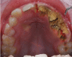
Jaw Osteonecrosis Image courtesy S Bhimji MD
References
- 1.
Shane E, Goldring S, Christakos S, Drezner M, Eisman J, Silverman S, Pendrys D. Osteonecrosis of the jaw: more research needed.
J Bone Miner Res. 2006 Oct;21(10):1503-5. [
PubMed: 16995804]
- 2.
McClung MR. Bisphosphonates.
Endocrinol Metab Clin North Am. 2003 Mar;32(1):253-71. [
PubMed: 12699302]
- 3.
Khosla S, Burr D, Cauley J, Dempster DW, Ebeling PR, Felsenberg D, Gagel RF, Gilsanz V, Guise T, Koka S, McCauley LK, McGowan J, McKee MD, Mohla S, Pendrys DG, Raisz LG, Ruggiero SL, Shafer DM, Shum L, Silverman SL, Van Poznak CH, Watts N, Woo SB, Shane E., American Society for Bone and Mineral Research. Bisphosphonate-associated osteonecrosis of the jaw: report of a task force of the American Society for Bone and Mineral Research.
J Bone Miner Res. 2007 Oct;22(10):1479-91. [
PubMed: 17663640]
- 4.
Ruggiero SL, Dodson TB, Fantasia J, Goodday R, Aghaloo T, Mehrotra B, O'Ryan F., American Association of Oral and Maxillofacial Surgeons. American Association of Oral and Maxillofacial Surgeons position paper on medication-related osteonecrosis of the jaw--2014 update.
J Oral Maxillofac Surg. 2014 Oct;72(10):1938-56. [
PubMed: 25234529]
- 5.
Rogers MJ, Gordon S, Benford HL, Coxon FP, Luckman SP, Monkkonen J, Frith JC. Cellular and molecular mechanisms of action of bisphosphonates.
Cancer. 2000 Jun 15;88(12 Suppl):2961-78. [
PubMed: 10898340]
- 6.
Reid IR, Bolland MJ, Grey AB. Is bisphosphonate-associated osteonecrosis of the jaw caused by soft tissue toxicity?
Bone. 2007 Sep;41(3):318-20. [
PubMed: 17572168]
- 7.
Rizzoli R, Burlet N, Cahall D, Delmas PD, Eriksen EF, Felsenberg D, Grbic J, Jontell M, Landesberg R, Laslop A, Wollenhaupt M, Papapoulos S, Sezer O, Sprafka M, Reginster JY. Osteonecrosis of the jaw and bisphosphonate treatment for osteoporosis.
Bone. 2008 May;42(5):841-7. [
PubMed: 18314405]
- 8.
Rosen LS, Gordon D, Kaminski M, Howell A, Belch A, Mackey J, Apffelstaedt J, Hussein M, Coleman RE, Reitsma DJ, Seaman JJ, Chen BL, Ambros Y. Zoledronic acid versus pamidronate in the treatment of skeletal metastases in patients with breast cancer or osteolytic lesions of multiple myeloma: a phase III, double-blind, comparative trial.
Cancer J. 2001 Sep-Oct;7(5):377-87. [
PubMed: 11693896]
- 9.
Marx RE, Cillo JE, Ulloa JJ. Oral bisphosphonate-induced osteonecrosis: risk factors, prediction of risk using serum CTX testing, prevention, and treatment.
J Oral Maxillofac Surg. 2007 Dec;65(12):2397-410. [
PubMed: 18022461]
- 10.
Cartsos VM, Zhu S, Zavras AI. Bisphosphonate use and the risk of adverse jaw outcomes: a medical claims study of 714,217 people.
J Am Dent Assoc. 2008 Jan;139(1):23-30. [
PubMed: 18167381]
- 11.
Fellows JL, Rindal DB, Barasch A, Gullion CM, Rush W, Pihlstrom DJ, Richman J., DPBRN Collaborative Group. ONJ in two dental practice-based research network regions.
J Dent Res. 2011 Apr;90(4):433-8. [
PMC free article: PMC3144130] [
PubMed: 21317245]
- 12.
Lyles KW, Colón-Emeric CS, Magaziner JS, Adachi JD, Pieper CF, Mautalen C, Hyldstrup L, Recknor C, Nordsletten L, Moore KA, Lavecchia C, Zhang J, Mesenbrink P, Hodgson PK, Abrams K, Orloff JJ, Horowitz Z, Eriksen EF, Boonen S., HORIZON Recurrent Fracture Trial. Zoledronic acid and clinical fractures and mortality after hip fracture.
N Engl J Med. 2007 Nov 01;357(18):1799-809. [
PMC free article: PMC2324066] [
PubMed: 17878149]
- 13.
Powell D, Bowler C, Roberts T, Garton M, Matthews C, McCall I, Davie M. Incidence of serious side effects with intravenous bisphosphonate: a clinical audit.
QJM. 2012 Oct;105(10):965-71. [
PubMed: 22753670]
- 14.
Sieber P, Lardelli P, Kraenzlin CA, Kraenzlin ME, Meier C. Intravenous bisphosphonates for postmenopausal osteoporosis: safety profiles of zoledronic acid and ibandronate in clinical practice.
Clin Drug Investig. 2013 Feb;33(2):117-22. [
PubMed: 23184667]
- 15.
Wang EP, Kaban LB, Strewler GJ, Raje N, Troulis MJ. Incidence of osteonecrosis of the jaw in patients with multiple myeloma and breast or prostate cancer on intravenous bisphosphonate therapy.
J Oral Maxillofac Surg. 2007 Jul;65(7):1328-31. [
PubMed: 17577497]
- 16.
Abu-Id MH, Warnke PH, Gottschalk J, Springer I, Wiltfang J, Acil Y, Russo PA, Kreusch T. "Bis-phossy jaws" - high and low risk factors for bisphosphonate-induced osteonecrosis of the jaw.
J Craniomaxillofac Surg. 2008 Mar;36(2):95-103. [
PubMed: 18234504]
- 17.
Marx RE, Sawatari Y, Fortin M, Broumand V. Bisphosphonate-induced exposed bone (osteonecrosis/osteopetrosis) of the jaws: risk factors, recognition, prevention, and treatment.
J Oral Maxillofac Surg. 2005 Nov;63(11):1567-75. [
PubMed: 16243172]
- 18.
Mellal A, Wiskott HW, Botsis J, Scherrer SS, Belser UC. Stimulating effect of implant loading on surrounding bone. Comparison of three numerical models and validation by in vivo data.
Clin Oral Implants Res. 2004 Apr;15(2):239-48. [
PubMed: 15008937]
- 19.
Hess LM, Jeter JM, Benham-Hutchins M, Alberts DS. Factors associated with osteonecrosis of the jaw among bisphosphonate users.
Am J Med. 2008 Jun;121(6):475-483.e3. [
PMC free article: PMC2601671] [
PubMed: 18501224]
- 20.
Jadu F, Lee L, Pharoah M, Reece D, Wang L. A retrospective study assessing the incidence, risk factors and comorbidities of pamidronate-related necrosis of the jaws in multiple myeloma patients.
Ann Oncol. 2007 Dec;18(12):2015-9. [
PubMed: 17804475]
- 21.
Sedghizadeh PP, Kumar SK, Gorur A, Schaudinn C, Shuler CF, Costerton JW. Microbial biofilms in osteomyelitis of the jaw and osteonecrosis of the jaw secondary to bisphosphonate therapy.
J Am Dent Assoc. 2009 Oct;140(10):1259-65. [
PubMed: 19797556]
- 22.
Lesclous P, Abi Najm S, Carrel JP, Baroukh B, Lombardi T, Willi JP, Rizzoli R, Saffar JL, Samson J. Bisphosphonate-associated osteonecrosis of the jaw: a key role of inflammation?
Bone. 2009 Nov;45(5):843-52. [
PubMed: 19631301]
- 23.
Hikita H, Miyazawa K, Tabuchi M, Kimura M, Goto S. Bisphosphonate administration prior to tooth extraction delays initial healing of the extraction socket in rats.
J Bone Miner Metab. 2009;27(6):663-72. [
PubMed: 19436946]
- 24.
Landesberg R, Woo V, Cremers S, Cozin M, Marolt D, Vunjak-Novakovic G, Kousteni S, Raghavan S. Potential pathophysiological mechanisms in osteonecrosis of the jaw.
Ann N Y Acad Sci. 2011 Feb;1218:62-79. [
PMC free article: PMC4477826] [
PubMed: 21291478]
- 25.
Ravosa MJ, Ning J, Liu Y, Stack MS. Bisphosphonate effects on the behaviour of oral epithelial cells and oral fibroblasts.
Arch Oral Biol. 2011 May;56(5):491-8. [
PubMed: 21146154]
- 26.
Marini F, Tonelli P, Cavalli L, Cavalli T, Masi L, Falchetti A, Brandi ML. Pharmacogenetics of bisphosphonate-associated osteonecrosis of the jaw.
Front Biosci (Elite Ed). 2011 Jan 01;3(1):364-70. [
PubMed: 21196316]
- 27.
English BC, Baum CE, Adelberg DE, Sissung TM, Kluetz PG, Dahut WL, Price DK, Figg WD. A SNP in CYP2C8 is not associated with the development of bisphosphonate-related osteonecrosis of the jaw in men with castrate-resistant prostate cancer.
Ther Clin Risk Manag. 2010 Nov 19;6:579-83. [
PMC free article: PMC2999510] [
PubMed: 21151627]
- 28.
Sarasquete ME, García-Sanz R, Marín L, Alcoceba M, Chillón MC, Balanzategui A, Santamaria C, Rosiñol L, de la Rubia J, Hernandez MT, Garcia-Navarro I, Lahuerta JJ, González M, San Miguel JF. Bisphosphonate-related osteonecrosis of the jaw is associated with polymorphisms of the cytochrome P450 CYP2C8 in multiple myeloma: a genome-wide single nucleotide polymorphism analysis.
Blood. 2008 Oct 01;112(7):2709-12. [
PubMed: 18594024]
- 29.
Allen MR, Ruggiero SL. Higher bone matrix density exists in only a subset of patients with bisphosphonate-related osteonecrosis of the jaw.
J Oral Maxillofac Surg. 2009 Jul;67(7):1373-7. [
PubMed: 19531405]
- 30.
Allen MR, Pandya B, Ruggiero SL. Lack of correlation between duration of osteonecrosis of the jaw and sequestra tissue morphology: what it tells us about the condition and what it means for future studies.
J Oral Maxillofac Surg. 2010 Nov;68(11):2730-4. [
PMC free article: PMC2963684] [
PubMed: 20869151]
- 31.
Sawatari Y, Marx RE. Bisphosphonates and bisphosphonate induced osteonecrosis.
Oral Maxillofac Surg Clin North Am. 2007 Nov;19(4):487-98, v-vi. [
PubMed: 18088900]
- 32.
Sharma D, Ivanovski S, Slevin M, Hamlet S, Pop TS, Brinzaniuc K, Petcu EB, Miroiu RI. Bisphosphonate-related osteonecrosis of jaw (BRONJ): diagnostic criteria and possible pathogenic mechanisms of an unexpected anti-angiogenic side effect.
Vasc Cell. 2013 Jan 14;5(1):1. [
PMC free article: PMC3606312] [
PubMed: 23316704]
- 33.
Fedele S, Porter SR, D'Aiuto F, Aljohani S, Vescovi P, Manfredi M, Arduino PG, Broccoletti R, Musciotto A, Di Fede O, Lazarovici TS, Campisi G, Yarom N. Nonexposed variant of bisphosphonate-associated osteonecrosis of the jaw: a case series.
Am J Med. 2010 Nov;123(11):1060-4. [
PubMed: 20851366]
- 34.
Otto S, Hafner S, Grötz KA. The role of inferior alveolar nerve involvement in bisphosphonate-related osteonecrosis of the jaw.
J Oral Maxillofac Surg. 2009 Mar;67(3):589-92. [
PubMed: 19231785]
- 35.
Pogrel MA. Bisphosphonates and bone necrosis.
J Oral Maxillofac Surg. 2004 Mar;62(3):391-2. [
PubMed: 15015179]
- 36.
Rosen HN, Moses AC, Garber J, Iloputaife ID, Ross DS, Lee SL, Greenspan SL. Serum CTX: a new marker of bone resorption that shows treatment effect more often than other markers because of low coefficient of variability and large changes with bisphosphonate therapy.
Calcif Tissue Int. 2000 Feb;66(2):100-3. [
PubMed: 10652955]
- 37.
Yamazaki T, Yamori M, Ishizaki T, Asai K, Goto K, Takahashi K, Nakayama T, Bessho K. Increased incidence of osteonecrosis of the jaw after tooth extraction in patients treated with bisphosphonates: a cohort study.
Int J Oral Maxillofac Surg. 2012 Nov;41(11):1397-403. [
PubMed: 22840716]
- 38.
Morag Y, Morag-Hezroni M, Jamadar DA, Ward BB, Jacobson JA, Zwetchkenbaum SR, Helman J. Bisphosphonate-related osteonecrosis of the jaw: a pictorial review.
Radiographics. 2009 Nov;29(7):1971-84. [
PubMed: 19926757]
- 39.
Store G, Larheim TA. Mandibular osteoradionecrosis: a comparison of computed tomography with panoramic radiography.
Dentomaxillofac Radiol. 1999 Sep;28(5):295-300. [
PubMed: 10490748]
- 40.
Bianchi SD, Scoletta M, Cassione FB, Migliaretti G, Mozzati M. Computerized tomographic findings in bisphosphonate-associated osteonecrosis of the jaw in patients with cancer.
Oral Surg Oral Med Oral Pathol Oral Radiol Endod. 2007 Aug;104(2):249-58. [
PubMed: 17560140]
- 41.
Stockmann P, Hinkmann FM, Lell MM, Fenner M, Vairaktaris E, Neukam FW, Nkenke E. Panoramic radiograph, computed tomography or magnetic resonance imaging. Which imaging technique should be preferred in bisphosphonate-associated osteonecrosis of the jaw? A prospective clinical study.
Clin Oral Investig. 2010 Jun;14(3):311-7. [
PubMed: 19513765]
- 42.
Bedogni A, Blandamura S, Lokmic Z, Palumbo C, Ragazzo M, Ferrari F, Tregnaghi A, Pietrogrande F, Procopio O, Saia G, Ferretti M, Bedogni G, Chiarini L, Ferronato G, Ninfo V, Lo Russo L, Lo Muzio L, Nocini PF. Bisphosphonate-associated jawbone osteonecrosis: a correlation between imaging techniques and histopathology.
Oral Surg Oral Med Oral Pathol Oral Radiol Endod. 2008 Mar;105(3):358-64. [
PubMed: 18280968]
- 43.
Elad S, Gomori MJ, Ben-Ami N, Friedlander-Barenboim S, Regev E, Lazarovici TS, Yarom N. Bisphosphonate-related osteonecrosis of the jaw: clinical correlations with computerized tomography presentation.
Clin Oral Investig. 2010 Feb;14(1):43-50. [
PubMed: 19603201]
- 44.
Phal PM, Myall RW, Assael LA, Weissman JL. Imaging findings of bisphosphonate-associated osteonecrosis of the jaws.
AJNR Am J Neuroradiol. 2007 Jun-Jul;28(6):1139-45. [
PMC free article: PMC8134141] [
PubMed: 17569974]
- 45.
Schulze D, Blessmann M, Pohlenz P, Wagner KW, Heiland M. Diagnostic criteria for the detection of mandibular osteomyelitis using cone-beam computed tomography.
Dentomaxillofac Radiol. 2006 Jul;35(4):232-5. [
PubMed: 16798917]
- 46.
Guerrero ME, Jacobs R, Loubele M, Schutyser F, Suetens P, van Steenberghe D. State-of-the-art on cone beam CT imaging for preoperative planning of implant placement.
Clin Oral Investig. 2006 Mar;10(1):1-7. [
PubMed: 16482455]
- 47.
Chiandussi S, Biasotto M, Dore F, Cavalli F, Cova MA, Di Lenarda R. Clinical and diagnostic imaging of bisphosphonate-associated osteonecrosis of the jaws.
Dentomaxillofac Radiol. 2006 Jul;35(4):236-43. [
PubMed: 16798918]
- 48.
Krishnan A, Arslanoglu A, Yildirm N, Silbergleit R, Aygun N. Imaging findings of bisphosphonate-related osteonecrosis of the jaw with emphasis on early magnetic resonance imaging findings.
J Comput Assist Tomogr. 2009 Mar-Apr;33(2):298-304. [
PubMed: 19346864]
- 49.
Popovic KS, Kocar M. Imaging findings in bisphosphonate-induced osteonecrosis of the jaws.
Radiol Oncol. 2010 Dec;44(4):215-9. [
PMC free article: PMC3423710] [
PubMed: 22933918]
- 50.
Ji X, Pushalkar S, Li Y, Glickman R, Fleisher K, Saxena D. Antibiotic effects on bacterial profile in osteonecrosis of the jaw.
Oral Dis. 2012 Jan;18(1):85-95. [
PMC free article: PMC3232327] [
PubMed: 21883710]
- 51.
Saad F, Brown JE, Van Poznak C, Ibrahim T, Stemmer SM, Stopeck AT, Diel IJ, Takahashi S, Shore N, Henry DH, Barrios CH, Facon T, Senecal F, Fizazi K, Zhou L, Daniels A, Carrière P, Dansey R. Incidence, risk factors, and outcomes of osteonecrosis of the jaw: integrated analysis from three blinded active-controlled phase III trials in cancer patients with bone metastases.
Ann Oncol. 2012 May;23(5):1341-1347. [
PubMed: 21986094]
- 52.
Woo SB, Hellstein JW, Kalmar JR. Narrative [corrected] review: bisphosphonates and osteonecrosis of the jaws.
Ann Intern Med. 2006 May 16;144(10):753-61. [
PubMed: 16702591]
- 53.
Ruggiero SL, Mehrotra B, Rosenberg TJ, Engroff SL. Osteonecrosis of the jaws associated with the use of bisphosphonates: a review of 63 cases.
J Oral Maxillofac Surg. 2004 May;62(5):527-34. [
PubMed: 15122554]
- 54.
Melo MD, Obeid G. Osteonecrosis of the jaws in patients with a history of receiving bisphosphonate therapy: strategies for prevention and early recognition.
J Am Dent Assoc. 2005 Dec;136(12):1675-81. [
PubMed: 16383049]
- 55.
Bashutski JD, Eber RM, Kinney JS, Benavides E, Maitra S, Braun TM, Giannobile WV, McCauley LK. Teriparatide and osseous regeneration in the oral cavity.
N Engl J Med. 2010 Dec 16;363(25):2396-405. [
PMC free article: PMC5695223] [
PubMed: 20950166]
- 56.
Harper RP, Fung E. Resolution of bisphosphonate-associated osteonecrosis of the mandible: possible application for intermittent low-dose parathyroid hormone [rhPTH(1-34)].
J Oral Maxillofac Surg. 2007 Mar;65(3):573-80. [
PubMed: 17307613]
- 57.
Lau AN, Adachi JD. Resolution of osteonecrosis of the jaw after teriparatide [recombinant human PTH-(1-34)] therapy.
J Rheumatol. 2009 Aug;36(8):1835-7. [
PubMed: 19671824]
- 58.
Cheung A, Seeman E. Teriparatide therapy for alendronate-associated osteonecrosis of the jaw.
N Engl J Med. 2010 Dec 16;363(25):2473-4. [
PubMed: 20950167]
- 59.
Wilde F, Heufelder M, Winter K, Hendricks J, Frerich B, Schramm A, Hemprich A. The role of surgical therapy in the management of intravenous bisphosphonates-related osteonecrosis of the jaw.
Oral Surg Oral Med Oral Pathol Oral Radiol Endod. 2011 Feb;111(2):153-63. [
PubMed: 20674411]
- 60.
Bedogni A, Saia G, Bettini G, Tronchet A, Totola A, Bedogni G, Ferronato G, Nocini PF, Blandamura S. Long-term outcomes of surgical resection of the jaws in cancer patients with bisphosphonate-related osteonecrosis.
Oral Oncol. 2011 May;47(5):420-4. [
PubMed: 21439892]
- 61.
Freiberger JJ, Padilla-Burgos R, McGraw T, Suliman HB, Kraft KH, Stolp BW, Moon RE, Piantadosi CA. What is the role of hyperbaric oxygen in the management of bisphosphonate-related osteonecrosis of the jaw: a randomized controlled trial of hyperbaric oxygen as an adjunct to surgery and antibiotics.
J Oral Maxillofac Surg. 2012 Jul;70(7):1573-83. [
PubMed: 22698292]
- 62.
Cella L, Oppici A, Arbasi M, Moretto M, Piepoli M, Vallisa D, Zangrandi A, Di Nunzio C, Cavanna L. Autologous bone marrow stem cell intralesional transplantation repairing bisphosphonate related osteonecrosis of the jaw.
Head Face Med. 2011 Aug 17;7:16. [
PMC free article: PMC3175443] [
PubMed: 21849044]
- 63.
Mozzati M, Gallesio G, Arata V, Pol R, Scoletta M. Platelet-rich therapies in the treatment of intravenous bisphosphonate-related osteonecrosis of the jaw: a report of 32 cases.
Oral Oncol. 2012 May;48(5):469-74. [
PubMed: 22265335]
- 64.
Vescovi P, Manfredi M, Merigo E, Meleti M, Fornaini C, Rocca JP, Nammour S. Surgical approach with Er:YAG laser on osteonecrosis of the jaws (ONJ) in patients under bisphosphonate therapy (BPT).
Lasers Med Sci. 2010 Jan;25(1):101-13. [
PubMed: 19543768]
- 65.
Vescovi P, Merigo E, Meleti M, Manfredi M, Guidotti R, Nammour S. Bisphosphonates-related osteonecrosis of the jaws: a concise review of the literature and a report of a single-centre experience with 151 patients.
J Oral Pathol Med. 2012 Mar;41(3):214-21. [
PubMed: 21958312]
- 66.
Adornato MC, Morcos I, Rozanski J. The treatment of bisphosphonate-associated osteonecrosis of the jaws with bone resection and autologous platelet-derived growth factors.
J Am Dent Assoc. 2007 Jul;138(7):971-7. [
PubMed: 17606496]
- 67.
Ruggiero SL, Fantasia J, Carlson E. Bisphosphonate-related osteonecrosis of the jaw: background and guidelines for diagnosis, staging and management.
Oral Surg Oral Med Oral Pathol Oral Radiol Endod. 2006 Oct;102(4):433-41. [
PubMed: 16997108]
- 68.
Ruggiero SL, Dodson TB, Assael LA, Landesberg R, Marx RE, Mehrotra B., Task Force on Bisphosphonate-Related Osteonecrosis of the Jaws, American Association of Oral and Maxillofacial Surgeons. American Association of Oral and Maxillofacial Surgeons position paper on bisphosphonate-related osteonecrosis of the jaw - 2009 update.
Aust Endod J. 2009 Dec;35(3):119-30. [
PubMed: 19961450]
- 69.
Advisory Task Force on Bisphosphonate-Related Ostenonecrosis of the Jaws, American Association of Oral and Maxillofacial Surgeons. American Association of Oral and Maxillofacial Surgeons position paper on bisphosphonate-related osteonecrosis of the jaws.
J Oral Maxillofac Surg. 2007 Mar;65(3):369-76. [
PubMed: 17307580]
- 70.
Khan AA, Sándor GK, Dore E, Morrison AD, Alsahli M, Amin F, Peters E, Hanley DA, Chaudry SR, Dempster DW, Glorieux FH, Neville AJ, Talwar RM, Clokie CM, Al Mardini M, Paul T, Khosla S, Josse RG, Sutherland S, Lam DK, Carmichael RP, Blanas N, Kendler D, Petak S, St-Marie LG, Brown J, Evans AW, Rios L, Compston JE., Canadian Association of Oral and Maxillofacial Surgeons. Canadian consensus practice guidelines for bisphosphonate associated osteonecrosis of the jaw.
J Rheumatol. 2008 Jul;35(7):1391-7. [
PubMed: 18528958]
- 71.
Hellstein JW, Adler RA, Edwards B, Jacobsen PL, Kalmar JR, Koka S, Migliorati CA, Ristic H., American Dental Association Council on Scientific Affairs Expert Panel on Antiresorptive Agents. Managing the care of patients receiving antiresorptive therapy for prevention and treatment of osteoporosis: executive summary of recommendations from the American Dental Association Council on Scientific Affairs.
J Am Dent Assoc. 2011 Nov;142(11):1243-51. [
PubMed: 22041409]
Disclosure: Mohit Gupta declares no relevant financial relationships with ineligible companies.
Disclosure: Neha Gupta declares no relevant financial relationships with ineligible companies.