Histology, Trichodysplasia Spinulosa

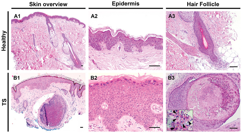
Histology, Trichodysplasia Spinulosa. The left column shows hematoxylin and eosin staining of healthy skin (A1) and trichodysplasia spinulosa lesional skin (B1) at low power. At high power, healthy (A2) and trichodysplasia spinulosa (B2) epidermis and hair follicles (A3 and B3) are depicted. An enlarged and dysmorphic hair follicle is evident in B3, containing eosinophilic granular protein deposits in the cytoplasm of cells (arrowheads in inset) with abrupt cornification in the follicle center. Bars represent 100 µm. Kazem S, van der Meijden E, Wang RC, et al. Polyomavirus-associated trichodysplasia spinulosa involves hyperproliferation, pRB phosphorylation and upregulation of p16 and p21. PLoS ONE. 2014;9(10):e108947. doi: 10.1371/journal.pone.0108947.

From: Anatomy, Skin (Integument), Epidermis

Cover of StatPearls
StatPearls [Internet].
Treasure Island (FL): StatPearls Publishing; 2024 Jan-.
Copyright © 2024, StatPearls Publishing LLC.

This book is distributed under the terms of the Creative Commons Attribution-NonCommercial-NoDerivatives 4.0 International (CC BY-NC-ND 4.0) ( http://creativecommons.org/licenses/by-nc-nd/4.0/ ), which permits others to distribute the work, provided that the article is not altered or used commercially. You are not required to obtain permission to distribute this article, provided that you credit the author and journal.

NCBI Bookshelf. A service of the National Library of Medicine, National Institutes of Health.