From: Chapter 36, Galectins

PDF files are not available for download.
NCBI Bookshelf. A service of the National Library of Medicine, National Institutes of Health.

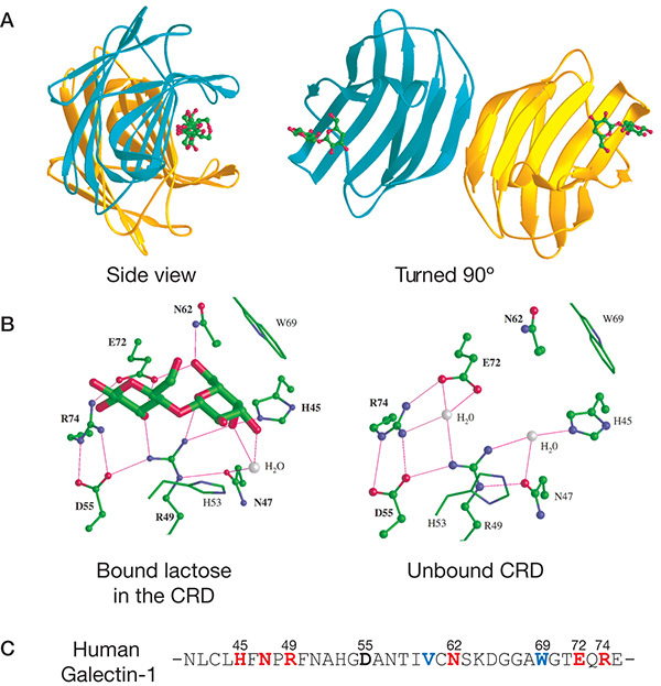
(A) Ribbon diagram of the crystal structure of human galectin-1 complexed with lactose. The homodimer is shown with each monomer colored differently and orthogonal views are presented. The subunit interface is based on interactions between the carboxy- and amino-terminal domains of each subunit. (B) A highlight of the interactions of key amino acid residues within the carbohydrate-recognition domain (CRD) with bound lactose, and the partial sequence of the CRD in human galectin-1. (C) Primary sequence of human galectin-1 with residues numbered and corresponding to those in the crystal structure. (Illustration kindly provided by Dr. Sean R. Stowell.)
Download Teaching Slide (PPTX, 2.0M)
From: Chapter 36, Galectins

PDF files are not available for download.
NCBI Bookshelf. A service of the National Library of Medicine, National Institutes of Health.